INTRODUCTION
A favorable long term result about osseointegrated implant has been reported since first Bra�nemark system report.1On the basis of these long term results, treatment of partial and total edentulism with dental implants has become an accepted treatment in dentistry.2-4Implant that was used in the beginning was mainly external connection type, but internal connection type that is represented by Astra Tech Implant System has been gradually increased. Numerous experimental and clinical studies have shown favorable results for the internal conical type.
The high success rate has been attributed to the formation of a direct bone-implant interface and maintenance of stable bone to implant contact after loading. So, many researchers devel- oped new implant surface and design. Some studies have reported that the microthread configuration offered improved conditions for osseointegration.5-7And, it has suggested that the rate and degree of osseointegration were superior for the rough surface compared with the turned one.8However, there are some biologic factors associated with failure of osseoin- tegration except implant design and surface property.9-11So, implant losses can occur, early or lately. In addition to above representing factors, demographic variables, health-status,
A 5-year retrospective clinical study of the Dentium implants
Jeong-Yol Lee1,2, DDS, MSc, PhD, Hyo-Jin Park2, DDS, MSc, Jong-Eun Kim2, DDS, MSc, Yong-Geun Choi2, DDS, MSc, Young-Soo Kim2,3, DDS, MSc, PhD, Jung-Bo Huh4, DDS, MSD, Sang-Wan Shin5*, DDS, MPH, PhD, MSc
1Department of Prosthodontics, Institute for Clinical Dental Research, Guro Hospital, Korea University Medical Center, Seoul,
2Advanced Prosthodontics, Postgraduate School of Clinical Dentistry, Institute for Clinical Dental Research, Korea University, Seoul,
3Department of Preventive Dentistry, Guro Hospital, Korea University Medical Center, Seoul,
4Department of Prosthodontics, School of Dentistry, Pusan National University, Pusan,
5Advanced Prosthodontics, Postgraduate School of Clinical Dentistry, Institute for Clinical Dental Research, Korea University Medical Center, Korea University, Seoul, Korea
PURPOSE. The aim of this retrospective study was to evaluate cumulative survival rate (CSR) of Implantium implants followed for 5 years and association between risk factors and the CSR. MATERIALS AND METHODS. A total of two hundred forty-nine Implantium Implants System (Dentium, Seoul, Korea) placed in ninety-five patients from 2004 to 2009 were investigated with several identified risk factors (sex, sys- temic disease, smoking, alchohol, reason of tooth loss, length, arch (maxilla or mandible), replace tooth type (incisor, canine, premolar or molar) Kennedy classification, prosthodontic type, prosthodontic design, opposite dentition, abutment type, occlusal material, occlusal unit, splint to tooth, cantilever, other surgery). Clinical examination (mobility, percussion, screw loosening, discomfort, etc.) and radiographic examination data were collected from patient records including all problems during follow-up period according to protocols described earlier. Life table analy- sis was undertaken to examine the CSR. Cox regression method was conducted to assess the association between potential risk factors and overall CSR. RESULTS. Five of 249 implants were failed. Four of these were lost before loading. The 5-year implant cumulative survival rate was 97.37%. Cox regression analysis demonstrated a significant predictive association between overall CSR and systemic disease, smok- ing, reason of tooth loss, arch, Kennedy classification and prosthodontic design (P<.05). The screw related complication was rare. Two abut- ment screw fractures were found. Another complications of prosthetic components were porcelain fracture, resin facing fracture and denture fracture (n=19). CONCLUSION. The 5-year CSR of Implantium implants was 97.37 %. Implant survival may be dependent upon systemic dis- ease, smoking reason of tooth loss, arch, Kennedy classification and prosthodontic design (P<.05). The presence of systemic diseases and combination of other surgical procedures may be associated with increased implant failure. [J Adv Prosthodont 2011;3:229-35]
KEY WORDS: Dental implant; Survival rate; Risk factors
Corresponding author: Sang-Wan Shin
Institute for Clinical Dental Research, Korea University, Guro Hospital 97 Gurodong-gil, Guro-gu, Seoul, 152-703, Korea
Tel. 82 2 2626 1922: e-mail, [email protected]
Received November 9, 2011 / Last Revison December 3, 2011 / Accepted December 8, 2011
ⓒ 2011 The Korean Academy of Prosthodontics
This is an Open Access article distributed under the terms of the Creative Commons Attribution Non-Commercial License (http://creativecommons.org/licenses/by- nc/3.0) which permits unrestricted non-commercial use, distribution, and reproduction in any medium, provided the original work is properly cited.
tobacco use, and anatomic location are identified as factors that increase the risk for implant losses. However some studies have shown that individual medical problems do not correlate with increased implant failure.12,13 Because of conflicting data from studies, clinicians are unable to provide concrete answer to questions posed by patients seeking dental implant treatment.
When new implant systems are introduced commercially, it is essential that the long-term clinical results added to the ani- mal studies are reported as scientific publications. In South Korea, although the development of dental implant is increased, few clinical studies have demonstrated the clinical performance of dental implants among South Korean population.14-16Also, in the studies of risk factor relation to implant failure, as the subject of previous studies were different races to Korean, accord- ing to arch form, dietary pattern, bone quality and clenching force, the reported results are different. Therefore, this study was aimed to evaluate the long-term clinical outcome of the implants for 5 years, and to determine the risk factors of implant failure in Korean population.
MATERIALS AND METHODS
During the period from 2004 to 2009, a total of 249 Dentium implants (Dentium Co., Seoul, Korea) were placed consecu- tively in 95 patients at the Korea University Guro Hospital. Data was collected using patient’s clinical chart. Implants loaded immediately after Stage I surgery and splinted with other implant system were ruled out as an exclusion criteria.
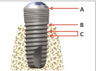
The Dentium implants’interface between abutment and fix- ture was mediated via a conical seal design that distributes the load to the surrounding cortical bone evenly and prevent screw loosening. The implants had a microthread configura- tion in the cervical portion, with SLA (Sandblasting with Large grit and Acid etching) surface (Fig. 1).
All patients were treated according to the Adell’s standard surgical protocol.17Additional surgical technique (Sinus aug- mentations, bone regeneration etc.) were used for 80 implants.
Patients were seen weekly or biweekly for the first 2 months after implant placement and the monthly for 4 months. Any pres- sure exerted by the temporary prosthesis was relieved. After 3 - 6 months, the implant was uncovered, healing abutments were placed at least for 1 month, and after that time, the final prosthetic procedure was performed.
Clinical examination (mobility, percussion, screw loosening, discomfort, etc.) and radiographic examination data were collected from patient’s record including all problems during follow-up period according to protocols described earlier.
The criteria for survival according to Albrektsson and Zarb18 were used to define implant performance. The survival rates were calculated by using a modification of the success crite- ria suggested by Albrektsson and colleagues.19So, implant fail- ure, defined as removal and sleeping of the implant was the pri-
mary outcome variable. The patients that moved or died were censored. The time between implant placement and the date of the last follow-up or implant failure was used to cal- culate the duration of implant survival in months.
The risk factors that could affect an implant survival rate were grouped into the following categories12,13:
1. The patient’s age at time of implant placement (years) and gender were recorded.
2. Health-status: Whether patients had systemic diseases (diabetes, hypertension, cardiovascular diseases etc), smoking status (non smoker, past smoker or current smoker) and alcohol history were recorded.
3. Implant variables: implant size (length, diameter), implant type and number of implants
4. Anatomic variables: Kennedy classification (tooth borne, distal extension), implant location (maxilla, mandible, ante- rior, posterior, tooth number)
5. Prosthesis variables: grouped according to prosthesis type, the kind of abutment, prosthetic design (single vs.
splinted implants), occlusal materials, opposing dentition, and number of occlusal units in the prosthesis.
6. The reasons of tooth loss were included.
Intraoral periapical radiographs were obtained annually using parallel technique after prosthesis placement. The images were evaluated for peri-implant radiolucency and vertical bone loss. To evaluate the marginal bone level, the dis- tance from a reference point at the implant to the most coro- nal point (a) where the marginal bone meets the implant (b) was measured in 0.1 mm increments. Measurements were made mesially and distally of each implant. For calibrating mag- nification, the ratio of real inter-thread width (0.6 mm) to inter- thread width (c) of radiograph was used (Fig. 2).
Kaplan-Meier estimate of implant survival was undertaken Fig. 1. Dentium implant (Dentium Co., Seoul, Korea).
to examine CSR. Multiple Cox proportional hazards regres- sion and log-rank test was used to assess the influence of poten- tial risk factors on implant survival after adjusting for confounders.
The level of statistical significance was set as α=.05.
RESULTS
The mean duration of clinical follow-up time of implants was 5.1 years. A total 95 patients were treated using 249 implants to support or retain dental prostheses. Patient ages ranged from 20 to 75 years (mean 56.8 years) (Fig. 3), and 42 patients report- ed general health problems (43.2%). The descriptive statistics are summarized in Table 1. The most frequently used implants lengths were 12 mm (43.0%) and 10 mm (35.7%). Diameters of the implants were mainly 3.8 mm and 4.3 mm. 135 (54.2%) implants were placed in the mandible and 114 (45.8%) in the maxilla. Regarding the prostheses used, most of the implants supported a fixed partial denture (67.5%) and 38 implants had a single crown (19.3%). The greater part of fixed bridge type was screw retained prosthesis (45.7%). Most implants (174) were opposed to natural teeth or implants (88.8%).
Implant Survival Rates and Loss
Of the 249 implants, 5 implants failed. Four of these were removed between placement and loading. Other one implant was lost after loading for 2 years as a result of a horizontal fix- ture fracture. Two of 4 failed implants were removed and replaced with new fixtures before loading forces were applied. And one failed implant after loading was replaced with a new one within 3 months. A life table analysis is present in Table 2 show- ing 97.37% cumulate survival rate after 5 years of follow up
(Table 2).
Risk Factors
Occlusal material, prosthesis design, Kennedy classification, arch, the reason of tooth loss, smoking and systemic dis- eases were statically associated with implant failure (Table 3).
The patient who had porcelain occlusal materials, single type prosthesis design, tooth borne case, implant in maxilla site, the tooth loss as a result of failed endodontic treatment, and sys- temic diseases is more likely to experience implant failure than the other patients. There was no significant association between other risk factors and failure.
Marginal Bone Level
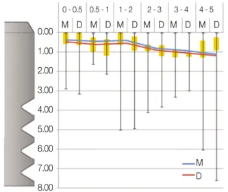
Implants remaining in the 95 patients at the 5-year revealed no signs of persisting peri-implant radiolucencies. Evaluation of the radiographs showed that an annual average bone loss each mesial and distal side was 0.18 mm and 0.19 mm after load- ing. The mean value in mm and standard deviations (SD) of the marginal bone changes from baseline up to the 1-year after loading were 0.41 (0.48) / 0.58 (0.65) (Table 4) (Fig. 4).
Complications
There were few complications related to peri-implantitis (2%).
The most common prosthetic complication encountered was fracture of the resin veneer and porcelain. One abutment screw was fractured in the case using single dual abutment. The other problem related to screw was loosening of gold and abut- ment screw (Table 5). All complications were easily solved.
Fig. 2. Reference point for measuring of marginal bone loss.
A: junction between implant machined collar bevel and rough sur- face, B: implant to marginal bone contact level, C: inter-thread distance.
Fig. 3. Age distribution of the patient at the implant installation.
50 40 30 20 10 0 N
20 30 40 50 60 70
Table 1. Distribution of implant according to variables
Variable Implant data (%)
Gender Male 57
Female 43
Medication status Healthy 56.8
Chronic conditions 43 .2
Reason of tooth loss Caries 10.8
Periodontal disease 67.9
Trauma 1.6
Congenital missing 1.2
Failed endo 3.2
Others 15.3
Length (mm) 8 6.0
10 35.7
12 43.0
14 15.3
Width (mm) 3.0 & 3.4 8.0
3.8 41.8
4.3 32.5
4.8 13.3
5.0 & 6.0 4.4
Arch Maxilla 45.8
Mandible 54.2
Kennedy classification Distal extension 65.5
Tooth bounded 24.0
Edentulous 10.5
Tooth type Incisor 12.0
Canine 5.6
Premolar 21.3
Molar 61.0
Variable Implant data (%)
Other surgery Yes 67.9
No 32.1
Prosthesis type Single type 19.3
Fixed partial denture 67.5 Removable partial denture 0.0
Overdenture 3.1
Hybrid type 6.1
Full fixed bridge 4.1
Prosthesis design Single type 19.2
Splinting type 80.8
Opposing dentition Natural or FPD 88.8 RPD or overdenture 7.1
CD 4.0
Abutment Combi abutment 2.0
Dual abutment 33.0
Screw abutment 45.7
Angled abutment 4.6
UCLA 10.7
Others 4.1
Occulsal material Gold 77.1
Porcelain 11.7
Resin 11.2
Splint to tooth Yes 99.0
No 1.0
Cantilever Yes 84.3
No 15.7
Table 2. Life table analysis showing cumulative survival rates
Time period Survived implants Adjusted denominator Censored implants Failed implant SR of interval Cumulative SR
at beginning (N) for Interval during interval (N) (N) (%) (%)
P - L 249 224.5 49 4 98.22 98.22
L - 1y 196 163 66 0 100 98.22
1 - 2 130 115.5 29 1 99.13 97.37
2 - 3 102 85.5 33 0 100 97.37
3 - 4 69 64 10 0 100 97.37
4 - 5 59 24.5 59 0 100 97.37
Table 3. Log-Rank test associated with implant failure
Exposure Risk ratios Prob > Chi Sq
Occlusal material Porcelain 188654.3 0.0447
Prosthesis design Single type 7236.3 0.0093
Kennedy classification Tooth bounded 281.0 0.0091
Distal extension 25.4
Arch Maxilla 2052.3 0.0222
Reason of tooth loss Failed endo 190046.6 <.0001
Periodontal disease 12018.8
Smoking Past smoker 155268.8 0.0004
Systemic disease Diseased patients 1790.2 0.0484
DISCUSSION
This study was to document the long-term clinical out- come of the Dentium implants for 5 years, to evaluate this sys- tem according to the criteria for survival according to Albrektsson and Zarb18and to determine the risk factors of implant failure in Korean population. In a total of 249 implants, four implants failed in the early postsurgical heal- ing period. One implant was removed after 2 years in function period due to implant fixture fracture. It would be probably asso- ciated with over load. The reasons of early failures of implants could be bone necrosis, infection of bacteria, bone quality, micro- movement after placement, early loading and weak initial sta- bility. Meanwhile, the late failure might be affected by a poor oral hygiene, excessive occlusal load, misfit of prosthesis and so on. Esposito et al. suggested that major etiologic fac- tors of late failure was excessive occlusal load related to parafunction such as bruxism and infection. Also patient’s immune reaction and implant surface properties could influ- ence a failure.9-11The overall 5-year cumulated survival rate in this study was reported 97.37%, which was similar to prior reports of TiUnite Implant system and Astra system.20,21Adell et al.
reported that failed rate in each interval was the highest within 1 to 2 years and was decreasing after that due to increasing stability.17
General health problems and past smoking history increased the risk of implant failure. This result was similar to other stud- ies. Moy et al.22reported that patients over age 60, smoked, had
a history of diabetes or postmenopausal, increased implant fail- ure compared with healthy patient. They suggested that smoking caused both systemic and local injury to tissues and is common contributor to decreased tissue oxygenation, which negatively affects wound healing. The diabetes could delay the wound healing response following-implant insertion and may be related to mechanical characteristics of the bone- to-implant contact.23
Among the reason of tooth loss, failed endodontic treatment and periodontitis increased risk of implant failures. Remaining infection might be the cause of the failure. There are no pre- vious studies about relationship between failed endodontic treat- ment and implant survival rate. However, some studies report- ed about periodontitis patients. The patient who had treated a periodontitis had a lower survival rate with increased marginal bone resorption than a patient in a healthy condition.24-26
All removed implants were initially placed in maxilla, so implants placed in maxilla were also associated with an increased risk for failure compared to implants placed in mandible. There is no significant difference between anteri- or part and posterior part. Many studies reported similar results. Recent research showed that average cumulative suc- cess rate in maxilla was higher than in mandible.22,27Tolstunov represented about implant location and related success rate. His findings were that implant location seemed to be significant factors in a success-analysis of dental implants.28
In present studies, implant length and width are not associated with risk factor. These results conflict with earlier findings. Some Table 4. Cumulative mean marginal bone loss (CMBL)
Follow-up after (Y) Number CMBL (mm)
[M/D] [M (SD)/D (SD)]
L - 0.5 100/100 0.35 (0.59)/0.44 (0.71) 0.5 - 1 23/23 0.41 (0.48)/0.58 (0.65) 1 - 2 46/47 0.37 (0.38)/0.53 (0.52) 2 - 3 27/27 0.79 (1.01)/0.82 (1.24) 3 - 4 28/29 0.92 (0.73)/1.03 (0.65)
4 - 5 8/8 1.13 (2.23)/1.20 (2.61)
L: Loading, M: Mesial, D: Distal, SD: Standard Deviation
Fig. 4. Box plot chart with the mean marginal bone level change (blue and red line). A black line shows a difference between the maximum and minimum bone level change.
0.00 1.00 2.00 3.00 4.00 5.00 6.00 7.00 8.00
M D M D M D M D M D M D M D 0 - 0.5 0.5 - 1 1 - 2 2 - 3 3 - 4 4 - 5
Table 5. Number of complications after loading
Complication type Number
None 163
Mucosa related Inflammation/Hyperplasia 6
Gingival recession 4
Screw loosening Abutment screw 2
Gold screw 5
Fractures Resin veneer 11
Porcelain 6
Abutment screw 1
studies reported that implant length influence the implant survival rate.29,30 And some studies discussed that implant diameter affected survival of implant.31-33These differences may be due to other variables affecting implant survival, including the implant surface, the surgeon’s learning curve, bone qual- ity and quantity.
Cumulative marginal bone level alteration adjacent to the fix- ture has been documented over the 5 years. Changes in mar- ginal bone level between baseline and annual follow-up were calculated. In most implants, the marginal bone level showed very small changes over the follow-up period. The mean difference of bone level changes in each mesial and distal side were 0.41 (0.48) / 0.58 (0.65) in the 1styear and 1.13 (2.23) / 1.20 (2.61) in the 5thyear as compared from the base line. An annual average bone loss each mesial and distal side was 0.18 mm and 0.19 mm after loading. It is lower than 0.2 mm per year, which is in accordance with Albrektsson’s criteria19, indicating acceptable bone level maintenance around implant. But some patients underwent an aggressive bone resorp- tion. It had to be considered with relation to risk factors.
Intraoral radiograph is still one of the most sensitive ways of identifying loss of bone support. In the present study, there are some limitations on accurate information about bone level changes. The parallel intraoral radiograph taking technique was used for projection angle of 90�to fixture axis, but a specially constructed film-holder was not used for this study. And intraoral radiographs disclose bone status from mesial and dis- tal aspects only.
The 5 year CSR of overall implants was 97.37%. However, there are a few limitations in this study. Because of retrospective study, the sampling was limited and the nature and quality of the predictor variables and confounding variables are depen- dent on the accuracy and quantity of the past record. In addi- tion, the number of patients and implants were not large enough and this study was undertaken in one hospital.
However, the results could be accepted for the clinical research, because it is essential to be supported with a mini- mum of 50 consecutive patients and in at least 5 years.
Further study is necessary to evaluate the relationship between patient oral hygiene and soft tissue reactions. If well-controlled prospective study is perform, more favor- able result might be achieved.
CONCLUSION
1. Of the 249 implants, 5 implants were failed and the 5-year CSR of Dentium implant system was 97.22%.
2. Implant survival may depend on systemic disease, smok- ing, reasons of tooth loss, arch, the edentulous site (by Kennedy classification) and prosthodontic design.
3. An annual average bone loss at each mesial and distal side was 0.18 mm and 0.19 mm.
4. There were few complications related to peri-implantitis and the most common prosthetic complication encountered was fracture of the resin veneer and porcelain.
REFERENCES
1. Bra�nemark PI, Hansson BO, Adell R, Breine U, Lindstro¨m J, Halle′n O, Ohman A. Osseointegrated implants in the treat- ment of the edentulous jaw. Experience from a 10-year period.
Scand J Plast Reconstr Surg Suppl 1977;16:1-132.
2. Fugazzotto PA, Gulbransen HJ, Wheeler SL, Lindsay JA. The use of IMZ osseointegrated implants in partially and com- pletely edentulous patients: success and failure rates of 2,023 im- plant cylinders up to 60+ months in function. Int J Oral Maxillofac Implants 1993;8:617-21.
3. Wedgwood D, Jennings KJ, Critchlow HA, Watkinson AC, Shepherd JP, Frame JW, Laird WR, Quayle AA. Experience with ITI osseointegrated implants at five centres in the UK. Br J Oral Maxillofac Surg 1992;30:377-81.
4. Leimola-Virtanen R, Peltola J, Oksala E, Helenius H, Happonen RP. ITI titanium plasma-sprayed screw implants in the treatment of edentulous mandibles: a follow-up study of 39 patients. Int J Oral Maxillofac Implants 1995;10:373-8.
5. Palmer RM, Palmer PJ, Smith BJ. A 5-year prospective study of Astra single tooth implants. Clin Oral Implants Res 2000;
11:179-82.
6. Wennstro¨m JL, Ekestubbe A, Gro¨ndahl K, Karlsson S, Lindhe J. Implant-supported single-tooth restorations: a 5-year prospec- tive study. J Clin Periodontol 2005;32:567-74.
7. Abrahamsson I, Berglundh T. Tissue characteristics at mi- crothreaded implants: an experimental study in dogs. Clin Implant Dent Relat Res 2006;8:107-13.
8. Abrahamsson I, Berglundh T, Linder E, Lang NP, Lindhe J. Early bone formation adjacent to rough and turned endosseous implant surfaces. An experimental study in the dog. Clin Oral Implants Res 2004;15:381-92.
9. Esposito M, Hirsch JM, Lekholm U, Thomsen P. Biological fac- tors contributing to failures of osseointegrated oral implants. (II).
Etiopathogenesis. Eur J Oral Sci 1998;106:721-64.
10. Esposito M, Hirsch JM, Lekholm U, Thomsen P. Biological fac- tors contributing to failures of osseointegrated oral implants. (I).
Success criteria and epidemiology. Eur J Oral Sci 1998;106:527- 51.
11. Esposito M, Thomsen P, Ericson LE, Sennerby L, Lekholm U.
Histopathologic observations on late oral implant failures. Clin Implant Dent Relat Res 2000;2:18-32.
12. Smith RA, Berger R, Dodson TB. Risk factors associated with dental implants in healthy and medically compromised pa- tients. Int J Oral Maxillofac Implants 1992;7:367-72.
13. Matukas VJ. Medical risks associated with dental implants. Int J Oral Implantol 1988;5:49-50.
14. Kim SH, Kim S, Lee KW, Han DH. The effects of local factors on the survival of dental implants: A 19 year retrospective study. J Korean Acad Prothodont 2010;48:28-40.
15. Ko SM, Lee JK, Eckert SE, Choi YG. Retrospective multicenter cohort study of the clinical performance of 2-stage implants in South Korean populations. Int J Oral Maxillofac Implants 2006;21:785-8.
16. Doh RM, Moon HS, Shin JS, Lee KW. Retrospective study of the implantium(R) implant with a SLA surface and internal con- nection with microthreads. J Korean Acad of Prothodont 2009;
47:136-47.
17. Adell R, Lekholm U, Rockler B, Bra�nemark PI. A 15-year study of osseointegrated implants in the treatment of the eden- tulous jaw. Int J Oral Surg 1981;10:387-416.
18. Albrektsson T, Zarb GA. Current interpretations of the os-
seointegrated response: clinical significance. Int J Prosthodont 1993;6:95-105.
19. Roos J, Sennerby L, Lekholm U, Jemt T, Gro¨ndahl K, Albrektsson T. A qualitative and quantitative method for eval- uating implant success: a 5-year retrospective analysis of the Bra�nemark implant. Int J Oral Maxillofac Implants 1997;12:504- 14.
20. Glauser R, Lundgren AK, Gottlow J, Sennerby L, Portmann M, Ruhstaller P, Ha¨mmerle CH. Immediate occlusal loading of Bra�nemark TiUnite implants placed predominantly in soft bone: 1-year results of a prospective clinical study. Clin Implant Dent Relat Res 2003;5:47-56.
21. Rasmusson L, Roos J, Bystedt H. A 10-year follow-up study of titanium dioxide-blasted implants. Clin Implant Dent Relat Res 2005;7:36-42.
22. Moy PK, Medina D, Shetty V, Aghaloo TL. Dental implant fail- ure rates and associated risk factors. Int J Oral Maxillofac Implants 2005;20:569-77.
23. Fiorellini JP, Nevins ML, Sekler J, Chung A, Oringer RJ.
Correlation of peri-implant health and aspartate aminotransferase levels: a cross-sectional clinical study. Int J Oral Maxillofac Implants 2000;15:500-4.
24. Stanford C. Dental implant outcomes may vary in patients with a history of periodontal disease. J Evid Based Dent Pract 2010;10:46-8.
25. Schou S, Holmstrup P, Worthington HV, Esposito M. Outcome of implant therapy in patients with previous tooth loss due to pe-
riodontitis. Clin Oral Implants Res 2006;17:104-23.
26. Ong CT, Ivanovski S, Needleman IG, Retzepi M, Moles DR, Tonetti MS, Donos N. Systematic review of implant outcomes in treated periodontitis subjects. J Clin Periodontol 2008;35:438- 62.
27. Bass SL, Triplett RG. The effects of preoperative resorption and jaw anatomy on implant success. A report of 303 cases. Clin Oral Implants Res 1991;2:193-8.
28. Tolstunov L. Implant zones of the jaws: implant location and re- lated success rate. J Oral Implantol 2007;33:211-20.
29. Wyatt CC, Zarb GA. Treatment outcomes of patients with im- plant-supported fixed partial prostheses. Int J Oral Maxillofac Implants 1998;13:204-11.
30. Jemt T, Lekholm U. Implant treatment in edentulous maxillae:
a 5-year follow-up report on patients with different degrees of jaw resorption. Int J Oral Maxillofac Implants 1995;10:303-11.
31. Shin SW, Bryant SR, Zarb GA. A retrospective study on the treat- ment outcome of wide-bodied implants. Int J Prosthodont 2004;17:52-8.
32. Eckert SE, Meraw SJ, Weaver AL, Lohse CM. Early experience with Wide-Platform Mk II implants. Part I: Implant survival. Part II: Evaluation of risk factors involving implant survival. Int J Oral Maxillofac Implants 2001;16:208-16.
33. Attard NJ, Zarb GA. Implant prosthodontic management of par- tially edentulous patients missing posterior teeth: the Toronto ex- perience. J Prosthet Dent 2003;89:352-9.