Introduction
The placement of endosseous implants in the poste- rior maxilla is often complicated by maxillary sinus pneumatization, resulting in lack of supporting bone.
Therefore grafting procedures have been developed to increase the amount of alveolar bone. The Sinus graft (previously designated“sinus lift”), introduced by Tatum1) and Boyne and James2), is a relatively new
procedure now being used with increasing frequency3). Numerous clinical studies have reported the clinical outcomes of placing implants in the augmented maxil- lary sinus3-8). In the grafted sinus, reports of implant survival under functional loading varied from 36% to 61.7%5,8), even reaching 100% in recent meta-analysis8).
Autogenous bone has been considered the gold stand- ard graft material because of its osteoinductive and os- teoconductive properties6). The healing period for sinuses grafted with autogenous bone can be as short as 3 to 4 months versus the 8 to 10 months often recommended for bone substitutes2,9,10). Adding autogenous bone to other graft materials also can shorten healing times9,11). But its use is limited by donor-site morbidity, sparse avail- ability, and uncontrolled resorption. Therefore many al-
Effect of maxillary sinus graft on the survival of endosseous implants: A 10-year retrospective study
Hye-Ran Jeon
1,2, Eun-Kyoung Pang
2, Ah-Ran Pae
3, Myung-Rae Kim
1,4, Na-Ra Kang
1,4*1. Ewha Womans University Graduate School of Clinical Dentistry Department of Implant Dentistry 2. Ewha Womans University School of Medicine Department of Periodontology
3. Ewha Womans University School of Medicine Department of Prosthodontics
4. Ewha Womans University School of Medicine Department of Oral and Maxillofacial Surgery
ABSTRACT
Purpose: The aim of this study was to determine the survival rates of implants placed in grafted maxillary sinuses and compare the results obtained with graft materials, implant surfaces and timing of implant placement.
Materials and Methods: Between January 1996 and December 2005, 391 implants were placed in 161 patients who underwent sinus grafting treatment simultaneously or separately at Ewha Womans University Hospital. According to inclusion criteria, 272 implants were placed in 102 patients with 112 sinus grafts (30 females, 72 males), aged 26 to 88 years (mean age 49.0±9.7). The follow-up period ranged from 12 to 134 months (mean F/U 47±32). Survival rates were evaluated according to graft material, implant surface and timing of implant placement. The Kaplan-Meier procedure and the log rank (Mantel-Cox) test were used to estimate survival rates and test for equality of survival rates between different groups of patients.
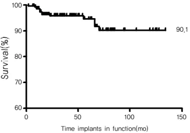
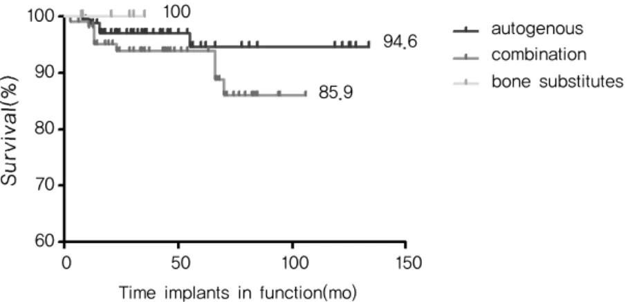
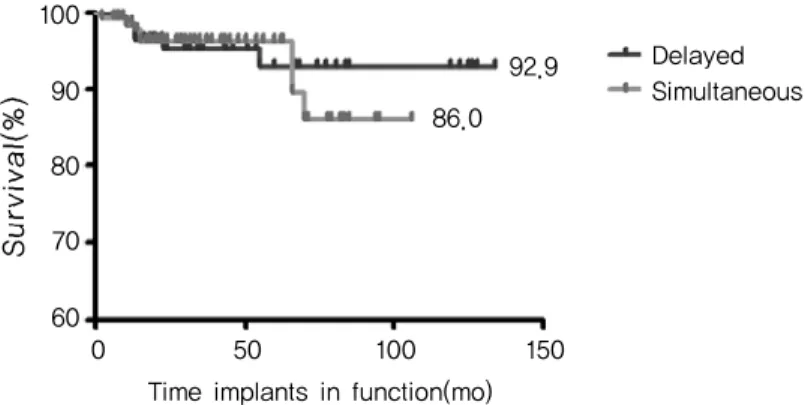
Results: Ten-year cumulative survival rate for implants placed in the grafted sinuses was 90.1%. The survival rates for autogenous bone, combination and bone substitutes were 94.6%, 85.9% and 100%, respectively (p > 0.05). According to implant surface, survival rates were 84.8% in machined group and 97.5% in rough group (p < 0.05). The survival rates were 92.9% in delayed group and 86.0% in simultaneous group (p > 0.05).
Conclusion: Ten-year cumulative survival rate for implants placed in the grafted sinuses was 90.1%. Rough-surfaced implants have a higher survival rate than machined-surfaced implants when placed in grafted sinuses (p < 0.05).
(J Korean Acad Periodontol 2008;38:309-316)
KEY WORDS: Sinus graft; implant; survival; surface characteristics.
Correspondence: Na-Ra Kang, DDS, Ph.D
Ewha Womans University, School of Medicine, Department of Oral and Maxillofacial Surgery, 204 Anyangcheon-gil, Yangcheon-gu, Seoul, 158-710, South Korea
e-mail: [email protected], Tel: 82-505-336-8478, Fax: 82-2-2650-5764
* The titanium disks were provided by Osstem (Pusan, Korea).
Received: Sep 5, 2007; Accepted: Dec 13, 2007
lografts, xenografts and alloplastic graft materials have been used alone or in combination with autogenous bone to graft maxillary sinuses. The advantage of using bone substitutes is that a second surgical site is not needed.
But it is not osteoinductive and does not contain osteo- progenitor cells. The 1996 Sinus Consensus Conference reported similar success rates for implants placed in si- nus grafts using different materials3).
Implant surface texture may influence the process of early bone formation around implants. Surface roughness increases the surface area for osseointegration.
A more favorable implant-bone interface is estab- lished on rough-surfaced implants than on implants with a machined surface12-14).
The sinus graft procedure is referred to as one-stage when the implants and graft are placed simultaneously, while in the two-stage procedure, implant placement is delayed for several months to allow for graft maturation.
The decision is made based on the amount of bone present at the alveolar crest. Less than 4mm is consid- ered insufficient endosteum to mechanically maintain the implants, and the two-stage procedure is recom- mended15). However, Peleg et al.16) reported 100% im- plant survival in simultaneous placements with 1 to 2mm of crestal bone. Implant success rates for one-stage procedures ranged between 64% to 98%17-19) and two-stage success rates were 92% to 100%20-22). In studies that did not differentiate between simultaneous and delayed implant placement, success rates were between 88% and 90%23,24).
The aim of this study was to determine the survival rates of implants placed in grafted maxillary sinuses and compare the results obtained with graft materials, implant surfaces and timing of implant placement.
Materials and Methods
Between January 1996 and December 2005, 391 im- plants were placed in 161 patients who underwent si- nus grafting treatment simultaneously or separately at Ewha Womans University Hospital. According to in- clusion criteria, 272 implants were placed in 102 pa-
tients with 112 sinus grafts (30 females, 72 males), aged 26 to 88 years (mean age 49.0±9.7). The follow -up period ranged from 12 to 134 months (mean F/U 47±32). The inclusion criteria were followed: (1) a minimum follow-up period of 1 year after functional loading; (2) sinus graft procedures were performed alone; (3) access to antrum occurred by the lateral window procedure; (4) graft material, type of implants used and timing of implant placement were clearly re- corded; (5) surgery and prosthodontics were performed within the same clinic.
Implant survival rates according to the following variables:
1. Type of graft material
Patients in this study could be allocated to at least one of three groups: (1) autogenous bone alone (n=151); (2) autogenous bone in combination with bone substitutes (n=106); or (3) bone substitutes alone (n=15). In most cases autogenous bone was used in particulate chips. Large autogenous cancellous bone grafts were harvested from the superior anterior me- dial part of the iliac crest (n=53). When smaller amounts of bone were sufficient for grafting, they were taken from the mandibular symphyseal area (n=34), mandibular retromolar area (n=87) or the maxillary tuberosity (n=125). In the group using com- bination grafts, two graft materials were used with autogenous bone: allograft (Dembone®, Pacific Coast Tissue Bank, Los Angeles, USA; 9 cases) and xeno- graft (Bio-Oss®, Geistlich Pharm AG, Wolhausen, Switzerland; 97 cases). In the group using only bone substitutes, previous two materials were used: allo -graft (2 cases) and xenograft (13 cases).
2. Type of implant surface
This category consisted of two groups: (1) machined surface; or (2) rough surface, regardless of degree and type of roughness.
3. Timing of implant placement
This category comprised two groups: (1) simulta- neous procedure; or (2) delayed procedure. In delayed procedure the bone graft was allowed to consolidate for 5~9 months.
Implant survival described as presence of implants.
The success proposed by Buser et al25,26) and Cochran et al27) were used at each recall. They included: (1) ab- sence of clinically detectable implant mobility, (2) ab- sence of pain or any subjective sensation, (3) absence of recurrent peri-implant infection and (4) absence of continuous radiolucency around the implant.
Statistical analysis
The Kaplan-Meier procedure was used to estimate survival rates. The log rank (Mantel-Cox) test was performed to test for equality of survival rates be- tween different groups of patients using GraphPad Prism version 5.00 for Windows (GraphPad Software, San Diego, USA).
Results
Out of 272 implants, 14 implants were failed. The 10-year cumulative survival rate was 90.1% (Fig. 1).
Most failures (10 out of 14) occurred within 2 years after implant placement. The failures were divided in- to six early failures before loading and eight late failures after loading. No failure occurred after 70 months. 13 patients (28 implants) were considered as drop-out because of moving without leaving changed phone numbers.
1. Type of graft material
The overall survival rates for autogenous bone, combination and bone substitutes alone were 94.6%
(59 patients, 151 implants, 5 failures), 85.9% (39 pa- tients, 106 implants, 9 failures) and 100% (6 patients, 15 implants, no failure), respectively (Fig. 2). The use of different filling materials apparently did not sig- nificantly influence survival rates of implants (p>0.05).
Figure 1. The 10-year cumulative survival rate: Kaplan-Meier analysis was performed to determine the im- plant survival curve for 272 implants placed between January 1996 and December 2005.
100
90
80
70
60
0 50 100 150
Time implants in function(mo)
90.1
2. Type of implant surface
Table 1 shows the distribution of implants according to surface characteristics. Implants with machined surfaces displayed a mean survival rate of 84.8% (30 patients, 76 implants, 10 failures); implants with
rough surfaces displayed a mean survival rate of 97.5% (77 patients, 196 implants, 4 failures). There was a statistically significant difference between the survival rates according to implant surface (p<0.05).
Table 1. Implant distribution according to surface
Machined surface Rough surface
Brånemark 60 Brånemark 76
3i 13 3i 36
Microvent 3 Osstem 28
Ankylos 22
Neoplant 19
Restore 12
CAMLOG 3
Figure 2. Overall implant survival rates according to graft material (p>0.05).
Figure 3. Overall implant survival rates according to implant surface (*p<0.05).
100
90
80
70
60
0 50 100 150
Time implants in function(mo)
autogenous combination bone substitutes 100
94.6
85.9
rough*
100
90
80
70
60
0 50 100 150
Time implants in function(mo)
machined 97.5*
84.8
3. Timing of implant placement
Fig. 4 presents the survival rates of implants placed according to either the simultaneous or delayed protocol. The overall implant survival rates were 92.9% (42 patients, 122 implants, 6 failures) for de- layed and 86.0% (67 patients, 150 implants, 8 failures) for simultaneous procedures (p>0.05).
Discussion
In this study, the 10-year cumulative survival rate was 90.1%. It is comparable with other studies. Baik7) reported that 6-year cumulative survival rates for machined-surfaced implants (Bårnemark, Nobel Biocare, Gӧteborg, Sweden) and acid-etched surfaced implants (Osseotite®, 3i Implant Innovations Inc., Florida, USA) in grafted maxillary sinuses were 82.3% and 86.7%, respectively. The 1996 Sinus Consensus Conference reported an overall survival rate of 90% at the 3-year time span3). This report included a meta-analysis of the data collected from 38 surgeons who performed 1,007 sinus grafts with 2,997 implants. Tong et al.4) published evidence- based reviews of the maxillary sinus grafts in their meta-analysis. The overall survival rate for the 1,096 implants included was 93% (follow-up: 6-60 months).
According to Del Fabbro et al.6), the overall implant survival rate was 91.49%. The database included 6,913
implants placed in 2,046 subjects with loaded follow-up time ranging from 12 to 75 months.
Wallace and Froum5) reported on 2,178 interventions and 5,267 placed implants with an overall survival rate of 91.8% utilizing only the lateral window technique.
In this study, different graft materials did not sig- nificantly influence on survival rates of implants (p>
0.05). Autogenous bone is considered the gold stand- ard for intraoral bone grafting. However, it has high- er morbidity including risk of neural disturbances due to possible lesions of the inferior alveolar nerve branches, and gait disturbances in case of harvesting from the iliac crest. Furthermore, autogenous bone grafts have been reported to have a history of greater than average resorption28), leading to subsequent sinus repneumatization and/or implant failure6). The use of non-resorbable or slowly resorbable grafting materials should prevent this phenomenon11,21). Bone substitutes appeared to be reliable for sinus floor elevation, with no significant differences in clinical outcomes and im- plant survival. In a study by Froum et al.9) implant survival rates for a xenograft when utilized with or without autogenous bone were similar. Several histo- logical studies9,29,30) showed that similar percentages of vital bone can be achieved in bone substitutes and in grafts with autogenous bone, provided the bone sub- stitutes are allowed to a longer maturation period.
Autogenous bone is the material of choice when sinus Figure 4. Overall implant survival rates according to timing of implant placement (p>0.05).
Time implants in function(mo) 100
90
80
70
60
0 50 100 150
Delayed Simultaneous 92.9
86.0
grafting procedures must be associated with onlay grafting of the maxilla in case of severe atrophy24,31).
In this study the survival rate of implants with rough surfaces is greater than that of implants with a machined surface (p<0.05). Clinical and histological studies show the superiority of implants with rough as compared with machined surfaces in the human poste- rior maxilla3,5,13,14). A rough implant surface may re- tain the blood clot in direct contact with the surface (contact osteogenesis), whereas the clot may retract away from a machined surface (distance osteo- genesis)32). Contact osteogenesis precedes and accel- erates osteogenic cell migration, and results in earlier bone formation on the implant surface. These phe- nomena lead to a more favorable implant-bone inter- face compared to distance osteogenesis. Thus the os- teoconductive nature of a rough implant surface may increase the rate at which bone forms on the implant surface, thereby allowing a reduction of the time in- terval between implant placement and functional load- ing33).
The implant survival rates for the simultaneous and delayed placement were 86.0% and 92.9%, respectively (p>0.05). Similar implant survival rates were reported with both procedures, in agreement with other stud- ies3,5). It is difficult to obtain reliable information concerning this topic. A previous review of the liter- ature concerning this topic showed lower survivals of implants when placed in conjunction with the grafting procedure3). On the other hand, it is also considered that the failure rate for delayed implants is influenced by the fact that delayed placement is more likely to be utilized in cases that had lesser height of residual crestal bone as opposed to simultaneous placements that are most likely to have a greater height of re- sidual crestal bone. In general residual crestal bone height is a primary consideration in choosing a simul- taneous over a delayed implant placement. Also sur- geonʼs skill and the length of delay may influence survival rates.
Ioannidou et al.15) suggested that the determination
of simultaneous or delayed procedure depends on the ability of the surgeon to place a fixed dental implant.
The distance between the threads of most threaded dental implants ranges from 0.65 to 0.80mm.
Therefore, in order to engage three threads, one must have at least 2.5mm of bone, and for five threads, about 4mm of bone. Most clinicians would prefer to have more than a few threads engaged in bone for a simultaneous sinus graft procedure. The 4- to 5-mm level is often suggested as a minimum by experienced sinus graft surgeons.
When comparing simultaneous grafting to implant placement with delayed approaches, the length of the delay also was found to be a factor34). A delay of 4 to 8 months compared with a delay of greater than 8 months demonstrated a much better success rate after 8 months with a 3-year survival rate of 97%. The shorter 4- to 8- month delay yielded a 3-year suc- cess rate of 84%3). These results suggest a clinical protocol for implant staging related to graft material selection.
There are so many variables - such as use of mem- brane, particulate versus block bone, smoking, sys- temic diseases, the variation among clinicians and so on - which could not be fully explored in data comparison. In most cases of present study particulate forms of graft materials and few or no membrane were used according to operators’ preference. A re- view by Wallace et al.5) found that the block grafting technique results in a statistically significant lower implant survival rate (83.3%) than do all particulate grafts combines (92.3%). Several studies supports the hypothesis that membrane utilization is a useful ad- junctive therapy that results in an increased survival rate for implants placed in sinus grafts9,35,36). For all possible questions, prospective studies with control of confounding factors are needed in future research be- cause retrospective studies are at a greater risk of bias.
In this study total 272 implants were evaluated ret- rospectively in 102 patients who underwent sinus
grafting treatment at Ewha Womans University Hospital between January 1996 and December 2005.
Survival rates of implants placed in grafted maxillary sinus were assessed according to graft materials, im- plant surfaces and timing of implant placement and several conclusions were drawn:
1. Ten-year cumulative survival rate for implants placed in the grafted sinuses was 90.1%.
2. Rough-surfaced implants have a higher survival rate than machined-surfaced implants when placed in grafted sinuses (p<0.05).
In this study, dental implants placed in the grafted sinuses were successful from surgical placement through long-term loading and function. This study presented that there was no statistically significant difference among the survival rates according to graft materials and timing of implant placement. Therefore more studies about implant design and surface char- acteristics are needed for improvement of survival rates for implants with sinus grafts. Also further prospective, well-controlled studies are needed to ac- count for the many variables related to sinus graft procedures.
References
1. Tatum H, Jr. Maxillary and sinus implant reconstructions.
Dent Clin North Am 1986;30:207-229.
2. Boyne PJ, James RA. Grafting of the maxillary sinus floor with autogenous marrow and bone. J Oral Surg 1980;38:
613-616.
3. Jensen OT, Shulman LB, Block MS, Iacono VJ. Report of the Sinus Consensus Conference of 1996. Int J Oral Maxillofac Implants 1998;13:11-45.
4. Tong DC, Rioux K, Drangsholt M, Beirne OR. A review of survival rates for implants placed in grafted maxillary sinuses using meta-analysis. Int J Oral Maxillofac Implants 1998;13:175-182.
5. Wallace SS, Froum SJ. Effect of maxillary sinus augmenta-
tion on the survival of endosseous dental implants. A sys- tematic review. Ann Periodontol 2003;8:328-343.
6. Del Fabbro M, Testori T, Francetti L, Weinstein R.
Systematic review of survival rates for implants placed in the grafted maxillary sinus. Int J Periodontics Restorative Dent 2004;24:565-577.
7. Joonhea Baik, Myungrae Kim. Effects of the sinus lift aug- mentation on the survival of the osseointegrated dental im- plants placed in the posterior maxilla. The Journal of Korean Academy of Oral and Maxillofacial Implantology 2001;5:10-24.
8. Graziani F, Donos N, Needleman I, Gabriele M, Tonetti M.
Comparison of implant survival following sinus floor aug- mentation procedures with implants placed in pristine pos- terior maxillary bone: a systematic review. Clin Oral Implants Res 2004;15:677-682.
9. Froum SJ, Tarnow DP, Wallace SS, Rohrer MD, Cho SC.
Sinus floor elevation using anorganic bovine bone matrix (OsteoGraf/N) with and without autogenous bone: a clin- ical, histologic, radiographic, and histomorphometric analy- sis--Part 2 of an ongoing prospective study. Int J Periodontics Restorative Dent 1998;18:528-543.
10. Wheeler SL, Holmes RE, Calhoun CJ. Six-year clinical and histologic study of sinus-lift grafts. Int J Oral Maxillofac Implants 1996;11:26-34.
11. Hallman M, Sennerby L, Lundgren S. A clinical and histo- logic evaluation of implant integration in the posterior maxilla after sinus floor augmentation with autogenous bone, bovine hydroxyapatite, or a 20:80 mixture. Int J Oral Maxillofac Implants 2002;17:635-643.
12. Buser D, Schenk RK, Steinemann S et al. Influence of sur- face characteristics on bone integration of titanium implants. A histomorphometric study in miniature pigs. J Biomed Mater Res 1991;25:889-902.
13. Lazzara RJ, Testori T, Trisi P, Porter SS, Weinstein RL. A human histologic analysis of osseotite and machined surfa- ces using implants with 2 opposing surfaces. Int J Periodontics Restorative Dent 1999;19:117-129.
14. Khang W, Feldman S, Hawley CE, Gunsolley J. A mul- ti-center study comparing dual acid-etched and ma- chined-surfaced implants in various bone qualities. J Periodontol 2001;72:1384-1390.
15. Ioannidou E, Dean JW. Osteotome sinus floor elevation and simultaneous, non-submerged implant placement: case report and literature review. J Periodontol 2000;71:1613-1619.
16. Peleg M, Mazor Z, Chaushu G, Garg AK. Sinus floor aug- mentation with simultaneous implant placement in the se- verely atrophic maxilla. J Periodontol 1998;69:1397-1403.
17. Misch CE, Dietsh F. Endosteal implants and iliac crest grafts to restore severely resorbed totally edentulous max- illae--a retrospective study. J Oral Implantol 1994;20:
100-110.
18. Keller EE, Eckert SE, Tolman DE. Maxillary antral and nasal one-stage inlay composite bone graft: preliminary re- port on 30 recipient sites. J Oral Maxillofac Surg 1994;52:438-447.
19. Williamson RA. Rehabilitation of the resorbed maxilla and mandible using autogenous bone grafts and osseointegrated implants. Int J Oral Maxillofac Implants 1996;11:476-488.
20. Smiler DG, Holmes RE. Sinus lift procedure using porous hydroxyapatite: a preliminary clinical report. J Oral Implantol 1987;13:239-253.
21. Tidwell JK, Blijdorp PA, Stoelinga PJ, Brouns JB, Hinderks F. Composite grafting of the maxillary sinus for placement of endosteal implants. A preliminary report of 48 patients. Int J Oral Maxillofac Surg 1992;21:204-209.
22. Coatoam GW, Krieger JT. A four-year study examining the results of indirect sinus augmentation procedures. J Oral Implantol 1997;23:117-127.
23. Misch CE. Maxillary sinus augmentation for endosteal im- plants: organized alternative treatment plans. Int J Oral Implantol 1987;4:49-58.
24. Jensen J, Sindet-Pedersen S, Oliver AJ. Varying treatment strategies for reconstruction of maxillary atrophy with im- plants: results in 98 patients. J Oral Maxillofac Surg 1994;52:210-216.
25. Buser D, Mericske-Stern R, Bernard JP et al. Long-term evaluation of non-submerged ITI implants. Part 1: 8-year life table analysis of a prospective multi-center study with 2359 implants. Clin Oral Implants Res 1997;8:161-172.
26. Buser D, Weber HP, Bragger U, Balsiger C. Tissue in- tegration of one-stage ITI implants: 3-year results of a lon- gitudinal study with Hollow-Cylinder and Hollow-Screw implants. Int J Oral Maxillofac Implants 1991;6:405-412.
27. Cochran DL, Buser D, ten Bruggenkate CM et al. The use of reduced healing times on ITI implants with a sand- blasted and acid-etched (SLA) surface: early results from clinical trials on ITI SLA implants. Clin Oral Implants Res 2002;13:144-153.
28. Moy PK, Lundgren S, Holmes RE. Maxillary sinus aug- mentation: histomorphometric analysis of graft materials for maxillary sinus floor augmentation. J Oral Maxillofac Surg 1993;51:857-862.
29. Valentini P, Abensur D, Wenz B, Peetz M, Schenk R.
Sinus grafting with porous bone mineral (Bio-Oss) for im- plant placement: a 5-year study on 15 patients. Int J Periodontics Restorative Dent 2000;20:245-253.
30. Wallace SS, Froum SJ, Tarnow DP. Histologic evaluation of a sinus elevation procedure: a clinical report. Int J Periodontics Restorative Dent 1996;16:46-51.
31. Keller EE, Tolman DE, Eckert S. Surgical-prosthodontic re- construction of advanced maxillary bone compromise with autogenous onlay block bone grafts and osseointegrated en- dosseous implants: a 12-year study of 32 consecutive patients. Int J Oral Maxillofac Implants 1999;14:197-209.
32. Davies JE. Mechanisms of endosseous integration. Int J Prosthodont 1998;11:391-401.
33. Testori T, Del Fabbro M, Feldman S et al. A multicenter prospective evaluation of 2-months loaded Osseotite im- plants placed in the posterior jaws: 3-year follow-up results. Clin Oral Implants Res 2002;13:154-161.
34. Jensen OT. The Sinus Bone Graft, 2nd ed. Chicago:
Quintessence Publishing Co.; 2006:56.
35. Tarnow DP, Wallace SS, Froum SJ, Rohrer MD, Cho SC.
Histologic and clinical comparison of bilateral sinus floor elevations with and without barrier membrane placement in 12 patients: Part 3 of an ongoing prospective study. Int J Periodontics Restorative Dent 2000;20:117-125.
36. Tawil G, Mawla M. Sinus floor elevation using a bovine bone mineral (Bio-Oss) with or without the concomitant use of a bilayered collagen barrier (Bio-Gide): a clinical re- port of immediate and delayed implant placement. Int J Oral Maxillofac Implants 2001;16:713-721.