Korean J Thorac Cardiovasc Surg 2014;47:43-46 □ Case Report □ http://dx.doi.org/10.5090/kjtcs.2014.47.1.43 ISSN: 2233-601X (Print) ISSN: 2093-6516 (Online)
− 43 −
1Department of Thoracic and Cardiovascular Surgery, Korea University Anam Hospital, 2Department of Thoracic and Cardiovascular Surgery, Kangwon National University Hospital, 3Department of Cardiovascular Surgery, Sejong Heart Institute, Sejong General Hospital
Received: June 24, 2013, Revised: September 4, 2013, Accepted: September 17, 2013
Corresponding author: Kilsoo Yie, Department of Cardiovascular Surgery, Sejong Heart Institute, Sejong General Hospital, 28 Hohyeon-ro 489beon-gil, Sosa-gu, Bucheon 422-711, Korea
(Tel) 82-32-340-1439 (Fax) 82-32-340-1236 (E-mail) [email protected]
C The Korean Society for Thoracic and Cardiovascular Surgery. 2014. All right reserved.
CC This is an open access article distributed under the terms of the Creative Commons Attribution Non-Commercial License (http://creative- commons.org/licenses/by-nc/3.0) which permits unrestricted non-commercial use, distribution, and reproduction in any medium, provided the original work is properly cited.
Phlegmasia Cerulea Dolens after Coronary Artery Bypass Surgery:
What Should We Know
Kang-Hoon Lee, M.D.1, Hyun-Suk Park, M.D.2, Kilsoo Yie, M.D.3
Phlegmasia cerulea dolens (PCD) is one of the most critical disorders of acute deep vein thrombosis in that it can cause permanent disability secondary to the compartment syndrome. Although several etiological factors have been proposed, PCD after coronary artery bypass surgery is extremely rare and its definitive pathophysiology is still under debate. We herein present a case of PCD that resulted in the compartment syndrome after coronary ar- tery bypass surgery. Early recognition and decompression of PCD are crucial for saving the affected limbs.
Key words: 1. Coronary artery bypass surgery 2. Complication
3. Compartment syndromes 4. Necrotizing fasciitis
CASE REPORT
A 67-year-old male with a history of diabetes (hemoglobin A1c, 9.4 mg/dL) and hypertension was admitted to the Cardiology Department after presenting with Canadian Cardiovascular Society class II angina. He had no symptoms or signs of claudication, deep vein thrombosis, or hypothyroidism. His cardiac function was unremarkable, with an ejection fraction of 65%. A coronary angiogram confirmed the presence of diffuse stenosis in the left anterior descending artery and right coronary artery branch vessels. Other labo- ratory findings were normal, including complete blood cell counts, electrolyte levels, chemistry results, coagulation factor assays, and the results of a urinalysis. He had no other comorbidities.
The patient underwent elective on-pump coronary artery
bypass grafting (CABG) under systemic heparinization (400 U/kg). A senior resident harvested two great saphenous veins (GSVs) from the bilateral medial calves with the thigh ab- ducted and rotated externally and with a small pillow kept under the knee. The harvested wound tissue was simply su- tured and wrapped loosely with an elastic bandage. The left internal thoracic artery was anastomosed to the left anterior descending artery following posterolateral and posterior de- scending artery bypass using two GSV grafts. The anesthesia, cardiopulmonary bypass (CPB), and aortic cross-clamping times were 312, 131, and 106 minutes, respectively. No ad- verse events occurred during the operation. The blood pres- sure, acid-base balance, and other parameters were all unremarkable.
On the first postoperative day, the patient complained of severe left calf pain immediately after extubation. His left leg
Kang-Hoon Lee, et al
− 44 −
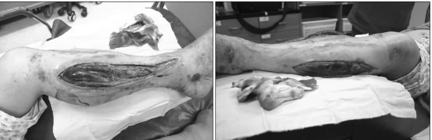
Fig. 1. Compartment syndrome after greater saphenous vein harvesting for coronary artery bypass surgery. Photo was taken on the 10 days after faciotomy. Note the muscular necrosis and tissue edema.
Fig. 2. T2 weighted coronal and ax- ial magnetic resonance images show diffuse edematous changes in the superficial and deep fascia of the left leg.
was markedly swollen, violet in color, tender, and warm up to the knee. Normal Doppler sounds were identified in the dorsalis pedis artery, lateral malleolar artery, and femoral vein. However, the venous flow below the popliteal vein was obscured in the color Doppler ultrasonography, and the pa- tient’s left foot was almost paralyzed. A decreased platelet count (85,000/μL) and an increased D-dimer level (355 μg/L) were noted. Because it was believed that anticoagulation ther- apy and infrapopliteal thrombectomy would be ineffective at that time, a prompt four-part fasciotomy was performed to prevent irreversible neurological and muscle damage.
Unfortunately, despite all of our efforts, the patient’s swollen muscles became infected with Acinetobacter baumannii after 1 week (Figs. 1, 2). A delayed skin graft was required after repeated aggressive debridement. The foot drop did not re-
cover, and the patient was discharged 5 months post- operatively with a disability.
DISCUSSION
In this case, based on the clinical and ultrasonography findings, we postulate that the sudden infrapopliteal venous occlusion led to the lower limb compartment syndrome and a neuromuscular disability. The compartment syndrome is caused by tissue compression within a closed space, which results in impaired tissue perfusion and threatens to com- promise the neurovascular system. Phlegmasia cerulea dolens (PCD), a rare form of acute deep vein thrombosis, creates sudden, severe venous hypertension and the resultant com- partment syndrome [1,2]. Most PCD is associated with a hy-
What Should We Know
− 45 − percoagulable state such as that occurring with malignancy, the postpartum period, immune disease, or drug reactions (e.g., heparin-induced thrombocytopenia). PCD can follow major surgery or trauma with serious dysregulation of the co- agulation-anticoagulation system. Acute deep vein thrombosis and increased intracompartmental pressure lead to a reduction in the arteriovenous pressure gradient with subsequent re- duced microvascular perfusion. Consequently, compartment phenomena such as nerve compression, tissue ischemia, or venous gangrene can occur.
Various clinical sequences other than hypercoagulability can also be related to the occurrence of PCD. For example, failure of venous return during extracorporeal circulation can induce PCD-type venous hypertension without thrombosis and lead to the abdominal compartment syndrome [3]. Similarly, several case reports of the lower limb compartment syndrome after CABG have been reported in the English-language liter- ature [4,5]. Because its rarity makes it difficult to determine the precise mechanism, previous authors have proposed only two hypotheses for the compartment syndrome after CABG.
A CPB-induced systemic inflammatory response has generally been considered to be the responsible cause. During CPB, vasoconstriction and increased capillary permeability can be triggered. In addition, hypotension during CPB can worsen the tissue perfusion in the extremity, which may induce an inflammatory injury after reperfusion [4]. The systemic in- flammatory aspects of its pathogenesis can lead not only to the lower limb compartment syndrome after conventional CABG but also to the abdominal compartment syndrome [6].
Local factors related to surgery are also believed to be important. The length of time that the leg position is main- tained, the procedure itself, inotropic drugs, and a com- pression dressing after vein harvesting can all impair tissue perfusion and cause hypoxemia. These factors may play a role in the development of the compartment syndrome. The occurrence of cases of the lower limb compartment syndrome without CPB-like off-pump CABG or varicose vein surgery supports the local-factor theory over the CPB effect [5,7].
Furthermore, it is remarkable that the reported compartment syndrome after CABG always involves the limb from which the graft vein was harvested [4,5-7]. Therefore, we postulate that superficial vein harvesting itself and the CPB-related sys-
temic inflammatory responses are both closely related to the compartment syndrome after CABG. Regardless, most pre- vious reports have described a warm compartment limb with no evidence of blocked arterial blood flow. These findings suggest that a considerable number of cases of the compart- ment syndrome after CABG originate from PCD pathology.
We should also emphasize that early recognition and de- compression are crucial for saving the affected limb. In gen- eral, PCD after surgery is predictable, in that it is largely as- sociated with a pre-existing hypercoagulable state and specific surgery types. Therefore, it should be preventable. However, unpredictable occurrences are not uncommon and not always preventable. In the setting of CABG, before the procedure has started, the leg position should be checked for excessive compression. The CPB time must be shortened, and hypo- xemia and hypotension during the operation should be prevented. After vein harvesting, the dressing should be ap- plied in a gentle manner, and any compression from the dressing should be minimized. Surgeons must have a high level of suspicion when symptoms develop. Because the nor- mal range of intramuscular pressure (range, 10 to 15 mmHg) does not always rule out PCD, the diagnosis should be based on a comprehensive clinical assessment [1]. If confirmed, management must commence as soon as possible. If possible, surgical thrombectomy and anticoagulation with or without thrombolytic therapy should be considered. A fasciotomy is the most effective way to relieve intramuscular pressure and can be performed as a bedside procedure under local anesthesia.
In conclusion, multiple factors appear to be involved in the compartment syndrome after CABG. PCD is thought to un- derlie this condition, and further studies are warranted.
Awareness is important for early recognition, and optimized management is essential for a disability-free return to normal life.
CONFLICT OF INTEREST
No potential conflict of interest relevant to this article was reported.
Kang-Hoon Lee, et al
− 46 − REFERENCES
1. Tiwari A, Haq AI, Myint F, Hamilton G. Acute compartment syndromes. Br J Surg 2002;89:397-412.
2. Mumoli N, Invernizzi C, Luschi R, Carmignani G, Camaiti A, Cei M. Phlegmasia cerulea dolens. Circulation 2012;125:
1056-7.
3. Wu ET, Huang SC, Chiu IS, Ko WJ. Abdominal compart- ment syndrome caused failure of venous return in a neonate during extracorporeal membrane oxygenation support.
Pediatr Crit Care Med 2006;7:400-1.
4. Pasic M, Carrel T, Tonz M, Vogt P, von Segesser L, Turina M. Acute compartment syndrome after aortocoronary bypass.
Lancet 1993;341:897.
5. Vaidyanathan KR, Sundaramoorthi T, Byalal JR, et al.
Lower extremity compartment syndrome after off-pump aor- tocoronary bypass. J Thorac Cardiovasc Surg 2006;131:
1173-4.
6. Rabbi JF, Valaulikar G, Appling NA, Bee TK, Ostrow BF, Weiman DS. Secondary abdominal compartment syndrome causing failure to wean from cardiopulmonary bypass. Ann Thorac Surg 2012;93:e99-100.
7. Shahin Y, Tapuria N, Chong PL. Compartment syndrome following short saphenous varicose vein surgery: a case report. Cases J 2009;2:7118.