Clinico-pathological Analysis of the Lungs from Patients with Lung Transplantation in a Single Institute in Korea
Recently, the numbers of lung transplantation (LT) has been increased in Korea. However, post-LT outcome has not been successful in all patients, which may be partially affected by the primary lung disease. Therefore comprehensive understanding in original pathological diagnosis of patients with LT would be needed for achieving better clinical outcome. To address this issue, we performed clinico-pathological analysis of the explanted lungs from 29 patients who underwent LT over a 9-yr period in Seoul National University Hospital.
Among them, 26 patients received single (1/26) or double (25/26) LT, while heart-lung transplantation was performed in 3 patients. The final clinico-pathological diagnoses were idiopathic pulmonary fibrosis/usual interstitial pneumonia (UIP) (n = 6), acute interstitial pneumonia (AIP)/diffuse alveolar damage (DAD) (n = 4), AIP/ non-specific interstitial pneumonia with DAD (n = 1), collagen vascular disease-related interstitial lung disease (CVD-ILD)/DAD (n = 3), CVD-ILD/UIP (n = 1), lymphangioleiomyomatosis (n = 1), bronchiectasis (n = 4), pulmonary arterial hypertension (n = 2), tuberculosis (n = 1), bronchiolitis obliterans (BO) (n = 1), and lung cancer (n = 1). Moreover, 4 patients who had chemotherapy and hematopoietic stem cell transplantation due to hematologic malignancy showed unclassifiable interstitial pneumonia with extensive fibrosis in the lungs. Our study demonstrates that pathology of the explanted lungs from Korean patients with LT is different from that of other countries except for interstitial lung disease and bronchiectasis, which may be helpful for optimization of selecting LT candidates for Korean patients.
Keywords: Histology; Lung Transplantation; Lung; Transplantation; Lung Disease, Interstitial; Bronchiectasis
Hyojin Kim,1 Yoon Kyung Jeon,1 Hyun Joo Lee,2 Young Tae Kim,2,3,4 and Doo Hyun Chung1,3,5
1Department of Pathology, 2Department of Thoracic Surgery, 3Xenotransplantation Research Center,
4Transplantation Research Institute, and
5Department of Biomedical Sciences, Seoul National University Hospital, Seoul National University College of Medicine, Seoul, Korea
Received: 5 January 2015 Accepted: 18 June 2015 Address for Correspondence:
Doo Hyun Chung, MD
Department of Pathology and Biomedical Sciences, Seoul National University College of Medicine, 103 Daehak-ro, Jongno-gu, Seoul 03080, Korea
Tel: +82.2-740-8915, Fax: +82.2-743-5530 E-mail: [email protected]
Funding: This research was supported by a grant of the Korea Health Technology R&D Project through the Korea Health Industry Development Institute (KHIDI), funded by the Ministry of Health & Welfare, Korea (grant No.: HI13C0954).
http://dx.doi.org/10.3346/jkms.2015.30.10.1439 • J Korean Med Sci 2015; 30: 1439-1445
INTRODUCTION
Lung transplantation (LT) prolongs survival and improves pul- monary function of patients who have end-stage lung disease, thereby being a life-saving treatment for these patients. Appro- ximately, 3,000 LTs per year are performed worldwide (1). Com- pared with western countries, Korea was not an early adopter for LT; the first LT was performed in July, 1996, and nine institu- tions have performed LT so far (2, 3). Recently, the number of LT has been increased in Korea. However, the number of lung donors is not enough for patients who need LT, indicating that extensive effort for optimization of LT should be made to achieve successful clinical outcome. Moreover, post-transplantation com- plications and clinical outcome may be closely related with the primary disease of the lung.
Therefore, comprehensive understanding of primary pathol- ogy of explanted lungs from LT patients could be helpful for im- proving success rates of LT. To address this issue, we reviewed and analyzed pathology of the lungs from LT recipients at our center over the subsequent decade (2006-2014).
MATERIALS AND METHODS
Twenty-nine consecutive patients who underwent single or double-lung, or heart-lung transplantation at the Seoul Nation- al University Hospital (SNUH) between January, 2006 and De- cember, 2014 were included in this study. Clinical information was collected from the medial records and included ages at time of transplantation, genders, underlying pulmonary diseases, referral diagnoses, types of transplantation, and post-transplan- tation clinical course.
The factors evaluated during perioperative period included intensive care unit (ICU) care, preoperative infection, depen- dence on mechanical ventilator (MV) or extracorporeal mem- brane oxygenation (ECMO) support, use of intraoperative sup- port such as cardiopulmonary bypass (CPB) and/or ECMO, operation time, total ischemic time, and lengths of ECMO, MV support and hospital stay. The surgical pathology reports were also reviewed. At least three representative sections were taken from each lobe of the explanted lungs along with hilar nodes and vessels of LT recipients. Routine hematoxylin and eosin- stain was prepared from formalin-fixed paraffin-embedded
sections and various special stains for microorganisms were performed when indicated. Three pathologists reviewed all mi- crosectioned slides of the lungs from LT recipients and compared with clinical information.
Statistical analysis
Survival analysis was performed using the Kaplan-Meier meth- od with the log-rank test. A P value < 0 .05 was considered sta- tistically significant. SPSS 18.0 software (SPSS Inc., Chicago, IL, USA) was used for statistical analyses.
Ethics statement
This study was approved by the institutional review board of SNUH (IRB No. 2014-12-29) and the need of informed consent was waived.
RESULTS
Clinical characterization of patients with lung transplantation
The clinico-pathological features of 29 patients who underwent
deceased-donor LT are summarized in Table 1. Of 29 patients, one patient received single LT, 25 patients did bilateral sequen- tial single LT, and three patients underwent heart-lung trans- plantation. These patients included eighteen males and eleven females with a median age of 49 yr (from 11 to 70 yr) at the time of transplantation. Table 2 showed perioperative clinical para- meters of 29 patients. Nineteen patients had ICU care before operation. About one third of patients (11/29) had an episode of infection during pre-operative period and the most common pathogens were Vancomycin-resistant Enterococcus (n = 5) and imipenem-resistant Acinetobacter baumannii (n = 5). For re- spiratory support in preoperative management, 12 and 18 pa- tients required tracheostomy and MV, respectively. During op- eration, majority of patients (27/29) got respiratory support us- ing CPB (n = 14), ECMO (n = 3), and both CPB and ECMO (n = 10). Mean operation time was 647.7 ± 136.8 min (range, 444-917 min), and total ischemic time was 627.2 ± 174.8 min (range, 283- 965 min) except one unavailable case. Mean ECMO and MV weaning times were 9.3 days (range, 0.5-80.1 days) and 14 days (range, 1-47 days) for 14 and 24 patients, respectively. Mean pe- riod of hospitalization was 88.3 days (range, 1-375 days). None
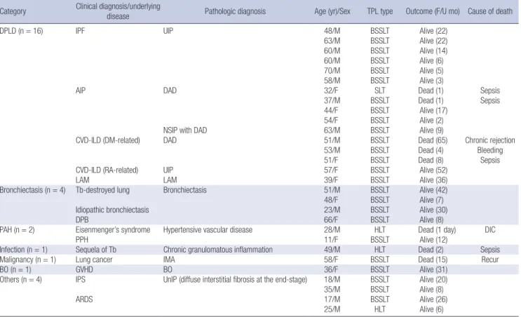
Table 1. Clinicopathologic features of patients with lung transplantation and the clinical outcome Category Clinical diagnosis/underlying
disease Pathologic diagnosis Age (yr)/Sex TPL type Outcome (F/U mo) Cause of death
DPLD (n = 16) IPF UIP 48/M BSSLT Alive (22)
63/M BSSLT Alive (22)
60/M BSSLT Alive (14)
60/M BSSLT Alive (6)
70/M BSSLT Alive (5)
58/M BSSLT Alive (3)
AIP DAD 32/F SLT Dead (1) Sepsis
37/M BSSLT Dead (1) Sepsis
44/F BSSLT Alive (17)
54/F BSSLT Alive (2)
NSIP with DAD 63/M BSSLT Alive (9)
CVD-ILD (DM-related) DAD 51/M BSSLT Dead (65) Chronic rejection
53/M BSSLT Dead (4) Bleeding
51/F BSSLT Dead (8) Sepsis
CVD-ILD (RA-related) UIP 57/F BSSLT Alive (52)
LAM LAM 39/F BSSLT Alive (36)
Bronchiectasis (n = 4) Tb-destroyed lung Bronchiectasis 51/M BSSLT Alive (42)
48/F BSSLT Alive (7)
Idiopathic bronchiectasis 23/M BSSLT Alive (30)
DPB 66/F BSSLT Alive (8)
PAH (n = 2) Eisenmenger’s syndrome Hypertensive vascular disease 28/M HLT Dead (1 day) DIC
PPH 11/F BSSLT Alive (12)
Infection (n = 1) Sequela of Tb Chronic granulomatous inflammation 49/M HLT Dead (2) Sepsis
Malignancy (n = 1) Lung cancer IMA 58/F BSSLT Dead (15) Recur
BO (n = 1) GVHD BO 36/F BSSLT Alive (31)
Others (n = 4) IPS UnIP (diffuse interstitial fibrosis at the end-stage) 18/M BSSLT Alive (20)
35/M BSSLT Alive (8)
ARDS 17/M BSSLT Alive (26)
25/M HLT Alive (6)
F/U, follow-up; mo, month; TPL, transplantation; SLT, single lung transplantations; BSSLT, bilateral sequential single lung transplantations; HLT, heart-lung transplantation; DPLDs, diffuse parenchymal lung diseases; IPF, idiopathic pulmonary fibrosis; UIP, usual interstitial pneumonia; AIP, acute interstitial pneumonia; DAD, diffuse alveolar damage; NSIP, non-specific interstitial pneumonia; CVD-ILD, collagen-vascular disease-related interstitial lung disease; DM, dermatomyositis; RA, rheumatoid arthritis; LAM, lymphangioleio- myomatosis; Tb, tuberculosis; DPB, diffuse panbronchiolitis; PAH, Pulmonary arterial hypertension; PPH, primary pulmonary hypertension; IMA, invasive mucinous adenocarci- noma; GVHD, graft-versus-host disease; BO, bronchiolitis obliterans; IPS, idiopathic pneumonia syndrome; ARDS, acute respiratory distress syndrome; UnIP, unclassifiable inter- stitial pneumonia; DIC, disseminated intravascular coagulation.
of them had re-transplantation.
Clinico-pathological analysis of primary pulmonary diseases of patients with LT
Diffuse parenchymal lung diseases (DPLDs) accounted for 55%
(16/29) of lung pathology of recipients with LT (Table 1). Clini- cal or referral diagnosis of these patients included idiopathic pulmonary fibrosis (IPF, n = 6), acute interstitial pneumonia (AIP, n = 5), collagen vascular disease-related interstitial lung disease (CVD-ILD, n = 4), and lymphangioleiomyomatosis (LAM, n = 1). The final pathologic diagnosis of explanted lungs in all patients with IPF was usual interstitial pneumonia (UIP) (n =
6), which was the most frequent underlying lung disease for LT in this cohort. Among five patients with clinical diagnosis of AIP, four cases showed diffuse alveolar damage (DAD) pattern, while one case demonstrated non-specific interstitial pneumo- nia (NSIP) with partial DAD pattern. Furthermore, patients with CVD-ILD exhibited either DAD (n = 3) or UIP (n = 1) pattern.
There was minor discrepancy between referral and final diag- nosis in one case; the patient was 63-yr old male with aggravat- ed productive cough and sputum for 3 months was referred to our hospital under the clinical diagnosis of AIP. In contrast to clinical diagnosis, the explanted lungs showed diffuse intersti- tial thickening and inflammatory cell infiltration, which was Table 2. Perioperative factors of recipients with lung transplantation
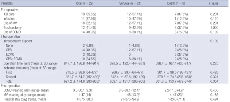
Variables Total (n = 29) Survival (n = 21) Death (n = 8) P value
Pre-operative
ICU care 19 (65.5%) 12 (57.1%) 7 (87.5%) 0.201
Infection 11 (37.9%) 10 (47.6%) 1 (12.5%) 0.110
Use of MV 18 (62.1%) 12 (57.1%) 7 (87.5%) 0.201
Tracheostomy 12 (41.4%) 9 (42.9%) 3 (37.5%) 1.000
Use of ECMO 14 (48.3%) 8 (38.1%) 6 (75.0%) 0.109
Intra-operative
Intraoperative support 0.109
None 2 (6.9%) 1 (4.8%) 1 (12.5%)
CPB 14 (48.3%) 12 (57.1%) 2 (25.0%)
ECMO 3 (10.3%) 0 3 (37.5%)
CPB+ECMO 10 (34.5%) 8 (38.1%) 2 (25.0%)
Operation time (min) (mean ± SD, range) 647.7 ± 136.8 (444-917) 628.5 ± 122.4 (444-881) 698.4 ± 167.4 (435-917) 0.225 Ischemic time (min) (mean ± SD, range)
First 275.5 ± 98.6 (64-477)a 266.7 ± 99.4 (64-477) 301.7 ± 98.5 (193-437)b 0.426
Second 351.7 ± 84.7 (192-499)a 342.4 ± 87.6 (192-499) 379.6 ± 74.3 (236-463)b 0.324
Total 627.2 ± 174.8 (283-965)a 609.1 ± 181.1 (283-965) 681.3 ± 153.7 (472-874)b 0.354
Post-operative
ECMO weaning (day) (range, mean) 0.5-80.1 (9.3)c 0.5-80.1 (12.1)d 2.2-11.5 (4.9)e 0.450
MV weaning (day) (range, mean) 1-47 (14)f 1-46 (13.9)g 4-47 (23)h 0.185
Hospital stay (day) (range, mean) 1-375 (88.3) 21-375 (94.9) 1-243 (71.1) 0.494
an = 28; bn = 7; cn = 14; dn = 8; en = 6; fn = 24; gn = 20; hn = 4. ICU, intensive care unit; MV, mechanical ventilator; ECMO, extracorporeal membrane oxygenation; CPB, car- diopulmonary bypass; min, minutes; SD, standard deviation.
A B
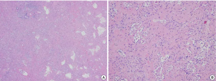
Fig. 1. Histologic features of explanted lungs showing non-specific interstitial pneumonia (NSIP) with diffuse alveolar damage (DAD) pattern in a patient who clinically suggested with acute interstitial pneumonia. Diffuse interstitial thickening and chronic inflammatory cell infiltration are observed in both lobes, suggesting NSIP (A). In addition, focal DAD pattern is also observed in the right lobe (B) (Hematoxylin and eosin stain; original magnification: × 200).
consistent with NSIP, and partially combined with DAD pattern (Fig. 1).
Bronchiectasis was one of common indications for LT (n = 4).
Two patients (51-yr-old male and 48-yr-old female) underwent LT under the clinical diagnosis of bronchiectasis, which might be due to the sequelae of tuberculosis (Tb). However, there were no pathologic findings such as granulomas, suggesting lack of active tuberculosis in explanted lungs. A 23-yr-old male who had suffered from recurrent upper respiratory infection since 5-yr old age had been clinically suggested for Katagener’s syn- drome, but the genetic test was negative for Katagener’s syn- drome. The other patients (F/66 yr) had been clinically diag- nosed with diffuse panbronchiolitis, which was confirmed by pathologic examination of explanted lungs.
Pulmonary arterial hypertension (PAH) (n = 2) was rare pri- mary indication for LT in our institute. An 11-yr-old female re- ceived bilateral sequential single LT due to primary pulmonary hypertension, while a 28-yr-old male underwent heart-lung trans- plantation for congenital heart disease and subsequent Eisen- menger’s syndrome. In microscopic examination, the walls of pulmonary arteries were thickened with plexiform intimal pro- liferation, being consistent with PAH.
Infection and malignancy were also rare indication for LT. A 49-yr-old male had a medication for 6 months due to tubercu- losis (Tb) pleurisy 15 yr ago, and one year later partial removal of pericardium was done due to constrictive pericarditis related with Tb complication. After surgery, generalized edema and re- current pneumonia developed due to refractory constrictive pericarditis and dyspnea with pneumonic infiltration was ag- gravated a few months before heart-lung transplantation. How- ever, examination of bacteria revealed no antibiotics-resistant strains in this patient. Microscopic examination revealed exten-
sive granulomatous inflammation with caseation necrosis, in- dicating tuberculosis.
A 58-yr-old female with localized adenocarcinoma underwent left lower lobectomy. Eight months later, wedge resection of the lung was performed due to multiple nodules newly detected in both lungs. Microscopic examination showed invasive muci- nous adenocarcinoma (i.e., mucinous bronchioloalveolar car- cinoma [BAC]). However, there was no evidence of extrapul- monary tumor metastasis based on systemic evaluation, and thus she underwent bilateral sequential single LT to eliminate residual tumor in the lungs, which was mucinous type of ade- nocarcinoma involving both lungs in the absence of lymph node metastasis. Molecular studies revealed KRAS mutation in the tumor. The tumor recurred in the lungs 10 months after LT and the patient died of disease progression 16 months after LT.
Bronchiolitis obliterans (BO) (n = 1) was rare indication for LT in our cohort. A 36-yr-old female who had undergone che- motherapy and allogenic peripheral blood stem cell transplan- tation (PBSCT) for T-cell prolymphocytic leukemia developed chronic graft-versus-host disease (GVHD) in the liver and lungs, which was attenuated by corticosteroids therapy. However, ap- proximately 4 yr after PBSCT, the patient presented with dys- pnea on exertion and further exacerbated due to influenza in- fection, thereby undergoing bilateral sequential single LT. The main histologic findings were luminal obstruction in a few small bronchioles with peribronchiolar lymphoplasmacytic infiltration, fibrosis, and foamy cell collection, being consistent with BO.
Four patients who received chemotherapy and autologous (n
= 1) or allogenic (n = 3) PBSCT due to hematolymphoid malig- nancy underwent LT. Among them, two patients were referred under the clinical diagnosis of idiopathic pneumonia syndrome, whereas other two patients presented with rapidly progressive
A B
Fig. 2. Histologic features of end-stage lung from patients who received chemotherapy and peripheral blood stem cell transplantation. Diffuse and marked interstitial fibrosis with nearly complete loss of alveolar spaces and shrinkage of the lung tissue around a bronchus are observed (Hematoxylin and eosin stain; original magnification: (A) × 100, (B) × 400).
acute respiratory distress syndrome (ARDS). Histological ex- amination of the lungs from four patients showed diffuse and marked pulmonary interstitial fibrosis with nearly complete loss or collapse of alveolar spaces and shrinkage of the lung tissue around a bronchus (Fig. 2).
Clinical outcome of patients with lung transplantation Clinical outcome of 29 recipients is briefly described in Table 1.
The overall survival rate was 72.4% during a follow-up from 1 day to 65 months (mean 16.03 months). The underlying pul- monary diseases of eight patients who died after LT included AIP/DAD (n = 2), CVD-ILD/DAD (n = 3), Eisenmenger’s syn- drome (n = 1), Tb (n = 1), and lung cancer (n = 1). Main causes of death for these patients were sepsis (n = 4), disseminated in- travascular coagulation (n = 1), uncontrolled massive hemop- tysis (n = 1), disease recurrence (n = 1), or chronic rejection (n
= 1). We compared the perioperative clinical factors of the pa- tient according to clinical outcome (Table 2). However, there was no difference in these clinical parameters between two groups.
The survival of patients who were pathologically diagnosed with DAD (8/29) was compared with the survival of those who were diagnosed with other disease (21/29), suggesting that DAD was one of factors to be associated with poor survival after LT (P = 0.034) (Fig. 3). To characterize effect of DAD on patient’s surviv- al further, we compared preoperative clinical parameters be- tween two groups (Table 3). All patients with DAD pattern took ICU care and used MV and ECMO during preoperative period.
On the other hand, 11 out of 21 in patients without DAD pat- tern were treated in ICU. Among them, 11 and 6 patients used MV and ECMO, respectively. PaO2/FiO2 ratio and arterial pCO2
in patient with DAD pattern were significantly lower than in with- out the pattern (P = 0.001 and P = 0.032, respectively). These
findings suggest that patients with DAD patterns showed worse clinical status in preoperative period than patient without DAD.
DISCUSSION
In this study, we performed clinic-pathological analysis of ex- planted lungs from patients with LT in our institute, although the number of LT is limited. In comparative analysis with clini- cal and pathology diagnosis in these patients with LT, there was a high concordance between the referral clinical diagnosis of patients before LT and the confirmed pathological diagnosis of explanted lung in our cohort. Three (10%) of 29 patients showed discrepancy between referral and final pathologic diagnosis, which was lower than those reported by two studies using large cohort (17% and 21%) (4, 5). Among three patients, one had AIP vs. NSIP with DAD pattern and two presented with ARDS vs. diffuse interstitial fibrosing disease pattern at the end-stage.
Furthermore, our data demonstrated that IPF/UIP, AIP/DAD, CVD-associated ILD, and bronchiectasis were the major indi- cations for LT, accounting for 66% of LT cases in our institute.
Pathologically, UIP (n = 7) in IPF, CVD-ILD and DAD (n = 8) in AIP, or CVD-ILD was frequently observed in explanted lungs from LT patients, indicating that IPF/UIP was the most common single disease of patients with LT in our cohort. In contrast to our results, The registry of the International Society for Heart and Lung Transplantation (ISHLT) reported that chronic ob- structive pulmonary disease (COPD) (including emphysema) is the most common indications (38%) for adult LT, followed by ILD (mostly IPF) (28%) and bronchiectasis (19%) worldwide (1). Moreover, bronchiectasis was caused mostly by cystic fibro- sis in ISHLT registry. Meanwhile, a Japanese cohort of LT recipi- ents showed that primary pulmonary hypertension, ILD, LAM, and BO were common indications (6). These results suggest that the discrepancy between various registries might be due to difference in ethnics, socio-economic status, or regions of pa- tients. Especially COPD is the rare indication in Asia compared with ISHLT data. There may be possible explanations for this discrepancy. First, incidence of α1-antitrypsin deficiency em- Table 3. Preoperative variables reflecting patients’ severity based on the presence of pathologic diffuse alveolar damage (DAD) pattern
Variables
Pathologic finding
P value DAD pattern
(n = 8) non-DAD pattern (n = 21)
ICU care 100% 52.4% 0.018
MV use 100% 52.4% 0.018
ECMO use 100% 28.6% 0.001
PaO2/FiO2 (mean ± SD) 58.1 ± 15.4 150.9 ± 103.2 0.001
Arterial pH (mean) 7.40 ± 0.08 7.31 ± 0.12 0.069
Arterial pCO2 (mean) 47.8 ± 8.9 60.3 ± 20.8 0.032 DAD, diffuse alveolar damage; ICU, intensive care unit; MV, mechanical ventilator;
ECMO, extracorporeal membrane oxygenation; SD, standard deviation.
Fig. 3. Survival curve based on the presence of pathologic diffuse alveolar damage (DAD) pattern using the Kaplan-Meier method with the log-rank test.
Overall survival
Months
0 20 40 60
1.0
0.8
0.6
0.4
0.2
0
Without DAD pattern
With DAD pattern
P = 0.034
physema, one of the causes of COPD, is extremely rare in Asian population. Second, many COPD patients are old age thus, phys- ical status of candidates deteriorated to receive LT. To achieve successful outcome in LT for old patients, preoperative respira- tory rehabilitation programs is indispensable. However, this pro- gram is limited in Korea compared with Western countries. These factors might affect indication and/or frequency of patients with COPD for LT in various countries.
It has been reported that unstable clinical condition of pati- ents (e.g., mechanical ventilation or extra-corporeal membrane oxygenation) is relative contraindication of LT (7), suggesting that pre-LT condition of patients including primary diseases of lungs might be critical factors to affect clinical outcomes of LT recipients. Consistent with this suggestion, patients with AIP has been rarely attempted for LT, especially during acute phase of disease because the outcome of these patients was poor (8, 9). In this study, survival analysis revealed that presence of DAD pattern in explanted lung was a poor prognostic factor for LT (P = 0.034) because patients with DAD frequently had poor pre- operative clinical status. These findings suggest that careful con- sideration on LT indication should be needed for patients with refractory AIP/DAD because these patients might be expected for poor clinical outcome after LT.
One patient with BAC underwent LT died of recurrence of lung cancer 10 months after LT. The presence of malignancy in recipients might be considered as contraindication for LT (7).
However, some clinic centers have performed LT for treatment of patients with pulmonary BAC. BAC, a pulmonary malignan- cy that does not widely invade stroma and spread outside of the lungs, has been thought as suitable disease for LT (10). Never- theless, LT for BAC still remains controversial in terms of poten- tial therapeutic modality for BAC. Garver et al. (11) reported that four of seven patients with BAC confined to lungs exhibited re- current BAC after LT. An international survey reported that the recurrence-free survival at 5 yr was 35% and the median time of recurrence was 12 months in 26 patients who received LT for BAC (12). These findings suggest that indication of LT should be carefully considered for patients with BAC although LT is cur- rently performed for patients with advanced BAC in a few centers.
Our study demonstrated that five patients who had been treat- ed with chemotherapy and PBSCT due to hematolymphoid ma- lignancy developed PBSCT-mediated complications such as BO, thereby undergoing LT in the course of treatment. Howev- er, microscopic examination revealed that destroyed lung pa- renchyma were found with marked interstitial fibrosis and thick- ening, and moderate interstitial inflammation in explanted lungs, which were unclassifiable into a specific histopathological cat- egory. We hypothesize that pathological processes in these pa- tients may be partly attributable to previous chemotherapy be- cause the pathologic findings in the lungs are similar to those observed in drug-induced pneumonitis (13). Furthermore, PB-
SCT has been known to be a condition to induce various pul- monary manifestations including chronic GVHD with BO, DPLD (e.g., NSIP), vasculopathies, veno-occlusive disease, and radia- tion fibrosis (14, 15). Thus, it is feasible that PBSCT may be one of factors to be related with complications mimicking BO, which led patients to undergo LT.
The strength of our study is that significant information of LT patients in single institute was provided by comprehensively analyzing clinicopathologic aspects in these patients. However, there is some limitation of our study for generalizing our results because the sample size of our study is too small.
In conclusion, current study demonstrated that pathology of the explanted lungs from patients with LT in our institute includ- ed idiopathic and CVD-associated IPF/UIP, AIP/DAD, and bron- chiectasis, which were different from those of other countries.
Thus, a nationwide registry on the lung pathology of patients with LT in Korea may provide important information further to achieve better outcome of LT in Korean patients.
DISCLOSURE
The authors have no conflicts of interest to disclose.
AUTHOR CONTRIBUTION
Study concepts & design: Chung DH, Jeon YK. Acquisition of data: Kim H, Lee HJ, Jeon YK, Kim YT, Chung DH. Analysis and interpretation of results: Kim H, Lee HJ, Jeon YK, Chung DH.
Manuscript preparation: Kim H, Jeon YK, Kim YT, Chung DH.
Manuscript approval: all authors.
ORCID
Hyojin Kim http://orcid.org/0000-0001-9201-8328 Yoon Kyung Jeon http://orcid.org/0000-0001-8466-9681 Hyun Joo Lee http://orcid.org/0000-0002-3092-2167 Young Tae Kim http:// orcid.org/0000-0001-9006-4881 Doo Hyun Chung http://orcid.org/0000-0002-9948-8485
REFERENCES
1. Yusen RD, Edwards LB, Kucheryavaya AY, Benden C, Dipchand AI, Dob- bels F, Goldfarb SB, Levvey BJ, Lund LH, Meiser B, et al.; International Society for Heart and Lung Transplantation. The registry of the Interna- tional Society for Heart and Lung Transplantation: thirty-first adult lung and heart-lung transplant report--2014; focus theme: retransplantation.
J Heart Lung Transplant 2014; 33: 1009-24.
2. Haam SJ, Lee DY, Paik HC. An overview of lung transplantation in Ko- rea. Transplant Proc 2008; 40: 2620-2.
3. Korean Network for Organ Sharing (KONOS). KONOS 2013 statistical yearbook. Available at http://www.konos.go.kr/konosis/ [accessed on 10 July 2014].
4. Stewart S, McNeil K, Nashef SA, Wells FC, Higenbottam TW, Wallwork J.
Audit of referral and explant diagnoses in lung transplantation: a patho- logic study of lungs removed for parenchymal disease. J Heart Lung Trans- plant 1995; 14: 1173-86.
5. Calabrese F, Alessandrini L, Loy M, Marulli G, Balestro E, Perissinotto E, Valente M, Rea F. Comparison between referral diagnosis of patients re- quiring transplantation and pathologic diagnosis of native lungs. J Heart Lung Transplant 2009; 28: 1135-40.
6. Sato M, Okada Y, Oto T, Minami M, Shiraishi T, Nagayasu T, Yoshino I, Chida M, Okumura M, Date H, et al.; Japanese Society of Lung and Heart- Lung Transplantation. Registry of the Japanese Society of Lung and Heart- Lung Transplantation: official Japanese lung transplantation report, 2014.
Gen Thorac Cardiovasc Surg 2014; 62: 594-601.
7. Orens JB, Estenne M, Arcasoy S, Conte JV, Corris P, Egan JJ, Egan T, Kes- havjee S, Knoop C, Kotloff R, et al.; Pulmonary Scientific Council of the International Society for Heart and Lung Transplantation. Internation- al guidelines for the selection of lung transplant candidates: 2006 update- -a consensus report from the Pulmonary Scientific Council of the Inter- national Society for Heart and Lung Transplantation. J Heart Lung Trans- plant 2006; 25: 745-55.
8. Avnon LS, Pikovsky O, Sion-Vardy N, Almog Y. Acute interstitial pneu- monia-Hamman-Rich syndrome: clinical characteristics and diagnostic and therapeutic considerations. Anesth Analg 2009; 108: 232-7.
9. Vourlekis JS, Brown KK, Cool CD, Young DA, Cherniack RM, King TE, Schwarz MI. Acute interstitial pneumonitis. Case series and review of the literature. Medicine (Baltimore) 2000; 79: 369-78.
10. Paloyan EB, Swinnen LJ, Montoya A, Lonchyna V, Sullivan HJ, Garrity E.
Lung transplantation for advanced bronchioloalveolar carcinoma con- fined to the lungs. Transplantation 2000; 69: 2446-8.
11. Garver RI Jr, Zorn GL, Wu X, McGiffin DC, Young KR Jr, Pinkard NB.
Recurrence of bronchioloalveolar carcinoma in transplanted lungs. N Engl J Med 1999; 340: 1071-4.
12. de Perrot M, Chernenko S, Waddell TK, Shargall Y, Pierre AF, Hutcheon M, Keshavjee S. Role of lung transplantation in the treatment of bron- chogenic carcinomas for patients with end-stage pulmonary disease. J Clin Oncol 2004; 22: 4351-6.
13. Camus P, Fanton A, Bonniaud P, Camus C, Foucher P. Interstitial lung disease induced by drugs and radiation. Respiration 2004; 71: 301-26.
14. Watkins TR, Chien JW, Crawford SW. Graft versus host-associated pul- monary disease and other idiopathic pulmonary complications after hematopoietic stem cell transplant. Semin Respir Crit Care Med 2005;
26: 482-9.
15. Miyagawa-Hayashino A, Sonobe M, Kubo T, Yoshizawa A, Date H, Ma- nabe T. Non-specific interstitial pneumonia as a manifestation of graft- versus-host disease following pediatric allogeneic hematopoietic stem cell transplantation. Pathol Int 2010; 60: 137-42.