흰쥐에서 SAL5의 알코올성 지방간 형성에 미치는 영향
김복규1#, 양원경1, 박양춘2, 정가영3, 신은주3, 도선길3, 김승형1*
1 : 대전대학교 동서생명과학연구원, 2 : 대전대학교 한의과대학 폐계내과학교실, 3 : ㈜ 유니베라
Effect of SAL5 on chronic ethanol-induced fatty liver model
Bok-Kyu Kim
1#, Won-Kyung Yang
1, Yang-Chun Park
2, Ga-Young Jung
3Eun-Ju Shin
3, Seon-Gil Do
3, Seung-Hyung Kim
1*1 : Institute of Traditional Medicine and Bioscience, Daejeon University, Daejeon 34520, Republic of Korea 2 : Division of Respiratory Systems, Department of Internal Medicine, College of Korean Medicine, Daejeon University
3 : Univera, Inc., Seoul 04782, Korea
ABSTRACT
Objective : In this study, we investigated the effect of SAL5(mixing extracts of
Schisandra chinensisBaillon,
Artemisia capillarisThunb., and
Aloe veraLinne) on chronic ethanol-induced fatty liver model.
Methods : Sprague-Dawley male rats were fed Liber-DeCarli (normal), ethanol liquid diet (control), SAL5 (200 ㎎/㎏).
We administrated the SAL5 on chronic ethanol-induced fatty liver model for 5 weeks. We measured alkaline phosphtase (ALP), alanine transminase (ALT), aspartate transminase (AST) and γ-glutamyl transpeptase (γ-GTP) in serum and triglyceride (TG), superoxide dismutase (SOD), catalase, glutathione (GSH) and malondialdehyde (MDA) level in liver.
Liver histopathology was examined by Hematoxylin-eosin and Oil red O staining of the fixed liver tissues. Real-time PCR was performed to measure the mRNA expression of inflammatory cytokines and MMP-2, MMP-9.
Results : SAL5 administration resulted in significantly decreased liver marker enzymes activities of alanine transminase (ALT), γ -glutamyl transpeptase (γ -GTP) in serum and triglyceride (TG) activities in liver. The control group decreased the activities of superoxide dismutase (SOD), catalase (CAT) with the reduced level of glutathione (GSH) in liver. On the other hand, SAL5 group increased the activities of SOD, CAT and the level of GSH. SAL5 delayed the development of an alcoholic fatty liver by reversing fat accumulation in the liver, as evidenced in histological observations. The gene expression of mRNA were significantly decreased at the IL-1β, TNF-α , NOS-II and MMP-2 by SAL5.
Conclusions : These results indicate that SAL5 might have protective effect chronic ethanol-induced fatty liver models.
1)
Key words : SAL5, chronic ethanol-induced fatty liver, ethanol liquid diet
Ⅰ. 서 론
현대사회에서 만성적인 음주로 인한 알코올 환자의 증가는 서구 선진국뿐 아니라 전 세계적으로 커다란 사회문제로 대두 되고 있다
1). 우리나라도 음주로 인한 환자 치료 및 교육비 부
담이 증가하는 추세이며
2), 음주 관련 질병을 억제하거나 도움이 되는 생리활성물질 연구가 어느 때보다 주목받고 있다. 알코올 성 지방간 (alcoholic fatty liver)은 과음으로 인해 중성지방이 간 조직에 축적되는 질환이다
3). 과량의 알코올을 만성적으로 섭취하면 세포내 NADH/NAD
+의 비율이 증가하여 탄수화물,
*Corresponding author : Seung-Hyung Kim. Institute of Traditional Medicine and Bioscience, Daejeon University, 62 Daehak-ro, Dong-gu, Daejeon 34520, Republic of Korea.
·Tel : +82-42-280-2642 ·Fax : +82-42-274-2600 ·E-mail : [email protected]
#First author : Bok-Kyu Kim. Institute of Traditional Medicine and Bioscience, Daejeon University, 62 Daehak-ro, Dong-gu, Daejeon 34520, Republic of Korea.
·Tel : +82-42-280-2642 ·Fax : +82-42-274-2600 ·E-mail : [email protected]
·Received : 5 December 2017 ·Revised : 20 December 2017 ·Accepted : 15 January 2018
단백질 및 지질대사의 장애가 일어나, 간의 지방산 산화가 감소 되고, 합성은 증가되어 간에 중성지질이 축적되며, 알코올 분해 과정의 중간생성물인 아세트알데히드의 독성에 의해 microtubule 의 손상이 일어나 지방간이 유발되고 심해지면 알코올성 간염 이나 간경화증이 유발될 수 있다
4). 또한, 알코올 대사작용이 촉진되어 산소 소비량이 증가함에 따라 간조직의 부분적인 저 산소증과 괴사를 초래하거나 알코올 대사 시 생성되는 유리 라디칼에 의해 지질과산화물의 반응이 촉진되어 간 조직을 손상 시킬 수 있으며
5), 간 조직의 중성지질 및 콜레스테롤 함량이 크게 증가하고, 활성산소가 증가하여 간세포 파괴를 유발 할 수 있다
6). 또한, 알코올 대사에 의해 유도되는 CYP 2E1, p450 reductase, NADPH oxidase, aldehyde oxidase, xanthine oxidase 등은 생체 내 반응산소종의 생성을 낳고 과량의 알코 올 대사에서 이러한 효소들은 다량의 자유유리기를 생성함으 로써 생체 내 산화스트레스를 유발하며 알코올성 간 손상의 원인이 된다
7). 만성적인 알코올 섭취는 글루타치온 등과 같이 자유유리기를 중화시키는 항산화제의 생성이나 작용을 저해 함으로써 산화스트레스를 촉진하여 간세포 손상을 일으킨다
8).
오미자 (
Schisandra chinensisBaillon)는 오미자과 (Schisandraceae)에 속하는 오미자나무 의 열매로 간 보호작 용이 있으며 정상상태의 쥐에게 투여했을 때 간기능 촉진작용이 있어 혈청 중의 중성지방을 저하시키고 간 지방질을 감소시키며 약물대사 효소활성을 촉진 시키며
9)10), 항산화효과가 있다고 알려져 있다
11)12). 사철쑥 (
Artemisia capillarisThunb.
)은 국화과 (Compositae)에 속하는 다년생 초본으로
13)사철쑥에 대한 연구로는 항산화 활성
14)및 소염진통 효과
15), 염증반응 조절 및 superoxide 생성 억제
16)등이 보고되어 있다.
알로에는 백합과 (Liliaceae)의 알로에속에 속하는 다년생 초본 열대 식물로 민간요법으로 동서양에서 널리 사용되어 왔다.
400여종의 알로에 중 알로에 베라 (
Aloe veraLinne)는 가장 널리 알려져 있으며, 상업 또는 치료목적으로 가장 많이 활용 되고 있다
17).
알로에 겔은 면역 증진 또는 조절에 의한 상처 치료 효과, 살균작용 및 항염증, 항암, 면역조절 및 소화계 보호 효과 등이 보고되고 있다
18-22).
SAL5는 오미자, 사철쑥, 알로에 베라 세 가지 식물 추출물로 구성되어 있으며, 페놀 화합물을 함유하는 몇 가지 식물이 활성 산소의 청소부 역할과 항산화 활동과 관련이 있는 것으로 알 려져 있어, 페놀 화합물을 함유하는 38종의 식물추출물 중에 스크리닝 과정을 거쳐 선발된 3가지 식물추출물을 혼합하여 항산화 작용과 활성 산소 억제 효능을 확인하였고
23), 알코올에 의해 유도된 지방간 모델에서도 효능이 있는지 확인해 보고자 하였다. 지방간 모델을 제작하기 위해 사용한 Lieber-DeCarli 액체 식이는 다른 만성 알코올 식이 모델과는 달리, 임상적으로 사람의 알코올성 간 질환의 초기 단계와 유사한 경미한 간 손 상을 일으킨다
24,25). 따라서 이번 연구에서는 SAL5의 알코올성 지방간에서의 효과를 평가하기 위하여 Lieber-DeCarli 액체 식이를 이용한 알코올성 지방간 모델을 제작하여 실험을 진행 하였다.
Ⅱ. 재료 및 방법
1. 실험물질
SAL5는
S. chinensis추출물,
A. capillaris추출물과 Aloe vera를 함유한 복합체이다.
S. chinensis추출물과
A. capillaris추출물은 70 % 에탄올 추출물로 제조하였으며, Aloe vera는
㈜유니베라 (서울)로 부터 제공 받았다. SAL5는
S. chinensis추출물,
A. capillaris추출물, Aloe vera를 4 : 8 : 3 비율로 섞은 고유의 혼합물이다.
2. 실험동물 사육 및 Lieber-DeCarli 액체식이 공급 방법
실험동물은 6주령 수컷 Sprague-Dawley rat (Harlan Sprague Dawley, Inc., ㈜중앙실험동물, Seoul, Korea)를 실험 기간 동안 평균 온도 23 ± 2 ℃, 습도 50 ± 2 %로 유지 하였으며, 밤낮 주기 (12시간 light/12시간 dark, light turn on 9 am)가 조절되는 환경에서 물과 기본사료를 자유롭게 섭취 하면서 2 주간 적응 시켰다. 동물사육에 사용된 식이는 미국 영양학회 (American Institute of Nutrition, AIN)가 추천 하는 Lieber-DeCarli 식이 (Bethlehem, PA, USA)를 사용 하였다. Lieber-DeCarli 액체식이 (중앙실험동물, Seoul, Korea)는 가루 형태로 구입하여 매일 아침 사료 공급 전 가루 상태에 증류수를 첨가하여 액상 상태로 만들어 사료 1 ㎖당 1 kcal가 되도록 제조하여 사용하였다. 실험동물은 난괴법에 의해 8마리씩 6군으로 나누어, 기본사료를 공급받는 SD-Normal 군, Lieber-DeCarli 액체 표준식이를 공급받는 LDC-정상군 (LDC-Normal), LDC-정상군 표준식이의 탄수화물 대신에 에탄올에 의한 열량 보충으로 LDC-정상군과 동일한 열량의 액체식이를 공급받는 대조군 (CT), 대조군과 동일한 액체식 이를 공급받는 시험군 SAL5 (200 ㎎/㎏)로 하였다. 약물희 석은 0.5 % CMC (Carboxymethyl Cellulose, Sigma Aldrich, USA)로 혼탁하여 제작하고, 대조군은 0.5 % CMC 용액을 동량 투여하였다. 약물투여는 경구 투여용 금속제 존대 (zonde)를 이용하여 위 내로 강제 경구 투여하였다. (승인번호 DJUARB 2014-042)
3. 혈청 생화학적 분석
실험 종료 후 실험동물을 ethyl ether으로 마취시킨 후 50
I.U heparin (APU8AF, 중외제약) 20 ㎕을 처리한 3 ㎖ 주사
기 (BD Emerald TM, USA)를 이용하여 심장천자법으로 채혈
하였다. 실험동물로부터 분리한 혈청에서 alkaline phosphtase
(ALP), alanine transminase (ALT), aspartate transminase
(AST) and γ-glutamyl transpeptase (γ-GTP)를 생화학자동
분석기 (Hitachi-720, Hitachi Medical, Japan)를 이용하여
측정하였다.
4. 간 triglyceride 함량 분석
실험 종료 후 실험동물의 간을 적출하여 무게를 측정하고 –70 ℃에 보관하였다. 간에서의 중성지방 (triglyceride)함량은 95 % EtoH 500 ㎕에 0.1 g 또는 0.2 g의 간을 넣은 뒤 그라 인더로 갈고 원심분리 후 상층액 200 ㎕ 사용하여 sodium chlorate (NaClO
3)와 Triton X-100을 1 : 1 : 1 비율로 섞은 뒤 이 용액으로 TG 측정을 하였다.
5. 간 조직학적 분석 (hepatic histology)
지방간 증상 정도를 확인하기 위하여 간을 절취하여 10 % neutral buffered formalin에 24 시간 동안 고정 시킨 다음 graded alcohol로 탈수시키고 파라핀으로 포매하여 block을 제작한 다음 microtome으로 4 ㎛ 두께의 조직절편을 제작하여 hematoxylin & eosin (H&E)염색 및 Oil red O 염색을 시행 한 뒤 xylene clearing을 거쳐 permount로 처리한 후 광학 현미경을 이용하여 특이 병변의 유무를 관찰하였다.
6. Glutathione (GSH)과 Malondialdehyde (MDA) 함량 측정
간 조직 중 reduced glutathione (GSH)는 5,5-Dithiobis (2-nitrobenzoic acid)(DTNB)와 NADPH가 반응하는 원리를 이용한 방법으로 측정하였다
26). 지질과산화물인 malondialdehyde (MDA)의 함량은 지질과산화물이 thiobarbituric acid (TBA)와 반응하는 원리를 이용하여 측정한 흡광도 값을 MDA 검량 표준 곡선에 의거하여 계산하였다
27).
7. 항산화효소 활성 측정
간 조직 중 catalase (CAT)활성은 20 mM 과산화수소를 기질로 균질액 내의 CAT에 의해 감소되는 과산화수소량을 측 정하는 방법
28)으로 측정하였고, superoxide dismutase (SOD)활성은 xanthin과 xanthine oxidase의 반응에서 형성된 superoxide anion radical이 tetrazolium blue와 formazan을 형성하는 원리를 이용한 방법
29)으로 측정하였다.
8. 간 조직에서 유전자 발현 분석
실험 종료 후 각 실험동물로부터 적출한 간 조직에서의 유 전자 발현 양상을 real-time PCR 증폭법을 사용하여 관찰하 였다. 간 조직 (0.05 g)을 RNAzolB (Tel-Test)용액으로 Total RNA를 추출한 뒤 cDNA 합성 및 real-time PCR 분 석을 하였다. 간 조직 (liver tissue)에 RNAzolB 500 ㎕를 넣고 homogenizer로 조직을 분쇄하여 여기에 chloroform (CHCl
3) 50 ㎕를 첨가한 후 15초간 다시 혼합하였다. 이를 얼음에 15 분간 방치한 후 13,000 rpm에서 원심 분리한 후 약 200 ㎕의 상층액을 회수하여 2-propanol 200 ㎕와 동량 혼합 후 천천히 흔들고 얼음에서 15분간 방치하였다. 이를 다시
13,000 rpm에서 원심 분리한 후 80 % EtOH로 수세하고 3 분간 vaccum pump에서 건조하여 RNA를 추출하였다. 추출한 RNA는 diethyl pyrocarbonate (DEPC)를 처리한 20 ㎕의 증류수에 녹여 heating block 75 ℃에서 불활성화 시킨 후 first strand cDNA합성에 사용하였다. 역전사 (reverse transcription)반응은 준비된 total RNA 3 ㎍을 DNase I (10 U/㎕) 2 U/tube를 37 ℃ heating block에서 30분간 반 응한 후 75 ℃에서 10분 동안 변성시키고, 이에 2.5 ㎕ 10 mM dNTPs mix, 1 ㎕ random sequence hexanucleotides (25 pmole/ 25 ㎕), RNA inhibitor로서 1 ㎕ RNase inhibitor (20 U/㎕), 1 ㎕ 100 mM DTT, 4.5 ㎕ 5× RT buffer (250 mM Tris-HCl, pH 8.3, 375 mM KCl, 15 mM MgCl
2)를 가한 후, 1 ㎕의 M-MLV RT (200 U/㎕)를 다시 가하고 DEPC 처리된 증류수로서 최종 부피가 20 ㎕가 되도록 하였다. 이 20 ㎕의 반응 혼합액을 잘 섞은 뒤 2,000 rpm에서 5초간 원심 침강하여 37 ℃ heating block에서 45분 동안 반응시켜 first-strand cDNA를 합성한 다음, 95 ℃에서 5분 동안 방치 하여 M-MLV RT를 불활성화 시킨 후 합성이 완료된 cDNA를 polymerase chain reaction (PCR)에 사용하였다. Real time quantitative PCR은 Applied Biosystems 7500 Real-Time PCR system (Applied Biosystems, USA)를 이용하여 수행 하였다(Table 1).
Gene type Primer sequence rat
IL-1β
forward reverse
5′-CCCTGCAGCTGGAGAGTGTGG -3′
5′-TGTGCTCTGCTTGAGAGGTGCT -3′
rat IL-6 forward reverse
5′-TTCCTACCCCAACTTCCAATG -3′
5′-ATGAGTTGGATGGTCTTGGTC -3′
rat TNF-α
forward reverse
5′-GACCCTCACACTCAGATCATCTTCT -3′
5′-TGCTACGACGTGGGCTACG -3′
rat NOS-II
forward reverse
5′-CTTTACGCCACTAACAGTGGCA -3′
5′-AGTCATGCTTCCCATCGCTC -3′
rat COX-2
forward reverse
5′-TGGTGCCGGGTCTGATGATG -3′
5′-GCAATGCGGTTCTGATACTG -3′
rat MMP-2
forward reverse
5′-CAGGGAATGAGTACTGGGTCTATT -3′
5′-ACTCCAGTTAAAGGCAGCATCTAC -3′
rat MMP-9
forward reverse
5′-AATCTCTTCTAGAGACTGGGAAGGAG -3′
5′-AGCTGATTGACTAAAGTAGCTGGA -3′
GAPDH forward reverse
5′-CCAAGGTCATCCATGACAAC -3′
5′-TGACAAAGTGGTCGTTGAGG -3′
Table 1. Rat Probe & Oligonucleotide primers for real time PCR amplification.
유전자 발현은 TaqMan probe (FAM dye-labeled, ABi, USA)를, internal standard를 Mouse GAPDH probe set;
Endogenous Control (VIC® / MGB Probe, Probe limited)
from Applied Biosystems (4352338E)을 사용하였고, primer
의 최종 농도가 200 nM이 되게 반응시켰다. Real time
quantitative PCR의 조건은: pre-denaturation은 2 min at
50 ℃, 10 min 94 ℃, 그리고 40 cycles을 0.15 min at 95 ℃,
1 min at 45 ℃에서 수행하였다. 실험군과 대조군은 internal
standard로 G3PDH를 사용하여 target group의 Quantitative PCR y = x(1+e)n x = starting quantity y = yield n = number of cycles e = efficiency 로 계산하여 RQ (relative quantitative)을 측정하였다.
9. 통계처리
실험 결과는 평균 (mean)과 표준편차 (SD)로 나타내었고, 일원배치 분산분석 (one way ANOVA)을 실시한 후, students t-test를 실시하였으며, p < 0.001, p < 0.01, p < 0.05 수 준일 때 유의성이 있는 것으로 판정하였다.
Ⅲ. 결 과
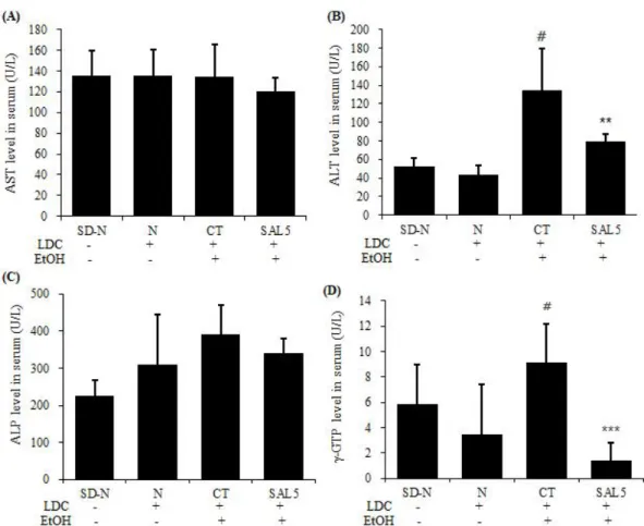
1. 혈청 중 AST, ALT, ALP, γ -GTP 활성 변화
혈청 내 AST (Fig. 1A)활성은 모든 실험군에서 큰 차이가 나타나지 않았고, ALT (Fig. 1B)와 γ-GTP (Fig. 1D)활성은 LDC-정상군에 비하여 대조군에서 유의성 있게 증가하였으며, SAL5 200 ㎎/㎏ 투여군에서는 대조군에 비하여 유의성 있게 감소하였다. ALP (Fig. 1C)활성은 LDC-정상군에 비하여 대 조군에서 증가하였지만 유의성은 없었고, SAL5 200 ㎎/㎏
투여군에서도 대조군에 비하여 감소하였지만 유의성은 없었다.
Fig. 1. Effects of SAL5 on the AST, ALT, ALP and γ-GTP activity in serum of chronic ethanol-induced fatty liver model.
The rats were orally administered with SAL5 for 5 weeks. Blood samples were collected using cardiac puncture method. SD-N: diet, N: LDC diet, CT: ethanol diet, SAL5: ethanol diet with 200 ㎎/㎏. The results were expressed the mean ± SD (N = 8). Statistically significant value compared with control group data by student`s t-test. (**p < 0.01, ***p < 0.001) # : p < 0.05 compared with LDC-normal group.
2. 간 중성지방 함량 변화
간에서 triglyceride (TG)함량은 LDC-정상군에 비하여 대 조군에서 유의성 있게 증가 하였으며, SAL5 200 ㎎/㎏ 투여 군에서는 유의적으로 감소하는 것으로 나타났다(Fig. 2).
3. 간 조직학적 분석
지방간 증상 확인을 위해서 조직학적 분석을 한 결과, LDC-정상군에 비하여 대조군에서는 간세포의 지방구의 크 기가 넓고 크게 분포하고 있는 것을 확인 하였고, SAL5 200
㎎/㎏ 투여군은 대조군에 비하여 간세포의 지방구의 크기가
비교적 작고 고르게 퍼져 있는 것을 확인 할 수 있었다(Fig. 3).
Fig. 2. Effects of SAL5 on the triglyceride (TG) in liver of chronic ethanol- induced fatty liver model.
The rats were orally administered with SAL5 for 5 weeks. After the animals were sacrificed, the liver was removed and TG concentration were measured.
SD-N: diet, N: LDC diet, CT: ethanol diet, SAL5: ethanol diet with 200 ㎎/㎏.
The results were expressed the mean ± SD (N = 8). Statistically significant value compared with control group data by student`s t-test. (*p < 0.05) # : p < 0.05 compared with LDC-normal group.
Fig. 3. Effect of SAL5 on fat accumulation in liver tissue of chronic ethanol-induced fatty liver model.
The rats were orally administered with SAL5 for 5 weeks. At the end experiment, the livers were fixed, and histologic analysis was performed.
These images were captured under a light microscope at x100 magnification.
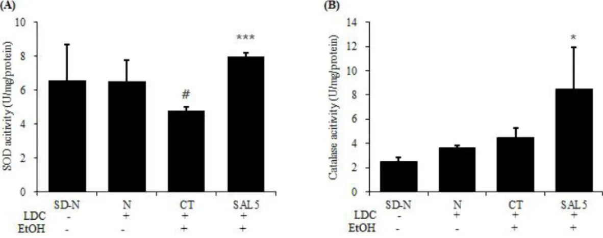
4. 간 조직 항산화효소 활성
SOD 활성 (Fig. 4A)은 LDC-정상군에 비하여 대조군에서 유의성 있게 감소하였으며, SAL5 200 ㎎/㎏ 투여군에서는 대조군에 비하여 유의성 있게 증가하였고, Catalase 활성 (Fig. 4B)은 SAL5 200 ㎎/㎏ 투여군에서 대조군에 비하여 유의성 있게 증가하였다.
Fig. 4. Effects of SAL5 on the SOD and catalase activity in liver tissue of chronic ethanol-induced fatty liver model.
The rats were orally administered with SAL5 for 5 weeks. After the animals were sacrificed, the liver was removed and SOD, catalase activity were measured. SD-N: diet, N: LDC diet, CT: ethanol diet, SAL5: ethanol diet with 200 ㎎/㎏. The results were expressed the mean ± SD (N = 8). Statistically significant value compared with control group data by Student`s t-test. (*p < 0.05, ***p < 0.001) # : p < 0.05 compared with LDC-normal group.
5. 간 조직 MDA, GSH 함량
간 조직내에서 MDA 함량 (Fig. 5A)은 LDC-정상군에 비하여 대조군에서 증가하였으며, SAL5 200 ㎎/㎏ 투여군에서 대조 군에 비하여 감소하였으나 유의성은 없었다. GSH 함량 (Fig. 5B)은 LDC-정상군에 비하여 대조군에서 감소하였으나 유의성은 없었고, SAL5 200 ㎎/㎏ 투여군에서 대조군에 비하여 유의성 있게 증가되었다.
Fig. 5. Effects of SAL5 on the MDA and GSH concentration in liver tissue of chronic ethanol-induced fatty liver model.
The rats were orally administered with SAL5 for 5 weeks. After the animals were sacrificed, the liver was removed and MDA, GSH concentration were measured. SD-N: diet, N: LDC diet, CT: ethanol diet, SAL5: ethanol diet with 200 ㎎/㎏. The results were expressed the mean ± SD (N = 8). Statistically significant value compared with control group data by student`s t-test. (*p < 0.05) # : p < 0.05 compared with LDC-normal group.
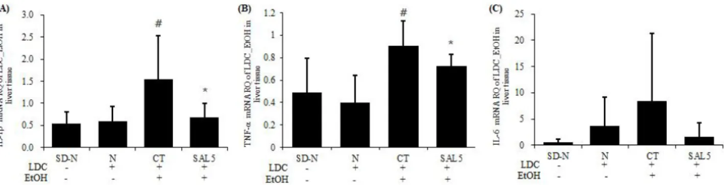
6. 간 조직 염증사이토카인 유전자 발현 분석
간 조직에서 유전자 발현은 대조군의 IL-1β, TNF-α, IL-6 mRNA 유전자 발현의 RQ값을 1로 했을 때 SAL5 200 ㎎/㎏ 투 여군의 상대정량 값을 분석하였다. IL-1β , TNF-α mRNA 유전자 발현 (Fig. 6A, B)은 LDC-정상군에 비하여 대조군에서 유의성 있게 증가하였으며, SAL5 200 ㎎/㎏ 투여군에서는 대조군에 비하여 유의성 있게 감소되었다. IL-6 mRNA 유전자 발현 (Fig. 6C)은 LDC-정상군에 비하여 대조군에서 증가하였지만 유의성은 없었고, SAL5 200 ㎎/㎏ 투여군에서는 대조군에 비하여 감소하였지만 유의성은 없었다.
Fig. 6. Effects of SAL5 on pro-inflammatory cytokines mRNA expression in liver tissue of chronic ethanol-induced fatty liver model.
The rats were orally administered with SAL5 for 5 weeks. After the animals were sacrificed, the liver was removed and IL-1β, TNF-α and IL-6 mRNA gene expression analysed quantitative real-time PCR at the end of the experiment. SD-N: diet, N: LDC diet, CT: ethanol diet, SAL5: ethanol diet with 200 ㎎/㎏. The results were expressed the mean ± SD (N = 8). Statistically significant value compared with control group data by student`s t-test. (*p < 0.05) # : p < 0.05 compared with LDC-normal group.
7. 간 조직 NOS-II, COX-2 mRNA 유전자 발현 분석
간 조직에서 유전자 발현은 대조군의 NOS-II, COX-2 mRNA 유전자 발현의 RQ값을 1로 했을 때 SAL5 200 ㎎/㎏ 투여군의
상대정량 값을 분석하였다. NOS-II mRNA 유전자 발현 (Fig. 7A)은 LDC-정상군에 비하여 대조군에서 유의성 있게 증가하였
으며, SAL5 200 ㎎/㎏ 투여군에서는 NOS-II mRNA 유전자발현이 대조군에 비하여 유의성 있게 감소하였다. 그리고, COX-2
mRNA 유전자 발현 (Fig. 7B)은 LDC-정상군에 비하여 대조군에서 증가하였지만 유의성은 없었고, SAL5 200 ㎎/㎏ 투여군에 서는 COX-2 mRNA 유전자발현이 대조군에 비하여 감소하였지만 유의성은 없었다.
Fig. 7. Effects of SAL5 on NOS-II, COX-2 mRNA expression in liver tissue of chronic ethanol-induced fatty liver model.
The rats were orally administered with SAL5 for 5 weeks. After the animals were sacrificed, the liver was removed and NOS-II, COX-2 mRNA gene expression analysed quantitative real-time PCR at the end of the experiment. SD-N: diet, N: LDC diet, CT: ethanol diet, SAL5:
ethanol diet with 200 ㎎/㎏. The results were expressed the mean ± SD (N = 8). Statistically significant value compared with control group data by student`s t-test. (**p < 0.01) # : p < 0.05 compared with LDC-normal group.
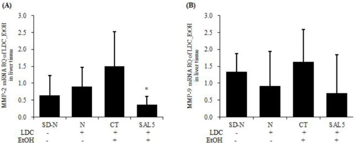
8. 간 조직 MMP-2, MMP-9 mRNA 유전자 발현 분석
간 조직에서 유전자 발현은 대조군의 MMP-2, MMP-9 mRNA 유전자 발현의 RQ값을 1로 했을 때 SAL5 200 ㎎/㎏ 투여군의 상대정량 값을 분석하였다. MMP-2, MMP-9 mRNA 유전자 발현 (Fig. 8A, B)은 LDC-정상군에 비하여 대조군에서 증가하였 지만 유의성은 없었고, SAL5 200 ㎎/㎏ 투여군에서는 MMP-2 mRNA 유전자발현 (Fig. 8A)이 대조군에 비하여 유의성 있게 감소되었다.
Fig. 8. Effects of SAL5 on MMP-2, MMP-9 mRNA expression in liver tissue of chronic ethanol-induced fatty liver model.
The rats were orally administered with SAL5 for 5 weeks. After the animals were sacrificed, the liver was removed and MMP-2, MMP-9 mRNA gene expression analysed quantitative real-time PCR at the end of the experiment. SD-N: diet, N: LDC diet, CT: ethanol diet, SAL5:
ethanol diet with 200 ㎎/㎏. The results were expressed the mean ± SD (N = 8). Statistically significant value compared with control group data by Student`s t-test. (*p < 0.05)
Ⅳ. 고 찰
본 연구는 만성적인 알코올 섭취 시 나타나는 알코올성 지방 간에서 SAL5 복합물이 알코올성 지방간의 형성 및 간 손상에 미치는 영향을 알아보고자 수행되었다. 알코올성 지방간 동물 모델은 Lieber-DeCarli 액체 표준식이 섭취를 통하여 유도 하였다. Lieber-DeCarli 액체 표준식이 사용할 경우 알코올
섭취량을 늘릴 수 있고, 이로 인해 지속적으로 혈중 알코올
농도를 높게 유지시킬 수 있다
30). 간은 유입되는 혈액의 지방
산을 이용하여 중성지방을 합성하고 필요시에는 중성지방을
혈액으로 방출하기도 하는데, 장기간의 알코올 섭취에 의한
간조직의 손상은 중성지방의 간외 유출을 억제하여 혈액순화
계의 중성지방 및 기타 지방의 농도를 낮추고 동시에 간의 조직
축적으로 유도하는 것으로 알려졌다
31). 혈청의 AST와 ALT의
활성은 고지방 식이, 고콜레스테롤 식이, 알코올 등으로 인한 간세포 독성 시 간세포에 장애가 발행하고, 간세포의 괴사가 진행되어 혈액으로 AST와 ALT가 유리됨에 따라 혈장 내에서 활성이 증가한다고 알려져 있다
32). 간세포의 모세관담측 융모, 담관상피 등에 주로 존재하고 골격 내 석회화를 촉진시키고, 장내 인 흡수 등에 관여하는 효소인 ALP는 골질환이나 간질환 등에서 활성이 높게 나타나는 것으로 알려져 있다
33). 또한, 혈중 γ -GTP는 지방간 유발시 활성이 증가하는데 특히, 알코 올에 민감하게 반응하고 간이나 담도에 질환이 있으면 다른 효소보다 높은 이상치를 보이는 것으로 보고되고 있다
34).
본 연구에서 SAL5 200 ㎎/㎏ 투여로 ALT, γ-GTP의 혈중 농도는 대조군에 비해 유의성 있게 감소하였고, ALP 농도는 대조군에 비해 감소하였지만 유의성은 없었다. 또한 간에서 triglyceride 활성을 유의적으로 감소시킴으로서 SAL5는 알 코올에 의한 독성으로부터 간을 보호하고 간세포 손상을 예방할 수 있다고 사료된다. 지방간은 과다한 알코올 섭취로 인한 간 독성 유발의 초기증상으로 간세포에 산소나 영양적인 측면에서 불균형을 초래하는 것으로 알려져 있다
35). 간세포 내의 지방 입자는 지방간이 유발됨으로서 수와 부피가 증가하게 된다
36). 지방간 증상을 판단하기 위해 H&E, Oil red O 염색을 실시 하여 조직학적 분석을 하였다. LDC-정상군에 비하여 대조군 에서 간세포의 지방조직이 넓고 크게 분포하고 있는 것을 확인 할 수 있었고, 대조군에 비하여 SAL5 200 ㎎/㎏ 투여군에서는 간세포에 축적된 지방구가 비교적 작고 고르게 퍼져 있는 것을 확인 할 수 있었다. 이는 SAL5 투여로 인해 알코올을 섭취한 동물의 간지질 함량을 개선하여 간 조직 지방질 축적에 효과를 나타낸 것으로 사료된다. 만성적인 또는 과량의 알코올 섭취는 NADH/NAD+의 비율을 증가시켜 탄수화물, 지방 및 단백질 대사 장애를 초래하는데 특히 간에서 지방산의 산화가 억제되는 동시에 지방산 합성이 증가되어 지방간을 초래한다
37,38). 또한 간세포의 마이크로좀 알코올 산화효소 (MEOS)를 유도하고 이 과정에서 유도되는 cytochrome P4502E1 (CYP2E1)은 reactive oxygen species (ROS)를 정상적인 조건에서보다 4~8배 정도 많이 생성한다. 이때 생성된 ROS가 생체막의 불포화지방산에 작용, 과산화지질을 생성하여 세포의 산화적 손상을 초래하고 여러 질환의 발생 및 노화를 촉진하게 된다
39-42). 인체에는 과잉 생성된 ROS를 제거하여 세포막과 세포내 물질을 보호하기 위한 항산화 기전이 존재하는데 그 중 한 가지 방법은 항산화 비타민이나 flavonoids와 같은 항산화제에 의해 제거되는 것 이고 다른 한 가지 방법은 superoxide dismutase (SOD), catalase, glutathione S-transferase (GST), glutathione peroxidase (GSH-px) 등과 같은 항산화 효소에 의해 제거 되는 것으로 이러한 기전이 산화적인 손상으로부터 신체를 방어 하는 역할을 한다
43,44).
본 연구에서 SAL5 200 ㎎/㎏ 투여로 SOD와 Catalase 활 성이 대조군에 비해 유의성 있게 증가된 것으로 보아 항산화 효소들을 효과적으로 활성화시켜 알코올성 산화스트레스로부터 간 보호 효과를 갖는 것으로 사료된다. 지질과산화는 활성산
소종들에 의해 매개된 기작으로서 여러 동물 인체실험들을 통 하여 다양한 종류의 간 손상을 일으키는 것으로 알려져 왔다
45). Malondialdehyde (MDA)는 지질과산화 과정 중 생성되는 대 표적 활성 알데히드로, 조직 내 알코올에 의해 유도된 지질과 산화물의 함량은 MDA 양으로 측정될 수 있다
46). 본 연구에서 MDA 함량은 LDC-정상군에 비해 대조군에서 유의성 있게 증가하였고, SAL5 200 ㎎/㎏ 투여군에서는 대조군에 비해 감소하였지만 유의성은 없었다. 하지만 MDA 함량이 감소된 것으로 보아 알코올에 의해 유도된 지질과산화 과정을 억제하 는데 효과가 있다고 사료된다. GSH는 생명체에 존재하는 주요 비단백 thiol로서 인체의 산화적 손상에 대한 방어체계에서 중요한 역할을 담당한다. 과산화물을 환원시키는 다양한 효소 과정에서 각종 효소들과 함께 세포 내외적으로 주요한 역할을 하는 비효소계 항산화제인 GSH는 redox 반응 및 해독작용을 통하여 정상 세포들의 구조 및 기능을 유지시켜준다
47). GSH 함량은 LDC-정상군에 비해 대조군에서 감소하였으며, SAL5 200 ㎎/㎏ 투여군에서 유의성 있게 증가된 것으로 보아 알코올 섭취로 인해 GSH 함량이 감소되는 것을 보호했을 것이라고 사료된다. 간세포의 염증과 관련된 cytokine 유전자인 TNF-α , TGF-β 1, IL-1β, IL-6, IL-8 등의 발현정도와 간 손상 정 도와는 상당한 연관성이 있다고 알려져 있으며
48-50), 모두 간 조직의 감염이 시작 또는 진행되는 과정에서 그 발현이 증가 한다. 염증 cytokine 유전자 발현 분석은 염증 인자인 NOS-II, COX-2, TNF-α, IL-1 β, IL-6을 통하여 mRNA 유전자발 현 분석을 하였으며, SAL5 200 ㎎/㎏ 투여군에서 대조군에 비해 IL-1β , TNF-α , NOS-II mRNA 유전자발현은 유의성 있게 감소되었으며, IL-6와 COX-2 mRNA 유전자 발현도 감소되는 것을 확인하였다. 또한 간성상세포와 쿠퍼세포에서 생성되는
51)간 섬유화 관련 유전자인 MMP-2, MMP-9 mRNA 유전자 발현은 SAL5 200 ㎎/㎏ 투여군에서 대조군에 비하여 감소되는 것으로 나타났다. 이러한 연구결과를 바탕으로 SAL5는 알코올에 의한 간 손상을 보호하는데 기여할 수 있을 것이라고 사료된다.
Ⅴ. 결 론
본 연구에서는 만성적 에탄올의 섭취에 의해 유도된 지방간 쥐 모델에서 SAL5의 알코올성 지방간 형성에 미치는 영향에 대해 관찰하여 다음과 같은 결과를 얻었다.
1. 혈중 ALT, γ -GTP 그리고, 간 triglyceride 함량은 SAL5 200 ㎎/㎏ 투여군에서 유의성 있게 감소하였다.
2. SAL5 200 ㎎/㎏ 투여군에서 알코올 섭취로 인해 증가 되는 지방축적을 억제 하였다.
3. SOD, catalase 활성은 SAL5 200 ㎎/㎏ 투여군에서
유의성 있게 증가 하였다.
4. SAL5 200 ㎎/㎏ 투여군에서 MDA 함량은 감소되었으며, GSH 함량은 유의성 있게 증가 하였다.
5. IL-1beta, TNF-α, NOS-II mRNA 유전자 발현은 SAL5 200 ㎎/㎏ 투여군에서 유의성 있게 감소되었고, IL-6, COX-2 mRNA 유전자 발현은 감소되었지만 유 의성은 없었다.
6. SAL5 200 ㎎/㎏ 투여군에서 MMP-2 mRNA 유전자 발현은 유의성 있게 감소되었고, MMP-9 mRNA 유전자 발현은 감소되었지만 유의성은 없었다.
결론적으로 간 기능 지표수준을 개선시키고, 지질과산화물 수준을 감소시키며, 항산화 효소 활성 증가와 GSH 함량 감소 억제, 간 조직에서 지방축적 감소효과 등을 통해 SAL5는 알 코올에 의해 형성된 지방간을 개선하는데 기여할 수 있을 것 으로 판단된다.
감사의 글
이 논문은 (주)유니베라의 지원으로 수행된 연구결과입니다.
References