Original Article
PET/CT 검사에서 Q.Clear 기법의 유용성에 대한 고찰연세의료원 세브란스병원 핵의학과
최용훈·김정열·최영숙·임한상·김재삼
The Usefulness of Q.Clear Technique in PET / CT
Yong Hoon Choi, Jung Yul Kim, Young Sook Choi, Han Sang Lim and Jae Sam Kim Department of Nuclear Medicine, Severance Hospital, Yonsei University Health System, Seoul, Korea
Purpose Recently, the performance of PET/CT scanner has been improved and various techniques have been developed to increase the image quality such as Sensitivity and Resolution. The purpose of this study is to evaluate the usefulness of Q.Clear (a fully convergent iterative reconstruction) technique of GE Discovery IQ equipment to enhance the image quality.
Materials and Methods All scans were acquired by Discovery IQ (GE Healthcare, MI, USA). In NEMA IEC Body Phantom test, Background to Hot-sphere (10 ㎜, 13 ㎜, 17 ㎜, 22 ㎜) ratio was 1:4 and scan time was 3 minutes. The images were reconstructed by VPHDs (VUE Point High-Definition + SharpIR) and Q.Clear to evaluate each Contrast.
We injected 18F-FDG 187 M㏃ to PET/SPECT Performance Phantom. And then it was scanned for 4 minutes to evaluate Resolution and Uniformity. T-test statistical analysis was performed on SUVmax of small lesions less than 2 ㎝ in 100 clinical patients regardless of disease type.
Results In the NEMA IEC Body Phantom, the Contrast was 63.6 ± 5.7% (VPHDs) and 75 ± 4.8% (Q.Clear). In the PET/SPECT Performance Phantom, the Resolution was 9.2 ㎜ (VPHDs) and 7.3 ㎜ (Q.Clear). Uniformity of Q.Clear was 10.8% better than VPHDs. T-test statistic of the clinical patients showed a significant difference of p value of 0.021.
Conclusion Both the phantom test and the clinical results showed that the quality of the image was improved in Q.Clear was applied. The SUVmax was highly measured in Q.Clear and the lesions were clearly distinguished visually.
Therefore Q.Clear can be useful in various aspects such as dose-reduction, patients evaluation and image analysis.
Key Words Q.Clear, VPHDs, SUVmax
서 론2)
시대의 발전에 따라 양전자방출단층촬영(Positron Emission Tomography, PET)은 전산화단층촬영술(Computed Tomography, CT)과 융합 되었고 지속적인 영상의 발전을 이루고 있다. 이 에 따라 해상도(resolution)와 민감도(sensitivity)가 향상 되었
·Received: September 29, 2017 Accepted: October 20, 2017
·Corresponding Author: Yong Hoon Choi
·Address for correspondence : Department of Nuclear Medicine, Severance Hospital, Yonsei University Health System 50-1 Yonsei-ro, Seodaemun-gu, Seoul, 120-749, Korea Tel: (82-2)2228-4852, Fax: +82-2-2227-7062 E-mail: [email protected]
고 피폭 저감, 검사 시간 단축 등의 다양한 이점을 얻게 되었다.
뿐만 아니라 영상의 재구성 알고리즘(Reconstruction algorithm) 의 발전으로 인하여 영상의 해상도를 높이고 더 정확한 재구 성 영상을 얻을 수 있게 되었다.1)
이러한 PET 영상의 문제점으로는 작은 병소에서의 계수율 감소를 들 수 있다. 부분 체적 효과(Partial Volume Effect, PVE)에 의하여 발생되는 작은 병소의 계수율 감소는 표준 섭 취 계수(Standardized Uptake Value, SUV)를 감소시킨다.2) 영 상의 재구성 Parameter 중 Iteration 횟수 증가로 이를 어느 정 도 보상 할 수 있으나 이에 따른 영상 잡음이 증가하며 결과적 으로 영상의 질이 떨어지게 된다.
이를 극복하기 위한 방법으로 GE사에서 개발한 Q.Clear는
영상의 재구성 알고리즘에서 영상 잡음을 조절할 수 있다. 그 에 따라 Iteration이 증가하여도 신호 대 잡음비(Signal to Noise Ratio, SNR)가 우수한 영상을 얻을 수 있다.
따라서 본 논문에서는 GE사의 Q.Clear가 팬텀과 환자에게 서 유용성이 있는지 알아보고자 한다.
실험재료 및 방법 1. 장비 및 대상
장비는 Discovery IQ(GE Healthcare, MI, USA)를 사용하였 고(Fig. 1), 팬텀 실험은 NEMA IEC Body Phantom(Fig. 2)과 PET/SPECT Performance Phantom(Fig. 3)을 사용하였다.
임상 영상은 2016년도 10월부터 2017년도 3월까지 18F-FDG PET/CT 검사를 시행한 환자들 중 질병에 상관없이 2 ㎝ 미만 의 국소 병변이 발견된 100명을 대상으로 무작위로 선택하여 분석 하였다.
Fig. 1. Discovery IQ PET/CT Scanner was used for acquisition.
Fig. 2. NEMA IEC Body Phantom was used.
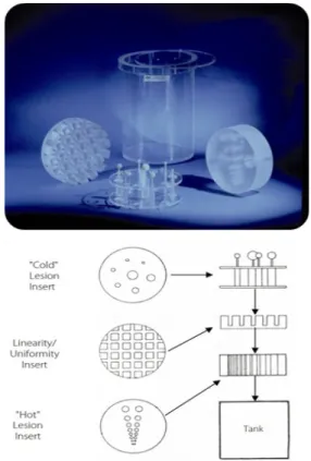
Fig. 3. PET/SPECT Performance Phantom was used.
2. 연구 방법
1) 팬텀 실험
NEMA IEC Body Phantom의 CT는 관전압 120 ㎸p, 관전류 40 ㎃, 절편 두께(Slice Thickness) 3.75 ㎜, 회전 시간(Rotation Time) 0.5 sec로 설정하였다. 배후방사능과 열소의 주사 선량
한 방법과 Q.Clear로 하였으며 Beta value는 400으로 설정하 였다.3)
PET/SPECT Performance Phantom의 조건은 CT가 관전압 120 ㎸p, 관전류 40 ㎃, 절편 두께(Slice Thickness) 3.75 ㎜, 회 전 시간(Rotation Time) 0.5 sec로 설정하였다. 18F-FDG 187 M
㏃을 주입 후 PET 방출 영상 획득 시간을 1 bed 당 4분으로 획 득하였다. 영상의 재구성은 OSEM기법인 VUE Point High- Definition,(VPHD)에 SharpIR을 추가한 방법과 Q.Clear로 하 였으며 Beta value는 400으로 설정하였다.
2) 임상 영상
환자는 검사 전 8시간 이상의 금식을 시행 하였고 500-1,000
㎖ 이상의 충분한 수분을 섭취한 상태로 검사를 진행하였다.
성별은 여성이 71명 남성이 29명이었고 나이는 58.7±12.7세 였다. 혈당은 96.3±9.7이었으며 표준섭취계수 최대값(SUVmax) 을 측정하여 분석하였다. 18F-FDG를 2.6M㏃/㎏ 주입하였고 1 bed 당 3분씩 획득하였으며 VPHDs(VUE Point High-Definition + SharpIR)와 Q.Clear로 각각 재구성하였다.
3. 영상 분석
1) 팬텀 실험
NEMA IEC Body Phantom의 Contrast결과는 6개의 sphere 와 배후 방사능영역에 관심영역(Region of Interest, ROI)을 3 회씩 설정하여 평균치로 비교 분석하였다. PET/SPECT Performance Phantom에서는 해상도와 균일도를 측정하였다.
영상 분석을 위하여 MIMvista workstation(software version 6.5) (MIM Software Inc., Cleveland, OH, USA)을 사용하였다.
2) 임상 영상
VPHDs 와 Q.Clear로 재구성한 임상 영상의 분석을 위 하여 팬텀 실험과 동일하게 MIMvista workstation (software version 6.5) (MIM Software Inc., Cleveland, OH, USA) 을 사용하였다. VPHDs와 Q.Clear로 재구성된 영상에서 병소 부위에 체적관심영역(Volume of Interest, VOI)을 설정하여 SUVmax를 각각 측정하여 비교 분석 하였다.
통계분석으로는 독립샘플 t-test를 진행하였으며 SPSS version 20.0을 사용하였다.
4. Q.Clear 원리
Q.Clear는 Bayesian penalized likelihood (PL) 재구성 알고리즘을 사용한다.4-5) 영상의 잡음이 증가하면 잡음이 많은 영역으로부터 멀리 떨어져서 영상을 최적화한다.
이를 통해 OSEM에서 발견되는 과도한 영상잡음의 악영 향 없이 최적의 영상에 도달할 수 있다.
Q.Clear는 또한 BSREM (Block Sequential Regularized Expectation Maximization) 알고리즘을 사용한다.6-7) BSREM 알고리즘은 단일 영상의 복셀이 부분적이거나 완전하거 나 혹은 과하게 나타나게 될지라도 이를 매번 100% 에 가깝게 구현할 수 있도록 하여 준다.
RDP (Relative Difference Penalty)를 사용하며 영상 잡음을 조절할 수 있는 이점이 있다.8)
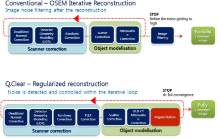
그리고 정규화에서 변조를 통하여 영상 품질과 정량간 의 균형을 최적으로 절충 해준다. 단점이었던 가장자리 가 보상이 되고 영상의 배후방사능에 의한 영상 잡음이 적게 유지되도록 설계되어 우수한 영상 품질을 제공한 다. Q.Clear에서 영상의 잡음은 정규화의 일부로 반복적 인 재구성 내에서 제어되기 때문에 사후 여과(Post filter) 가 필요하지 않게 되며 영상 잡음이 최소화된 최적의 영 상을 얻게 해준다(Fig. 3).
Fig. 4. Process flow maps for conventional OSEM iterative reconstruction and Q.Clear. Q.Clear is a fully convergent iterative reconstruction method. Unlike OSEM, Q.Clear controls noise as part of the regularization process inside iterative reconstruction.
Diameter Hot Spheres Cold Spheres
10 ㎜ 13 ㎜ 17 ㎜ 22 ㎜ 28 ㎜ 37 ㎜
1st Measured Contrast 42.1 51.6 66.1 78.2 67.8 77.6
2nd Measured Contrast 47.2 47.6 65.6 72 69.2 77.6
3rd Measured Contrast 33.7 60.8 66.5 72.4 71.1 78
Average 41.0 53.3 66.1 74.2 69.4 77.7
1st Measured Background 11.3 9.4 7 4.9 4 3.8
2nd Measured Background 13 10 7.6 5.6 4.4 3.3
3rd Measured Background 11.7 9.2 6.9 5.3 4 2.9
Average 12.0 9.5 7.2 5.3 4.1 3.3
Fig. 5. The ROIs in the NEMA IEC Body Phantom background area and at the 6 spheres for Contrast results.
Table 1. Contrast of NEMA IEC Body Phantom
결 과 1. 팬텀 실험
NEMA IEC Body Phantom의 4개의 열소와 2개의 냉소에 체 적관심영역을 설정하여 대조도(contrast)를 측정하였다(Fig.
5). VPHDs보다 Q.Clear에서 모든 영역의 결과가 높았으며, 평
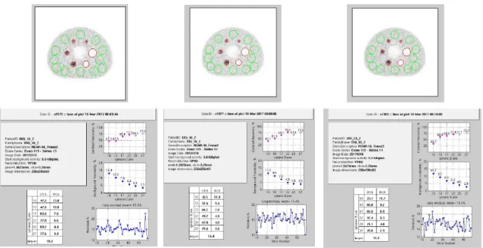
균적으로 약 12% 높게 나왔다(Table 1). Fig. 6. The resolution was 9.2 ㎜ and 7.3 ㎜, and the uniformity was 10.8% higher in Q.Clear.
2. 임상 영상
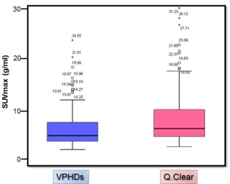
두 영상의 그룹에 대하여 SUVmax의 분포를 Box plot으로 분석한 결과 VPHDs보다 Q.Clear에서 평균 29.5% 가량 높게 측정 되었다(Fig. 7).
그리고 t-test결과 p-value가 0.021로 통계적으로 두 대조군 간의 유의한 차이가 있었다.
Fig. 7. This is a box plot that represents SUVmax of the two reconstruction.
임상영상에서는 VPHDs 영상 보다 Q.Clear에서 병소의 SUVmax가 높게 측정 되었고 육안상으로 병변의 구별이 용이 하였다(Fig. 8).
Fig. 8. PET/CT images of Clinical patient are individually reconstructed by OSEM and Q.Clear (A) OSEM SUVmax : 3.29 g/㎖
(B) Q.Clear SUVmax : 4.59 g/㎖
고 찰
핵의학과에 근무하는 직원이라면 미세 병변에서의 섭 취 감소나 심한 영상 잡음 등을 경험 하였을 것이다. 작
은 병변에서의 계수율 감소를 영상 잡음의 영향 없이 정상적으로 구현하기에는 어려움이 있다. 그래서 다양한 기법을 통해 영상의 품질을 올리려는 시도가 계속 되고 있다. 본 연구에서의 Q.Clear는 GE사의 최신 기법으로 써 영상 잡음을 줄이면서도 미세 병변을 정확하게 구현 하였고 팬텀실험에서도 기존의 재구성 영상보다 우수한 영상 품질을 보여 주었다. 이를 통하여 병변을 더욱 정 확하게 구별할 수 있게 되었고 환자의 치료평가와 예후 에 보다 정확하게 접근할 수 있게 되었다. Q.Clear는 단 순히 부분체적효과의 보상만이 아니라 전체적인 영상 품 질 향상에 그 목적이 있다. 작은 병소를 정확하게 구별 할 수 있을 뿐만 아니라 체중이 많거나 동위원소의 섭 취 불 충족 등 다양한 원인으로 발생하는 영상 품질 저 하를 보상하고 개선하여 준다. 하지만 본원 핵의학과 판 독의들은 Q.Clear의 새로운 재구성 방식과 OSEM 영상 과의 차이점을 고려하여 비교 판독을 통해 안정적인 결 과를 내는 것에 중점을 두고 있다. 따라서 Q.Clear의 적 용 시에는 이러한 부분을 고려해야 하며, 추후 다방면으 로의 유용성 입증을 위해서는 보다 다양한 연구를 통한 검증이 추가되어야 할 것으로 판단한다.
결 론
본 연구에서 Q.Clear를 적용한 팬텀과 임상 영상의 분석 결과 해상도, 균일도 및 대조도가 모두 우수하게 나왔다. 특히 본 연구에서 임상 영상은 직경 2㎝ 미만의 작은 병소를 대상으로 비교 분석을 하였는데 Q.Clear에 서 SUVmax가 높게 나왔고 육안상에서도 더욱 선명하게 나타났다. 이러한 영상의 질 향상은 선량 저감과 환자평 가 그리고 영상 분석 등 다양한 방면에서 활용할 수 있 을 것으로 사료된다.
요 약
최근 PET/CT 장비의 성능의 발전과 다양한 기법의 개발로 민감도와 해상도등 영상 품질을 개선할 수 있게 되었다. 본 논문에서는 GE사의 Discovery IQ 장비의 Q.Clear (a fully convergent iterative reconstruction) 기법 을 이용하여 영상의 질 향상에 유용성이 있는지 알아보 고자 한다.
장비는 Discovery IQ (GE Healthcare, MI, USA)를 사 용하였다. NEMA IEC Body Phantom의 배후방사능과
열소 체적(10 ㎜, 13 ㎜, 17 ㎜, 22 ㎜)의 비를 1:4로 하 고 3분간 촬영하여 VPHDs (VUE Point High-Definition SharpIR)와 Q.Clear의 대조도를 비교 분석하였다. PET/SPECT Performance Phantom에 18F-FDG를 187 MBq을 주입 후 4분간 촬영하여 해상도와 균일도를 비교 분석하였다. 그 리고 100명의 임상 환자에서 질환의 종류와 상관없이 2
㎝미만의 작은 병소의 SUVmax를 측정하여 t-test 통계 분석하였다.
NEMA IEC Body Phantom에서 VPHDs와 Q.Clear의 대조도가 63.6±5.7%, 75±4.8%로 나왔고 PET/SPECT Performance Phantom에서 해상도는 VPHDs가 9.2 ㎜, Q.Clear가 7.3 ㎜로 나왔다. 균일도는 Q.Clear가 10.8% 더 우수하였다. 임상 환자의 t-test 통계 결과 p-value가 0.021 로 유의한 차이가 있었다.
임상환자에서 SUVmax는 Q.Clear에서 높게 측정 되었 으며, 신호대 잡음 비도 우수하였다. 이는 부분체적효과 의 영향을 줄였기 때문으로 볼 수 있다. Phantom test와 임상 환자의 결과 모두 Q.Clear를 적용 하였을 때 영상 품질이 향상된 것을 확인하였다. 이러한 영상 품질 향상 은 병소를 더욱 정확하게 발견할 수 있고 나아가 선량 저감과 환자평가 그리고 영상 분석 등 다양한 방면에서 활용할 수 있을 것으로 사료된다.
참고문헌
1. 고창순. 고창순 핵의학. 제3판. 고려의학. 2008. P95-104.
2. Marine Soret, Stephen L. Bacharach, and Irène Buvat.
Partial-Volume Effect in PET Tumor Imaging. J Nucl Med.
2007;48:932-945.
3. Steve Ross. Q.Clear†. Available at:
http://www3.gehealthcare.com/en/search?q=Qclear&u=ht tp://www3.gehealthcare.com/en/search?q=steve%20ross&u
=http://www3.gehealthcare.com/en. 2013 General Electric Company-All rights reserved.
4. C-T.Chen, V. E. Johnson, W. H. Wong, X. Hul, and C. E.
Metz, Bayesian Image Reconstruction in Positron Emission Tomography, IEEE Transactions on Nuclear Science.
1990;37(2):636-641.
5. E.U. Mumcuoglu, R. Leahy, S.R.Cherry, Z. Zhou, Fast gradient-based methods for Bayesian reconstruction of transmission and emission PET images, IEEE Transactions on Medical. Imaging. 1994;13(4):687-701.
6. A. R. De Pierro and M. E. B. Yamagishi, Fast EM-like methods for maximum ‘a posteriori’ estimates in emission tomography, IEEE Transactions on Medical Imaging.
2001;20:280–288.
7. S. Ahn and J.A. Fessler, Globally convergent image reconstruction for emission tomography using relaxed ordered subsets algorithms, IEEE Transactions on Medical Imaging. 2003;22(5):613–26.
8. E. Asma, S. Ahn, S. Ross, A. Chen, and R. Manjeshwar, Accurate and consistent lesion quantitation with clinically acceptable penalized likelihood images, IEEE Nuclear Science Symposium Conference Record. 2012;23(7):
4062-4066