Assessment of PET-CT as a
Supplement to CT/MRI for Detection
of Nodal Metastasis in Hypopharyngeal
Squamous Cell Carcinoma Patients
with Palpably Negative Neck
Ho-Joon Lee
Department of Medicine
Assessment of PET-CT as a
Supplement to CT/MRI for Detection
of Nodal Metastasis in Hypopharyngeal
Squamous Cell Carcinoma Patients
with Palpably Negative Neck
Directed by Professor Jinna Kim
The Master's Thesis
submitted to the Department of Medicine,
the Graduate School of Yonsei University
in partial fulfillment of the requirements for the
degree of Master of Medical Science
Ho-Joon Lee
June 2014
This certifies that the Master's Thesis
of Ho-Joon Lee is approved.
Thesis Supervisor : Jinna Kim
Thesis Committee Member#1 : Yoon Woo Koh
Thesis Committee Member#2 : Won Jun Kang
The Graduate School
Yonsei University
ACKNOWLEDGEMENTS
I acknowledge my deepest gratitude to Professor Jinna
Kim, who is my thesis director, for supporting my efforts with
endless perseverance and commitment, facilitating every step
of the process. My appreciation for her guidance and
encouragement is tremendous. I am also indebted to Professor
Yoon Woo Koh and Won Jun Kang, for their help for pertinent
advice to assure the superior quality of this paper. I also thank
Jung Hwa Hong and Hye Sun Lee (Biostatistics Collaboration
Unit, Yonsei University College of Medicine, Seoul, Korea)
for their help in the statistical analyses.
<TABLE OF CONTENTS>
ABSTRACT
···1
I. INTRODUCTION
···3
II. MATERIALS AND METHODS
···4
1. Patient population
···4
2. Image protocol
···5
3. Surgery and histopathology
···7
4. Image interpretation
···7
5. Statistical analysis
···8
III. RESULTS
···10
IV. DISCUSSION
···15
V. CONCLUSION
···17
REFERENCES
···18
LIST OF FIGURES
Figure 1. Receiver operating characteristic (ROC) curves of
PET-CT, CT/MR, and their combined interpretation for
detecting nodal metastasis in hypopharyngeal squamous cell
carcinoma patients with palpably negative neck.···12
Figure 2. A 82-year-old man with left pyriform sinus cancer
(T1N1) with nodal metastasis at left level II who showed
false negative results on CT/MR but correctly diagnosed as
metastasis by PET-CT. ···14
Figure 3. A 55-year-old man with right posterior pharyngeal
wall cancer (T2N1) with nodal metastasis at right level II,
with occult nodal metastasis, missed by CT/MR and
PET-CT···14
LIST OF TABLES
Table 1. Patient characteristics.···10
Table 2. Diagnostic performance of PET-CT, CT/MR, and
their combined interpretation for detecting nodal metastasis
in hypopharyngeal squamous cell carcinoma patients with
palpably negative neck.···11
Table 3. PET-CT results for detecting nodal metastasis in the
clinically negative and positive neck groups of
ABSTRACT
Assessment of PET-CT as a Supplement to CT/MRI for Detection of
Nodal Metastasis in Hypopharyngeal Squamous Cell Carcinoma Patients
with Palpably Negative Neck
Ho-Joon Lee
Department of Medicine
The Graduate School, Yonsei University
(Directed by Professor Jinna Kim)
PURPOSE: To determine the diagnostic value of 18F-fluorodeoxyglucose (FDG) positron emission tomography-computed tomography (PET-CT) compared with CT or magnetic resonance (MR) imaging for detecting nodal metastasis in hypopharyngeal squamous cell carcinoma (SCC) patients with palpably negative neck and to assess the supplementary role of PET-CT to CT/MR.
MATERIALS AND METHODS: A total of 39 patients with palpably negative neck (36 men and 3 women; average age 65.4 years) underwent tumor resection and neck dissection as a primary treatment. Preoperatively evaluated PET-CT, CT and/or MR, were retrospectively reviewed. The diagnostic performance of PET-CT, CT/MR and a combination of PET-CT and CT/MR was assessed with
2 histopathologic results as a gold standard.
RESULTS: Twenty (51.3%) of our 39 patients were found to have neck metastases. On a level based analysis, the sensitivity of PET-CT, CT/MR, and combined interpretation of the two was 65.7%, 57.1%, and 65.7%, respectively, but without statistical significance. After the patients were categorized as cN0 neck based on CT/MR findings as well as palpation, 4 of the 6 patients who were falsely diagnosed as negative on CT/MR were still missed on PET-CT.
CONCLUSION: The addition of PET-CT examination to the anatomic imaging including CT and MR could not provide additional benefit for the preoperative evaluation of cervical nodal metastasis in hypopharyngeal SCC patients with non-palpable neck, showing insufficient results to spare elective neck dissection.
---
Key words: hypopharyngeal carcinoma, neck node metastasis, CT, MR,
Assessment of PET-CT as a Supplement to CT/MRI for Detection of
Nodal Metastasis in Hypopharyngeal Squamous Cell Carcinoma
Patients with Palpably Negative Neck
Ho-Joon Lee
Department of Medicine
The Graduate School, Yonsei University
(Directed by Professor Jinna Kim)
I. INTRODUCTION
Squamous cell carcinoma (SCC) of the hypopharyngeal region, which comprises 3-5% of all head and neck SCC, often present at advanced state with poor prognosis. The frequency of cervical lymph node metastasis and occult metastasis is very high in patients with hypopharyngeal SCC1, and its presence
is one of the most important prognostic factors, associated with the significant reduction of patient survival2.
For hypopharyngeal SCC patients with clinically positive neck, radical or modified radical neck dissection is usually performed as a standard approach for neck management. On the other hand, for patients with clinically negative neck, possibility of occult neck metastasis and unsatisfactory result of preoperative test have led surgeons to perform less morbid procedures such as selective neck dissection, as a diagnostic and elective therapeutic procedure3-5. Therefore, if a
4
preoperative imaging modality could predict negative neck with reasonable accuracy, it would help patients avoid associated morbidity as well as unnecessary treatment such as elective neck dissection and radiotherapy.
Computed tomography (CT) and magnetic resonance (MR) imaging are widely used for preoperative assessment of cervical lymph nodes as well as primary tumor in head and neck cancer, and has been implemented to routine clinical staging6. In addition, functional imaging with 18F-fluorodeoxyglucose
(FDG) positron emission tomography (PET)-CT has been recently introduced, and several studies have shown improved diagnostic performance compared to CT/MR in assessment of cervical nodal status in patients with head and neck cancer7-12, but whether addition of PET to the routinely performed anatomical
imaging would be beneficial to the patient, and when to add is uncertain. Therefore, the purpose of this study were to determine the diagnostic value of PET-CT and CT/MR for neck management in hypopharyngeal SCC patients with palpably negative neck, to assess the additive diagnostic values of PET-CT in patients with negative neck findings on CT/MR, and to provide relevant information for practical clinical workflow in these patients.
II. MATERIALS AND METHODS
1. Patient Population
We retrospectively reviewed 73 consecutive patients with pathologically diagnosed hypopharyngeal SCC who underwent curative resection of primary
tumor and neck dissection at a single institution during January 2006 to December 2012. This study was approved by the institutional review board, and considering the retrospective nature of this study, informed consent was waived. Among these patients, excluding 3 patients who received chemotherapy prior to surgery, a total of 39 patients who had no palpable lymph nodes in the neck and underwent PET-CT, CT and/or MR preoperatively were enrolled in our study (36 men and 3 women; average age 65.4 years, range 39 to 82 years). Patients with a history of prior treatment to the head and neck region, known distant metastasis, or second primary tumors were excluded.
2. Image Protocol
Contrast material-enhanced CT examinations were performed on one of two CT scanners (Somatom Sensation 16 or 64; Siemens, Erlangen, Germany). Contiguous 3-mm scans of the neck were acquired in the axial plane from the skull base to the carina, and then coronal images were reformatted at 2- to 3-mm increments. A total of 80-100 mL of contrast agent, iopromide (Ultravist 300; Bayer Schering Pharma, Berlin, Germany), was administered intravenously at a rate of 3 mL/sec using an automated injector. Contrast material-enhanced images were obtained 40–60 seconds after the initiation of contrast agent administration.
MR imaging was performed on a 1.5-Tesla or 3.0-Tesla magnet system (Intera or Achieva; Philips Medical Systems, Best, the Netherlands) with a
6
dedicated head and neck coil. Conventional MR imaging consisting of axial spin-echo (SE) T1-weighted and fat-saturated fast SE T2-weighted imaging was performed with a repetition time (TR; msec)/echo time (TE; msec) of 560/10 and 6480/70, respectively. All images were obtained with a 22-25 cm field of view and a section thickness of 3-5 mm. Gadopentetate dimeglumine (Magnevist; Bayer Schering Pharma, Berlin, Germany) was then administered intravenously at a dose of 0.2 mL/kg body weight and a rate of 2 mL/sec. Forty seconds after administering contrast material, fat-saturated axial and coronal SE T1-weighted images were obtained.
PET-CT was performed on a PET-CT scanner (Discovery STE; GE Healthcare, Milwaukee, WI, USA or Biograph TruePoint 40; Siemens Medical Systems, Knoxville, TN, USA) equipped with 16- or 40-slice CT, respectively. Patients were fasted for at least 6 hours before imaging, and the glucose level in peripheral blood was confirmed to be 140 mg/dL or less prior to the injection of FDG. A FDG dose of approximately 5.5 MBq/kg of body weight was administered intravenously 1 hour before image acquisition. After the initial low-dose CT study (Discovery STE, 30 mA, 130 kVp or Biograph TruePoint 40, 36 mA, 120 kVp), a standard PET protocol was used to scan from the neck to the proximal thighs with an acquisition time of 3 minutes per bed position in the three-dimensional mode. Images were reconstructed using ordered subset expectation maximization (2 iterations, 20 subsets).
3. Surgery and Histopathology
Patients underwent modified radical neck dissection or selective neck dissection (level I-V, II-V, II-IV, and II-III) based on preoperative clinical and radiologic findings. Level VI dissection was done in selected patients. For the ipsilateral neck, 9 patients underwent level I-V dissection, 4 patients underwent II-V dissection, 25 patients underwent II-IV dissection, and 1 patient underwent II-III dissection. For the contralateral neck, 1 patient underwent I-V dissection, 1 patient underwent II-V dissection, 20 patients underwent II-IV dissection and 1 patient underwent II-III dissection. Eight patients underwent additional level VI dissection.
The lymph nodes were dissected and labeled by the surgeons according to the imaging-based nodal classification13 and stained with hematoxylin and eosin
for histologic analysis. The results were given as a total number of the dissected lymph nodes and metastatic lymph nodes according to the neck levels.
4. Image Interpretation
The neck was divided into six levels based on the imaging-based nodal classification13. Level-by-level and patient-by-patient analyses were performed
for each imaging modality.
CT and MR were interpreted by two head and neck radiologists, and any disagreement was resolved by consensus. Nodes were considered metastatic when one of the following criteria was fulfilled: shortest axial diameter >11 mm
8
in the jugulodigastric region or >10 mm in other cervical regions, round shape (length/width <2), central necrosis, absence of fatty hilum or hilar vessel enhancement, or grouping of three or more nodes of borderline size. If patient underwent both of the CT and MR, the interpretation of CT and MR were combined. If there was discordance between the results of CT and MR, both were re-evaluated simultaneously and assessed in combination.
PET-CT images were interpreted on interactive workstations by two board-certified nuclear medicine physicians and any equivocal case was resolved by consensus. Readers were blinded to the results of the other imaging modalities and to the pathologic results at the time of the review. Image interpretation was based on visual and semiquantitative analysis using the attenuation-corrected PET emission images. For visual analysis, any focal FDG uptake greater than background activity and corresponding to nodular structures on CT, regardless of lymph node size, was considered abnormal. For semiquantitative analysis, a region of interest (ROI) was drawn at the lymph node of focal FDG uptake on the transverse section. The intensity of focal FDG uptake was expressed as the maximum standardized uptake value (SUVmax). Only hypermetabolic lesions
with strong focal uptake (SUVmax ≥2.5) were considered malignant.
For the combined result of PET-CT with CT/MR, any positive result on either PET-CT or CT/MR was considered positive.
The results of each imaging interpretation (PET-CT, CT/MR, and combined) were compared with histopathologic results from the neck dissection specimen as the gold standard for presence or absence of metastatic lymph nodes, on a level-by-level, and patient-by-patient basis.
Statistical analyses were performed using the Statistical Analysis System (version 9.2, SAS Institute Inc.; Cary, NC, USA) and MedCalc version 12.7.0 (MedCalc Software, Ostend, Belgium). The sensitivity, specificity, positive and negative predictive values (PPV and NPV), and accuracy were calculated for each imaging modalities and their diagnostic performance were compared using weighted least squares regression. Receiver operating characteristics (ROC) curves were generated and the area under the curve (AUC) was calculated. The package pROC14 for R statistical package (version 3.0.3, R foundation for
Statistical Computing; Vienna, Austria) was used for ROC curve plotting, and comparison of AUC was done with DeLong method.
In this study, patients were also categorized as clinically negative (cN0) and positive (cN+) neck, to assess the diagnostic value of PET-CT in the two groups. cN0 was defined as having no cervical lymph nodes palpated on physical examination and showing negative findings on CT/MR, since all patients undergo CT or MR preoperatively. Diagnostic values of PET-CT were analyzed, and Chi-square (Fisher’s exact test) method was used for the comparison of diagnostic performance and z test for the comparison of AUC. P values of less than .05 were considered to be statistically significant in all analyses.
10 III. RESULTS
Patient characteristics are summarized in Table 1.
Twenty-three patients (59.0%) underwent bilateral neck dissection and 16 patients (41.0%) underwent unilateral neck dissection. Of the 62 neck sides and 210 neck levels (1,706 lymph nodes) resected in all 39 patients, 24 neck sides and 35 neck levels (58 nodes) in 20 patients (51.3%) contained metastatic disease. Among the 24 affected neck sides (38.7%), 19 (30.6%) occurred at the ipsilateral side and 5 (8.0%) at the contralateral side, respectively. Of the 35 affected neck levels, 14 (40.0%) occurred at the ipsilateral level II, 9 (25.7%) at the ipsilateral level III, 5 (14.3%) at ipsilateral level IV, 5 (14.3%) at contralateral level II, and 2 (5.6%) at contralateral level III. The median number of lymph nodes removed per patient was 42 (range, 1-111), and that of metastatic lymph nodes was 1 (range, 0-10).
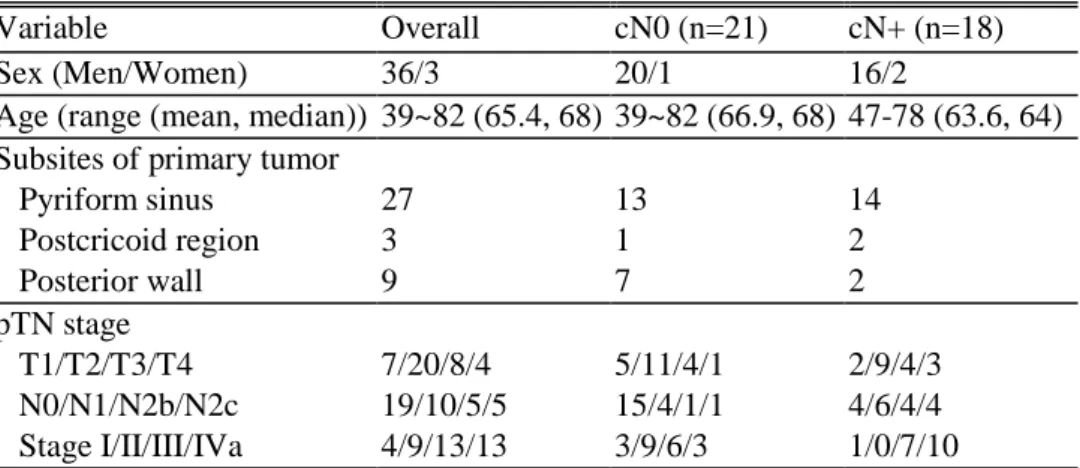
Table 1. Patient characteristics
Variable Overall cN0 (n=21) cN+ (n=18)
Sex (Men/Women) 36/3 20/1 16/2
Age (range (mean, median)) 39~82 (65.4, 68) 39~82 (66.9, 68) 47-78 (63.6, 64) Subsites of primary tumor
Pyriform sinus 27 13 14 Postcricoid region 3 1 2 Posterior wall 9 7 2 pTN stage T1/T2/T3/T4 7/20/8/4 5/11/4/1 2/9/4/3 N0/N1/N2b/N2c 19/10/5/5 15/4/1/1 4/6/4/4 Stage I/II/III/IVa 4/9/13/13 3/9/6/3 1/0/7/10 Abbreviations: cN0= negative neck based on palpation and CT/MR, cN+=positive neck based on palpation and CT/MR
The results of CT, CT/MR, and combined result of CT/MR and PET-CT in identifying metastatic neck nodes are summarized in Table 2. In the level based analyses, the sensitivity of PET-CT was higher than that of CT/MR, but without statistical significance (65.7% vs. 57.1%, P = 0.084). The combined interpretation of PET-CT and CT/MR (65.7%) showed also higher sensitivity than CT/MR alone, but did not yield different sensitivity from that of PET-CT alone. The specificity of combined interpretation was significantly lower than PET-CT alone (94.9% vs. 97.7%, P = 0.025) and CT/MR alone (94.9% vs. 97.1%, P = 0.046), respectively.
Table 2. Diagnostic performance of PET-CT, CT/MR, and their combined interpretation for detecting nodal metastasis in hypopharyngeal squamous cell carcinoma patients with palpably negative neck
Group Method TN FN FP TP Sensitivity (%) Specificity (%) Accuracy (%) PPV (%) NPV (%) Patient PET-CT 18 5 1 15 75.0 (56.0-94.0) 94.7* (84.7-100) 84.6 (73.3-96.0) 93.8 (81.9-100) 78.3 (61.4-95.1) (n=39) CT/MR 15 6 4 14 70.0 (49.9-90.1) 79.0 (60.6-97.3) 74.4 (60.7-88.1) 77.8 (58.6-97.0) 71.4 (52.1-90.8) Combined 14 4 5 16 80.0 (62.5-97.5) 73.7* (53.9-93.5) 76.9 (63.7-90.2) 76.2 (58.0-94.4) 77.8 (58.6-97.0) Level PET-CT 171 12 4 23 65.7 (50.0-81.4) 97.7* (95.5-99.9) 92.4* (88.8-96.0) 85.2* (71.8-98.6) 93.4 (89.9-97.0) (n=210) CT/MR 170 15 5 20 57.1 (40.8-73.5) 97.1† (94.7-99.6) 90.5 (86.5-94.5) 80.0 (64.3-95.7) 91.9 (88.0-95.8) Combined 166 12 9 23 65.7 (50.0-81.4) 94.9*† (91.6-98.1) 90.0* (86.0-94.1) 71.9* (56.3-87.5) 93.3 (89.6-96.9) Abbreviations: TN=true negative, FN=false negative, FP=false positive, TP=true positive, PPV=positive predictive value, NPV=negative predictive value
P-value<0.05; *†Determined with weighted least squares regression method Numbers within the parentheses are 95% confidence intervals.
12
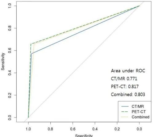
The AUC showed that the diagnostic performance of the combined interpretation was better than that of CT/MR alone, but poorer than PET-CT alone (Figure 1.). The difference between PET-CT and combined interpretation was statistically significant (P=0.024), but otherwise not significant.
Figure 1. Receiver operating characteristic (ROC) curves of PET-CT, CT/MR, and their combined interpretation for detecting nodal metastasis in
In the patient based analyses, the sensitivity, specificity, and accuracy of PET-CT for neck metastasis were slightly higher than those of CT/MR (75.0% vs. 70.0%, respectively, P = 0.564; 94.7% vs. 79.0%, respectively, P = 0.180; and 84.6% vs. 74.4%, respectively, P = 0.158). Combined interpretation of PET-CT and PET-CT/MR also yielded slightly higher sensitivity than PET-PET-CT alone (80.0%
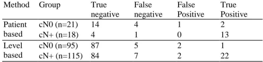
After the patients were categorized as cN0 and cN+ neck, we assessed the diagnostic value of PET-CT in the two groups (Table 3). Of the 21 patients with cN0 based on CT/MR findings as well as palpation, 6 patients (28.6%) revealed metastatic lymph nodes on final histopathologic analysis after surgery. Of the 6 patients who were falsely diagnosed as negative on CT/MR, 2 patients were correctly diagnosed as positive (Figure 2.), however, 4 patients were still missed on PET-CT (Figure 3.).
Table 3. PET-CT results for detecting nodal metastasis in the clinically nega-tive and posinega-tive neck groups of hypopharyngeal squamous cell carcinoma.
Method Group True
negative False negative False Positive True Positive Patient based cN0 (n=21) 14 4 1 2 cN+ (n=18) 4 1 0 13 Level based cN0 (n=95) 87 5 2 1 cN+ (n=115) 84 7 2 22
cN0= negative neck based on palpation and CT/MR, cN+=positive neck based on palpation and CT/MR
14
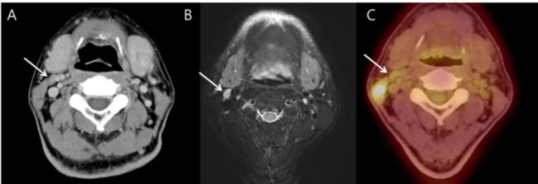
Figure 2. A 82-year-old man with left pyriform sinus cancer (T1N1) with nodal metastasis at left level II who showed false negative results on CT/MR but correctly diagnosed as metastasis by PET-CT. A and B. CT (A) and MR (B) images show small LN at level II (7-mm; arrows), falsely suggesting benign node based on size and shape. C. PET-CT shows focal intense uptake suggesting metastasis (arrow).
Figure 3. A 55-year-old man with right posterior pharyngeal wall cancer (T2N1) and nodal metastasis at right level II, which is occult nodal metastasis missed by CT/MR and PET-CT. A and B. CT (A) and MR (B) images show small LN at level II (arrows), with no features suggesting malignancy. C. PET-CT shows no
IV. DISCUSSION
This is the first correlation study of imaging interpretation results for cervical nodal metastasis with histopathology in patients with hypopharyngeal SCC and N0 neck who underwent neck dissection as a primary elective neck treatment. In our study, more than half of the patients (20 of 39 patients, 51.3%) with negative neck on clinical palpation had micrometastasis on histopathology after surgery, and 6 of 21 patients (28.6%) had occult lymph nodes on histopathologic analysis despite negative results on preoperative anatomic imaging.
In patients with head and neck SCC, if the risk of occult neck metastasis is judged to be greater than 15-20%, elective neck treatment such as selective neck dissection or irradiation should be considered as a standard strategy. However, although CT and MR are widely used for preoperative assessment of cervical lymph nodes in head and neck cancer, our study revealed that CT/MR could not decrease subclinical metastasis to less than 20%, which were not sufficiently sensitive to replace elective neck treatment in hypopharyngeal SCC patients.
Several previous studies have reported the value of PET-CT as well as CT/MR to detect lymph node metastases, although there is controversy regarding their clinical implications. Theoretically, PET-CT provides additional information to anatomical imaging, but previous collective studies have not focused on palpably or clinically negative neck, or even if they did, included SCCs of heterogenous primary sites, thus not fully reflecting the characterisctics
16 of hypopharyngeal SCC7,9,10,15,16.
Therefore, in our study, we analyzed whether the combination of PET-CT with CT/MR yields an additional benefit in diagnostic performance for the evaluation of subclinical metastasis in hypopharyngeal cancer patients with no palpable neck nodes because this consideration is more clinically relevant due to routine preoperative use of anatomic imaging studies. However, our study showed that, although the sensitivity of PET-CT was slightly than that of CT/MR, the addition of PET-CT to CT/MR could not provide more satisfactory diagnostic performance (NPV<80%) over CT/MR alone or PET-CT alone. In addition, of the 21 patients with cN0 based on CT/MR findings as well as palpation, 4 patients were still falsely diagnosed as negative on CT. PET-CT still showed a relatively high false negative rate even in the patients with cN0, which is unnegligible in clinical practice. Therefore, the addition of PET-CT examination to the anatomic imaging should be carefully applied because it could not provide additional benefit for the preoperative evaluation of cervical nodal metastasis in hypopharyngeal SCC patients with non-palpable neck. And this may imply a need for a scanner with higher resolution and a more dedicated head and neck imaging protocol, as reported by Rodrigues et al9.
There are potential limitations in this study. First, only 39 hypopharyngeal patients were included in this study, but large scale prospective studies are currently being carried out by the American College of Radiology Imaging Network (ACRIN)17 and may help overcome these limitations. Second, since
many of the patients are early staged, and selective neck dissection was performed, sampling of the neck may have been biased. Third, we did not collect data such as nodal recurrence after surgery, which would have revealed missed micrometastases, although the number is speculated to be small and insignificant.
V. CONCLUSION
The addition of PET-CT examination to the anatomic imaging including CT and MR could not provide additional benefit for the preoperative evaluation of cervical nodal metastasis in hypopharyngeal SCC patients with non-palpable neck, showing insufficient results to spare elective neck dissection.
18 REFERENCES
1. Psychogios G, Mantsopoulos K, Bohr C, Koch M, Zenk J, Iro H. Incidence of occult cervical metastasis in head and neck carcinomas: development over time. Journal of surgical oncology 2013;107:384-7. 2. Layland MK, Sessions DG, Lenox J. The influence of lymph node
metastasis in the treatment of squamous cell carcinoma of the oral cavity, oropharynx, larynx, and hypopharynx: N0 versus N+. Laryngoscope 2005;115:629-39.
3. Ferlito A, Robbins KT, Silver CE, Hasegawa Y, Rinaldo A. Classification of neck dissections: An evolving system. Auris Nasus Larynx; 2009. p.127-34.
4. Chan JY, Wei WI. Current management strategy of hypopharyngeal carcinoma. Auris Nasus Larynx 2013;40:2-6.
5. Bar Ad V, Chalian A. Management of clinically negative neck for the patients with head and neck squamous cell carcinomas in the modern era. Oral Oncol 2008;44:817-22.
6. Edge SB, American Joint Committee on Cancer. AJCC cancer staging manual. 7th ed. New York: Springer; 2010.
7. Roh JL, Park JP, Kim JS, Lee JH, Cho KJ, Choi SH, et al. (18)F Fluorodeoxyglucose PET/CT in Head and Neck Squamous Cell Carcinoma with Negative Neck Palpation Findings: A Prospective Study.
Radiology 2014;271:153-61.
8. Murakami R, Uozumi H, Hirai T, Nishimura R, Shiraishi S, Ota K, et al. Impact of FDG-PET/CT imaging on nodal staging for head-and-neck squamous cell carcinoma. Int J Radiat Oncol Biol Phys 2007;68:377-82. 9. Rodrigues RS, Bozza FA, Christian PE, Hoffman JM, Butterfield RI,
Christensen CR, et al. Comparison of whole-body PET/CT, dedicated high-resolution head and neck PET/CT, and contrast-enhanced CT in preoperative staging of clinically M0 squamous cell carcinoma of the head and neck. J Nucl Med 2009;50:1205-13.
10. Kyzas PA, Evangelou E, Denaxa-Kyza D, Ioannidis JP. 18F-fluorodeoxyglucose positron emission tomography to evaluate cervical node metastases in patients with head and neck squamous cell carcinoma: a meta-analysis. J Natl Cancer Inst 2008;100:712-20.
11. Branstetter BFt, Blodgett TM, Zimmer LA, Snyderman CH, Johnson JT, Raman S, et al. Head and neck malignancy: is PET/CT more accurate than PET or CT alone? Radiology 2005;235:580-6.
12. Kim JM, Kyung Tae. Value of 18-Fluorodeoxyglucose Position Emission Tomography/Computed Tomography in Diagnosis of Cervical Lymph Node Metastasis of Head and Neck Squamous Cell Carcinoma: Comparison with Computed Tomography and Magnetic Resonance Imaging. Korean J Otorhinolaryngol-Head Neck Surg 2012:364-8. 13. Som PM, Curtin HD, Mancuso AA. Imaging-based nodal classification
20
for evaluation of neck metastatic adenopathy. AJR. American journal of roentgenology 2000;174:837-44.
14. Robin X, Turck N, Hainard A, Tiberti N, Lisacek F, Sanchez JC, et al. pROC: an open-source package for R and S+ to analyze and compare ROC curves. BMC Bioinformatics 2011;12:77.
15. Ozer E, Naiboglu B, Meacham R, Ryoo C, Agrawal A, Schuller DE. The value of PET/CT to assess clinically negative necks. Eur Arch Otorhinolaryngol 2012;269:2411-4.
16. Schoder H, Carlson DL, Kraus DH, Stambuk HE, Gonen M, Erdi YE, et al. 18F-FDG PET/CT for detecting nodal metastases in patients with oral cancer staged N0 by clinical examination and CT/MRI. J Nucl Med 2006;47:755-62.
17. American College of Radiology Imaging Network; National Cancer Institute (NCI). FDG-PET/CT in Assessing the Tumor and Planning Neck Surgery in Patients With Newly Diagnosed H&N Cancer (ACRIN 6685). In: ClinicalTrials.gov [Internet]. Bethesda (MD): National Library of Medicine (US). 2000- [cited 2014 May 23]. Available from: http://clinicaltrials.gov/show/NCT00983697 NLM Identifier: NCT00983697.
ABSTRACT(IN KOREAN)
림프절이 촉진되지 않는 하인두 편평상피세포암종 환자에서
림프절 전이의 발견에서 CT/MR에 대한 PET-CT의 추가적인
가치의 평가
<지도교수 김진아>
연세대학교 대학원 의학과
이호준
목적: 림프절이 촉진되지 않는 하인두 편평상피세포암종 환자에서 림프절 전이의 발견하는 데 있어 양전자방출 단층촬영술-컴퓨터 단층촬영의 진단적 가치를 컴퓨터 단층촬영 또는 자기공명 영상(컴퓨터 단층촬영/자기공명 영상)과 비교하고, 해부학적 영상에 도움이 되는지를 평가한다. 대상 및 방법: 총 39명의 림프절이 촉지되지 않으며, 양전자방출 단층촬영술-컴퓨터 단층촬영, 컴퓨터 단층촬영/자기공명 영상을 시행 받았으며, 하인두 종양 절제술과 경부 림프절 곽청술을 시행받은 환자를 후향적으로 분석하였다. 개별/복합적 판단의 진단적 가치를 병리결과를 기준으로 평가하였다.22 결과: 총 39명의 환자 중 20명(51.3%)에서 경부 림프절 전이가 발견되었다. 림프절 층별 분석에서, 양전자방출 단층촬영술-컴퓨터 단층촬영, 컴퓨터 단층촬영/자기공명 영상, 복합적 판단시 민감도는 각각 65.7%, 57.1%, 65.7%였으며 통계학적으로 유의미한 차이는 없었다. 경부 촉진뿐 아니라 컴퓨터 단층촬영/자기공명 영상을 바탕으로 한 임상적 경부 림프절 병기 0기인 환자군 가운데 컴퓨터 단층촬영/자기공명 영상에서 위음성으로 판단한 6명중 4명은 양전자방출 단층촬영술-컴퓨터 단층촬영에서 여전히 위음성 소견을 보였다. 결론: 컴퓨터 단층촬영/자기공명 영상을 포함한 해부학적 영상에 양전자방출 단층촬영술-컴퓨터 단층촬영의 추가는 림프절이 촉지되지 않는 하인두 편평상피세포암종 환자의 경부 림프절 전이의 수술 전 평가에서 경부치료를 생략할 정도의 높은 정확도를 보이지는 못하여, 이득이 없는 것으로 나타났다.