∙ Received: June 29, 2012. Accepted: October 2, 2012.
∙ Corresponding author : Min Soo Park
Department of Nuclear Medicine, Yonsei University Health System, 134 Shinchon-Dong, Seodaemun-Gu, Seoul 120-749, Korea Tel: +82-2-2228-6064, Fax: +82-2-2227-7062 E-mail: [email protected]
Original Article
PET/CT에서의 고관절 삽입물에 의한 인공물과 Metal Artifact Reduction Algorithm의 유용성에 대한 고찰연세의료원 세브란스병원 핵의학과1, 신구대학교 방사선과2
박민수1 ․ 함준철1 ․ 조용인1 ․ 강천구1 ․ 박훈희2 ․ 임한상1 ․ 이창호1
The Study of Usefulness of Metal Artifact Reduction
Algorithm and Artifacts Caused by Metallic Hip Prosthesis on PET/CT
Min Soo Park1, Jun Cheol Ham1, Yong In Cho1, Chun Goo Kang1, Hoon-Hee Park2, Han Sang Lim1 and Chang Ho Lee1
1Department of Nuclear Medicine, Severance Hospital, Yonsei University Health System, Seoul, Korea,
2Department of Radiology, Shingu University, Seongnam, Korea
Purpose : PET/CT performed CT-based attenuation correction generates the beam hardening artifact by metallic implant. The attenuation correction causes over or underestimate of the area adjacent to metallic hip prosthetic material and change of 18F-FDG uptake. Also, the image quality and the diagnosability on genitourinary disease are reduced. Therefore, this study will evaluate the usefulness of MAR (Metal Artifact Reduction) algorithm method to improve the image quality on PET/CT. Materials and Methods : PET/CT was performed by fixing hip prosthesis in SPECT/PET phantom. In PET images with and Without MAR algorithm, the Bright streak, Dark streak, Metal region and Background area that appeared on CT were confirmed, and the change of each SUV (standardized uptake value) was analyzed. Also, in 15 patients who underwent total hip arthroplasty, each MAR algorithm and Without MAR algorithm and non attenuation correction was evaluated. Results : In PET image Without MAR algorithm, SUV of Bright streak region was 0.98±0.48 g/ml; Dark streak region was 0.88±0.02 g/ml; Metal region was 0.24±0.16 g/ml, Background area was 0.91±0.18 g/ml. In SUV of PET image with MAR algorithm, Bright streak region was 0.88±0.49 g/ml, Dark streak region was 0.63±0.21 g/ml, Metal region was 0.06±0.07 g/ml, Background was 0.90±0.02 g/ml. SUV generally decreased when applying MAR algorithm. In PET image Without MAR algorithm, SUVs of Bright region were higher than those measured in the Background, and it was false positive uptake. But, in PET image with MAR algorithm, SUVs of Bright region were similar to the Background, and false positive uptake disappeared. Conclusion : MAR algorithm could reduce an increase of 18F-FDG uptake due to attenuation correction in the hip surrounding tissue. However, decrease of SUV in Dark streak region should be considered in the future. Therefore, this study propose that the diagnostic accuracy can be improved in genitourinary diseases adjacent to metallic hip prosthesis, if provided PET images with and Without MAR algorithm, and non attenuation correction images at the same time. (Korean J Nucl Med Technol 2012;16(2):35-43)
Key Words : MAR (Metal Artifact Reduction) algorithm, Hip Prosthesis, Beam Hardening Artifact
서 론
PET/CT (Positron Emission Tomography/Computed Tomography) 는 암의 진단과 병리학적인 측면에서도 유용성이 입증되면서 임상 분야에서의 중요성이 강조되고 있다.1) 현재 PET/CT 검 사는 환자의 편리성 증대와 질적 향상의 해부학적 영상을 제공
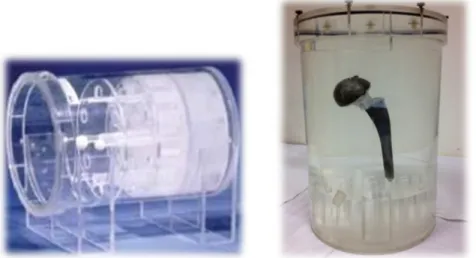
Fig. 1. NEMA (National Electrical Manufactures Association) PET Phantom™ (NU2-1994). A cylindrical Clear Lucite, Hot Sphere inserts, and Solid Teflon insert are fixed inside the phantom.
Fig. 2. A metallic hip prosthetic implant was fixed to SPECT/PET phantom.
PET/CT scan was performed, and the artifacts that generated by the metal was reproduced.
하기 위해 컴퓨터 전산화 단층 촬영 (Computed Tomography, CT)이 감쇠 보정으로 사용되고 있다.2) 이처럼 CT의 도입으 로 암의 병기 결정과 치료의 예후, 평가 등 향상된 진단적 가 치를 제공해 주고 있다. 그러나 CT를 기반으로 감쇠 보정이 시행되는 PET/CT의 경우 금속 삽입물로 인한 X선의 강한 흡수 차이로 인공물이 발생하게 된다.3-6) 이처럼 금속 삽입물 로 인해 CT HU (Hounsfield Unit)이 높게 측정되는 Bright Streak 영역과 HU가 낮게 평가되는 Dark Streak 영역이 발 생하게 된다.7-9)
최근 인구의 고령화가 진행되면서 많은 금속 인공물 삽입 술이 시행되고 있다. 특히 국민건강보험 공단 통계에 따르면 고관절 전치환술을 시행한 환자는 5년 동안 매년 평균 12%
가 증가하고 있는 추세이다. 고관절 전치환술은 퇴행성 관절
염과 괴사성 질환을 가진 환자에게 보다 편리한 생활을 제공 해주지만 CT를 기반으로 감쇠 보정이 시행되는 PET/CT 검 사에서는 고관절 금속 삽입물에 의한 선속 경화 현상(Beam Hardening Artifact) 으로 인공물이 발생된다. 이는 주변 부 위에 과대 혹은 과소 평가를 유발하는 감쇠 보정으로
18F-FDG의 섭취 변화를 일으키며10-14), 진단능을 저하시키는 원인이 된다.15-17) 따라서 본 연구에서는 고관절 삽입물에 의 해 유발되는 인공물을 phantom을 통해 재현하고 선속 경화 현상으로 생성되는 인공물의 형태와 저감에 대한 고찰, 영상 질 향상을 위한 방안을 연구했다. 더불어 MAR (Metal Artifact Reduction) algorithm 재구성 방법에 대한 유용성을 평가하여 고관절 삽입물 인근 주변 조직의 FDG에 대한 섭 취의 변화를 줄이고 고관절 삽입물과 인접해 있는 비뇨 생식 기와 삽입물 주변의 염증성 질환에 대한 영상 질 향상과 진 단능을 높이고자 하였다.
대상 및 방법
1. Phantom 연구
1) NEMA-1994 PET Phantom
MAR 적용 유무에 따른 SUV와 육안적 농도 변화를 알아 보기 위해 NEMA-1994 PET Phantom을 이용했다(Fig. 1).
Phantom 내부는 총 6047 ml이며, Phantom 내부에는 Clear Lucite, Hot Sphere, Solid Teflon 원통이 고정되어 있다. 이 때 배후 방사능은 4.29 kBq/ml (1.15 μCi/ml), Hot Sphere 원 통 안에는 16.49 kBq/ml (4.42 μCi/ml)가 되도록 물과 희석했 다.
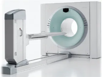
Fig. 3. Biograph Trupoint 40 (Siemens Medical System, CTI, Konxville, TN, USA). For image reconstruction methods, iteration was 3, subset was 8. Each image was analyzed that was with MAR algorithm, Without MAR algorithm, and non- attenuation correction.
A B-1 B-2 Fig. 4. ROIs were drawn on Clear
Lucite, Hot Sphere, Solid Teflon re- gions from CT image (A) through ac- quired NEMA-1994 PET Phantom, re- spectively, and each SUV of W/O MAR algorithm (B-1) and MAR algo- rithm (B-2) was compared. (a) is Solid Teflon, (b) is Hot Sphere, (c) is Clear Lucite region, (d) is Background.
A B
Fig. 5. (A) was CT artifact image caused by total hip prosthesis, (B) was attenuation corrected PET/CT image using (A). In Bight streak and Dark streak regions, the most adjacent to Metal were B_1, D_1, respectively, and the regions randomly set at the end of the influence of artifacts were B_2 and D_2. Each SUV was compared.
2) SPECT/PET Phantom
고관절 삽입물에 의한 인공물을 재현하기 위해 SPECT/
PET Phantom을 이용했다(Fig. 2). 총 6300 ml의 내부에 배후 방사능을 5.3 kBq/ml (1.42 μCi/ml)가 되도록 물과 희석하여 채우고 고관절 전치환술에 사용하고 있는 인공 삽입물을 Phantom 내부에 고정하여 PET/CT를 진행했다. 이 때 인체 의 연부조직과 구조는 고려되지 않았다.
2. 환자 정보 및 영상 획득
2011년 1월부터 2012년 2월까지 본원에 내원한 인공 고관 절 전치환술을 시행한 15명의 환자를 대상으로 했으며, 평균 나이는 65±3.5세였다. 사용 된 장비는 Biograph Truepoint 40 (Siemens Medical System, CTI, Konxville, TN, USA)이고 (Fig. 3), 감쇠 보정으로 이용 될 CT의 최대 관전압을 80 kVp 부터 140 kVp로 변환하여 선속 경화 현상의 감소 방안을 위 한 Phantom 실험을 진행했다(Fig. 4). 영상 재구성 방법으로 는 Iterations은 3, Subsets은 8이었으며, MAR algorithm을 적
용했을 때와 적용하지 않았을 때, 그리고 비 감쇠보정 영상 을 비교 분석했다. 이미지 획득으로는 Phantom 연구는 bed 당 15분, 환자 검사는 bed 당 3분으로 획득했다.
3. 영상 분석
NEMA-1994 PET Phantom을 통해 획득 된 Clear Lucite, Hot Sphere, Solid Teflon 영역에 각각 관심영역을 그려 SUV 의 변화를 분석했고(Fig. 4), 고관절을 고정 시킨 SPECT/PET Phantom의 최대 관전압 별 CT 영상에서의 HU와 Noise를 비교했다. 또한 W/O MAR (Without Metal Artifact Reduction) algorithm과 MAR algorithm, 비 감쇠 보정 영상 에서 각 인공물 영역에 따른 SUV 변화를 비교했다. 이 때 관 심영역은 CT를 기준으로 금속에 의해 생성되는 Bright streak, Dark streak, Metal, Background 영역에 3명의 방사선 사가 5회 이상 반복하여 측정했다. 특히 Bright와 Dark streak 영역에서는 금속으로부터 가장 근접한 영역인 B_1과 D_1을, 인공물의 영향이 있는 끝부분에 임의로 설정 된 영역 인 B_2와 D_2에서의 SUV를 비교했다(Fig. 5). 사용 된 통계 는 Paired t-test이며 SPSS 버전 17.0을 사용했다.
Table 1. As a result of comparing SUVs in the 3 spheres according to the presence of MAR algorithm using NEMA-1994 phantom, when applying MAR algorithm, there were no significant differences in Background region, however, Hot Sphere and Solid Teflon regions were statistically significant.
W/O MAR algorithm MAR algorithm p-value Solid Teflon 0.09±0.01 g/ml 0.00±0.00 g/ml p<0.05 Hot Sphere 3.85±0.04 g/ml 3.59±0.03 g/ml p<0.05 Clear Lucite 0.17±0.01 g/ml 0.17±0.26 g/ml p>0.01 Background 0.96±0.04 g/ml 0.95±0.02 g/ml p>0.05
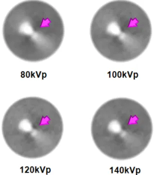
Fig. 6. Through the maximum tube voltage of CT used as attenuation correction was varied from 80 kVp to 140 kVp, the phantom experiments were conducted for reducing beam hardening artifact. On CT images, artifacts were slightly reduced as the maximum voltage rises up. And HU was measured by drawing ROIs, and in 140 kVp, Bright streak region decreased approximately 56%; Metal region, about 7%;
Background region, about 90% compared to 80 kVp. But, Dark streak region increased about 55%.
Fig. 7. Even if the maximum tube voltage rises, false positive uptake of Bright region didn't decreased (arrow). But, in Dark region, false negative uptake was decreased by increasing SUV, as the maximum voltage rises.
결 과
1. Phantom 연구 결과
NEMA-1994 Phantom을 이용하여 MAR algorithm 적용 유무에 따른 3개의 sphere에서의 SUV를 비교 평가한 결과 Clear Lucite 영역에서 W/O MAR algorithm 적용 시에는 0.17±0.01 g/ml, MAR algorithm 적용 시에서는 0.17±0.26 g/ml였고, Background 영역에서는 각각 0.96±0.04 g/ml와 0.95±0.02 g/ml로 통계적으로 유의한 SUV 차이를 보이지 않 았다. 하지만 Hot Sphere와 Solid Teflon 영역에서는 MAR algorithm을 적용했을 때에는 각각 3.59±0.03 g/ml와 0.00±
0.00 g/ml으로 SUV는 통계적으로 유의한 차이를 보였다 (Table 1). 육안적인 평가에서도 Solid Teflon 영역에 섭취 농 도가 MAR algorithm 적용 시 감소했다. 또한 고관절을 고정 시킨 SPECT/PET Phantom의 최대 관전압 변화에 따른 HU 와 Noise를 평가한 결과 80 kVp에 비해 140 kVp에서 HU는 Bright streak 영역에서는 약 56%, Metal 영역은 약 7%, Background 영역에서는 약 90%가 감소했고 Dark streak 영 역에서는 55%가 증가했다. Noise 변화는 Bright와 Dark, Background 영역에서는 감소하였고 Metal 영역에서는 증가 했다(Fig. 6, Table 2). 관전압 변화에 따른 CT를 기반으로 감 쇠보정 된 W/O MAR algorithm PET영상에서는 육안적으로 평가한 결과 큰 차이를 보이지 않았으며, Bright streak 영역 에서의 false positive uptake도 사라지지 않았다. 다른 영역에 서의 SUV 변화 또한 통계적으로 유의한 차이가 나타나지 않 았지만, Dark streak 영역에서는 최대 관전압이 상승 할수록 SUV는 증가했다(Fig. 7, Table 2).
SPECT/PET Phantom에 인공 고관절을 삽입하여 획득 된 MAR algorithm 영상과 W/O MAR algorithm, 비 감쇠 보정 영상을 비교한 결과 W/O MAR algorithm 적용 시 고관절 삽입물 주변의 Bright streak 영역에서 uptake가 증가했지만, MAR algorithm과 비 감쇠 보정 영상에서는 false positive uptake가 관찰되지 않았다. 하지만 Fusion영상과 비교한 결
Table 2. This shows the change of the HU, noise, and SUVs of attenuation corrected PET according to the variation of maximum tube voltage.
80 kVp 100 kVp 120 kVp 140 kVp
HU 295.56 201.05 157 128.45
Bright Noise 93.7 45.51 30.61 24.51
SUV 1.02±0.12 g/mL 1.00±0.08 g/mL 0.99±0.09 g/mL 0.99±0.04 g/mL
HU -332.95 -255.7 -196.65 -148.69
Dark Noise 87.22 54.46 43.01 32.69
SUV 0.63±0.11 g/mL 0.70±0.16 g/mL 0.77±0.15 g/mL 0.80±0.11 g/mL
HU 2669.23 2575.13 2514.07 2466.56
Metal Noise 100.14 149.07 177.9 195.47
SUV 0.16±0.09 g/mL 0.14±0.06 g/mL 0.15±0.12 g/mL 0.15±0.20 g/mL
HU 19.22 7.93 4.82 1.81
Background Noise 50.13 26.53 15 10.1
SUV 0.93±0.15 g/mL 0.92±0.12 g/mL 0.90±0.10 g/mL 0.91±0.06 g/mL
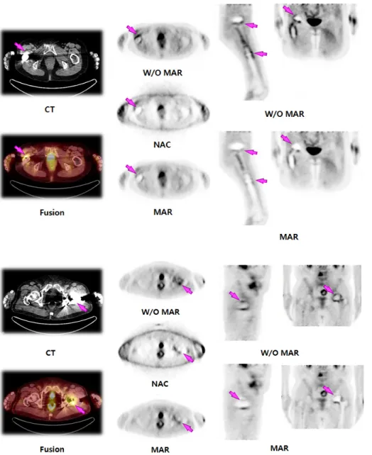
Fig. 8. It showed comparison of with MAR algorithm, W/O MAR al- gorithm, and non-attenuation cor- rection (NAC) PET images. In W/O MAR algorithm image, FDG uptake was increased in Bright streak re- gion around Metal, but, when ap- plying MAR algorithm and non at- tenuation correction, false positive uptake was disappeared. However, in MAR algorithm and non attenu- ation corrected PET images, the shade of metal was magnified.
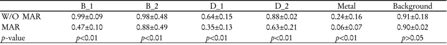
Table 3. When applied MAR algorithm, SUV of B_1 reduced about 50%, but, B_2 was not significantly different from Background.
However, all SUVs of B_1 and B_2 were higher than Background, and showed false positive uptake.
B_1 B_2 D_1 D_2 Metal Background
W/O MAR 0.99±0.09 0.98±0.48 0.64±0.15 0.88±0.02 0.24±0.16 0.91±0.18
MAR 0.47±0.10 0.88±0.49 0.35±0.13 0.63±0.21 0.06±0.07 0.90±0.02
p-value p<0.01 p<0.01 p<0.01 p<0.01 p<0.01 p>0.05
과 MAR algorithm과 비 감쇠 보정 영상에서는 고관절 삽입 물 주변이 더욱 과소 평가되어 금속의 음영이 크게 나타났으 며, 비 감쇠 보정 영상에서 금속 주변 결손 음영이 더욱 증가 했다(Fig. 8). 또한 MAR algorithm과 W/O MAR algorithm 에서 Bright, Dark, Metal, Background 영역에서의 SUV를 비 교한 결과 Background 영역을 제외하고 모든 영역에서 통계 적으로 유의한 차이를 나타냈다. 특히 금속에 가장 근접 영 역인 B_1에서 MAR algorithm 적용 시 SUV가 약 50% 감소 하였지만 B_2영역에서는 0.88±0.49 g/ml로 Background 영역 인 0.90±0.02 g/ml과 통계적으로 유의한 차이를 나타내지 않 았다. 하지만 W/O MAR algorithm 에서는 B_1, B_2 영역 모
두 0.99±0.09 g/ml와 0.98±0.48 g/ml로 Background 영역인 0.91±0.18 g/ml 보다 높게 SUV가 평가됐으며, 육안적으로 평 가 시에도 Bright streak 영역에서 false positive uptake가 나 타났다. Dark streak 영역에서는 W/O MAR algorithm과 MAR algorithm 모두 과소 평가되어 SUV가 낮게 측정됐으 며, 특히 MAR algorithm 적용 시 높은 SUV 감소를 보였다 (Table 3, Fig. 9).
2. 임상 영상 평가
인공 고관절 전치환술을 시행한 15명의 모든 환자에게 고
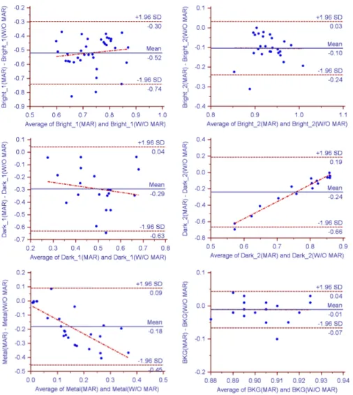
Fig. 9. This is Bland Altman graph of the change of SUVs due to artifacts in W/O MAR algorithm and MAR al- gorithm PET images. When applied MAR algorithm, SUVs were gen- erally decreased. In the case of D_2, the difference between the two methods became similar as increas- ing SUV; in Metal region, the differ- ence grew. In B_2 and Background region, the reduction rate was mean 0.01, and the two methods were similar.
관절 삽입물 주변에서 false positive uptake가 관찰 됐다. 이 러한 FDG 섭취 증가는 고관절 삽입물 주변의 Bright streak 영역에서 나타났으며, Dark streak 영역에서는 FDG uptake 의 결손으로 나타났다. 하지만 MAR algorithm 적용하여 영 상을 비교한 결과 Bright streak 영역에서의 false positive up- take가 관찰되지 않았다.
고 찰
최대 관전압의 변화로 CT영상에서 인공물과 HU는 변화 했지만 최대 관전압 별 감쇠보정된 PET 영상의 Bright streak 영역에서는 여전히 Background보다 SUV가 높게 평가되었 다. 육안적인 평가에서도 Bright streak 영역에서의 FDG 섭 취 증가가 관찰되었다. 이는 인공 고관절 삽입물 주변 조직 의 염증성 질환의 판별을 어렵게 할 뿐 아니라 CT상의 인공 물로 인해 비뇨 생식기 질환의 진단능을 저하시키는 원인이
될 것이다. Phantom 실험과 임상 영상 평가를 통해 MAR al- gorithm을 적용하여 재구성 시 Bright streak 영역에서의 FDG 섭취 증가를 없앨 수 있었다. 하지만 인공 고관절과 근 접한 영역에서의 SUV는 현저하게 저하되었으며, Dark streak 영역에서의 SUV 저하되는 문제점도 앞으로 더 많은 연구를 통해 보완되어야 할 것이다. 최근 이러한 금속 물질 로 인해 발생되는 CT의 인공물을 줄이기 위한 연구가 활발 히 진행 중이며, 특히 듀얼 에너지를 사용하여 금속의 인공 물을 감소시키는 방법 들이 고안되고 있는 중이다.18-19) 듀얼 에너지 CT에서는 서로 다른 X선원이 물질을 투과하고 감약 한 에너지를 선택적으로 재구성하여 에너지 파형에 따른 단 일 에너지 파형의 CT영상 (Monochromatic image)획득이 가 능해졌다.20) 금속 인공물 감약 방법은 금속 데이터가 있는 원 시 데이터에(금속 정보는 역치 값으로 설정되어 자동으로 인 식됨) 다차원 데이터 공간을 이용한 대리 감약 지수 (surrogate attenuation value)를 사용하는 방식으로 금속 물질
Fig. 10. Comparison image when applied MAR algorithm, W/O MAR algorithm, and NAC of 69- year-old patient who had ovarian cancer and underwent total right hip arthroplasty. In W/O MAR al- gorithm, the false positive uptake of Bright streak region was in- creased (arrow). But, in image applied MAR algorithm and NAC, the increased uptake of FDG dis- appeared that caused by metallic hip prosthesis.
Fig. 11. In 62-year-old patient had cervical cancer, the false positive uptake of Bright streak region was increased in CT im- age (arrow). However, the false positive uptake was not observed, when applied MAR algorithm.
로 유발되는 인공물을 줄일 수 있다.21) 이처럼 감쇠 보정 지 도로 듀얼 에너지를 이용한 기법들이 이용된다면 PET/CT에 서 MAR algorithm의 단점으로 지적 되었던 금속으로 인한 FDG의 false negative uptake의 확대를 줄이고 CT 자체의 진 단능을 높임과 동시에 Bright streak 영역에서의 false positive uptake를 방지하는데 도움이 될 것으로 생각된다. 앞으로 국 내의 PET/CT 장비 도입 시 CT의 algorithm 변환으로 금속 의 인공물을 감소시키고 이를 감쇠 보정 지도로 이용할 수 있는 소프트웨어와 하드웨어에 관한 연구가 필요할 것으로 생각된다. 다만 듀얼 에너지의 경우 환자의 피폭과 장비 과 부하 등의 문제점은 앞으로도 더욱 많은 논의와 연구가 이뤄 져야 할 것이다.
결 론
MAR algorithm 적용을 통해 인공 고관절 삽입물 주변 조 직의 감쇠 보정으로 인한 FDG 섭취 증가를 줄일 수 있었다.
하지만 MAR algorithm 적용으로 인한 고관절 삽입물 근접 영역에서의 결손과 Dark streak 영역에서의 SUV 저하에 관 한 문제점은 앞으로도 더욱 많은 연구가 진행되어야 할 것이 다. 고관절 삽입 인공물로 인해 발생하는 Bright streak 영역 에서의 false positive uptake를 감소시키고 이와 인접해 있는 비뇨 생식기 질환과 고관절 주변의 염증성 질환 판별에 W/O MAR algorithm과 MAR algorithm, 비 감쇠 보정 영상 이 동시에 제공된다면 더욱 진단능을 향상 시킬 수 있으리라 생각된다.
요 약
CT를 기반으로 감쇠 보정이 시행되는 PET/CT 검사에서 는 금속 삽입물에 의한 선속 경화 현상 (Beam Hardening Artifact)으로 인공물이 발생 된다. 이는 인접한 부위의 과대 혹은 과소 평가를 유발하는 감쇠 보정으로 인해 18F-FDG의 섭취 변화를 가져오며 영상의 질과 비뇨 생식기 질환의 진단 능을 저하시킨다. 따라서 본 연구에서는 PET/CT 영상의 질 향상을 위한 금속 인공물 저감 (Metal Artifact Reduction, MAR) algorithm 재구성방법의 유용성을 평가하고자 한다.
인공 고관절 삽입물에 대한 인공물을 평가하기 위해 SPECT/
PET Phantom에 고관절 삽입물을 고정 하여 PET/CT를 진 행했다. W/O MAR algorithm 영상과 MAR algorithm 영상 에서 CT상에 나타나는 Bright streak, Dark streak, Metal과 Background의 영역에서의 표준화 섭취 계수(Standardized Uptake Value)의 변화를 분석하였다. 또한 본원에 내원한 고 관절 전치환술을 시행한 15명의 환자에게서 W/O MAR al- gorithm과 MAR algorithm, 비 감쇠 보정 영상을 비교 평가 하였다. W/O MAR algorithm 영상에서 SUV는 Bright streak 영역에서는 0.98±0.48 g/ml, Dark streak 영역에서는 0.88±
0.02 g/ml, Metal 영역에는 0.24±0.16 g/ml, Background 영역 은 0.91±0.18 g/ml로 측정되었다. 하지만 MAR algorithm 영 상에서는 Bright streak 영역에서는 0.88±0.49 g/ml, Dark streak 영역은 0.63±0.21 g/ml, Metal 영역에서는 0.06±0.07 g/ml, Background는 0.90±0.02 g/ml로 측정되었다. MAR al- gorithm 적용 시 SUV가 평균적으로 감소 하였으며, W/O MAR algorithm 영상에서는 Bright streak영역에서 Background 보다 높게 측정되어 false positive uptake로 나타났으나 MAR algorithm 영상에서는 Background와 비슷한 농도와 SUV가 나타났고, false positive uptake가 관찰되지 않았다.
따라서 MAR algorithm을 적용하여 인공 고관절 삽입물 주 변 조직의 과대 혹은 과소 감쇠 보정으로 인한 섭취의 증가 를 줄일 수 있었다. 하지만 Dark streak 영역에서의 SUV 저 하를 줄이기 위한 방안은 더욱 많은 연구가 이뤄져야 할 것 이다. 고관절 삽입 인공물로 인해 발생하는 Bright streak 영 역에서의 false positive uptake를 감소시키고 이와 인접해 있 는 비뇨 생식기 질환에 W/O MAR algorithm과 MAR algo- rithm, 비 감쇠 보정 영상이 동시에 제공된다면 더욱 진단능 을 향상 시킬 수 있으리라 생각된다.
REFERENCES
1. Bar-Shalom R, Guralnik L, Tsalic M, Leiderman M, Frenkel A, Gaitini D, Ben-Nun A, Keidar Z, Israel O. The additional value of PET/CT over PET in FDG imaging of oesophageal cancer.
Eur J Nucl Med Mol Imaging 2005;32(8):918-924.
2. Ay M, Zaidi H. Computed Tomography-based attenuation correction in neurological positron emission tomography: eval- uation of the effect of x-ray tube voltage on quantitative analysis. NuclMed Commun 2006;27:339-346.
3. Bujenovic S, Mannting F, Chakrabarti R, Ladnier D. Artifactual 2-deoxy-2-[(18)F]fluoro-D-glucose localization surrounding metallic objects in a PET/CT scanner using CT-based attenu- ation correction. Mol Imaging Biol 2003;5(1):20-22.
4. DiFilippo FP, Brunken RC. Do implanted pacemaker leads and ICD leads cause metal-related artifact in cardiac PET/CT? J Nucl Med 2005;46(3):436-443.
5. Goerres G, Hany T, Kamel E, von Schulthess GK, Buck A.
Head and neck imaging with PET and PET/CT: artifacts from dental metallic implants. Eur J Nucl Med 2002;29:367-370.
6. Nuyts J, Stroobants S. Reduction of attenuation correction arti- facts in PET-CT. Conf Rec of the 2005 IEEE Nuc Sci Symp 2005;4:1895-1899.
7. Kak AC, Slaney M (2001) Principles of computerized tomo- graphic imaging. SIAM, New York, pp 118-124.
8. Hsieh J (1995) Image artifacts, causes, and correction. In:
Goldman LW, Fowlkes JB (eds) Medical CT and ultrasound, current technology and applications. Advanced Medical Publishing, Madison, p 487-518.
9. Oliver Watzke Willi A. Kalender, A pragmatic approach to metal artifact reduction in CT: merging of metal artifact reduced images, Eur Radiol 2004;14:849-856.
10. John A. Kennedy, Ora I, Alex F, Rachel BS, Haim A. The re- duction of artifacts due to metal hip implantsin CT-attenuation corrected PET images from hybrid PET/CT scanners. Med Bio Eng Comput 2007;45:553-562.
11. Goerres GW, Ziegler SI, Burger C, Berthold T, Von Schulthess GK, Buck A. Artifacts at PET and PET/CT caused by metallic hip prosthetic material. Radiology 2003;226:577-584.
12. Hiroaki Shimamoto, Naoya Lalimoto, Kouichi Fujino, Seiki Hamada, Eku Shimosegawa, Shumei Murakami. Metalic arti- facts cause by dental metal prostheses on PET images: a PET/CT Phantom study using different PET/CT scanners.
Ann Nucl Med 2009;23:443-449.
13. Sandra J. Rosenbaum, Thomas Lind, Gerald Antoch, Andreas Bockisch. False-Positive FDG PET Uptake-the Role of PET/CT. Eur Radiol 2006;16:1054-1064.
14. Waheeda Sureshbabu, Osama Mawlaqwi. PET/CT Imaging Artifacts. J Nucl Med Technol 2005;33:156-161.
15. Griffiths HJ, Priest DR, Kushner DM, Kushner D. Total hip re- placement and other orthopedic hip procedures. Radiol Clin North Am 1995;33:267-87.
16. Cyteval C, Hamm V, Sarrabère MP, Lopez FM, Maury P,
Taourel P. Painful infection at the site of hip prosthesis: CT imaging. Radiology 2002;224:477-83.
17. Stumpe KD, Nötzli HP, Zanetti M, Kamel EM, Hany TF, Görres GW, et al. FDG PET for differentiation of infection and aseptic loosening in total hip replacements: comparison with conventional radiography and three-phase bone scintigraphy.
Radiology 2004;231:333-41.
18. Guy MJ, Castellano-Smith IA, Flower MA, Flux GD, Ott RJ, Visvikis D. DETECT-dual energy transmission estimation CT-for improved attenuation correction in SPECT and PET.
IEEE Trans Nucl Sci 1998;45:1261-1267.
19. Kinahan PE, Alessio AM, Fessler JA. Dual energy CT attenu-
ation correction methods for quantitative assessment of re- sponse to cancer therapy with PET/CT imaging. Technol Cancer Res Treat 2006;5:319-327.
20. Brown CL, Hartman RP, Dzyubak OP, Takahashi N, Kawashima A, McCollough CH, Bruesewitz MR, Primak AM, Fletcher JG. Dual-energy CT iodine overlay technique for char- acterization of renal masses as cyst or solid a phantom feasibility study. European Journal of Radiology 2009;19(5):1289-1295.
21. Prell D, Kalender WA, Kyriakou Y. Development, im- plementation and evaluation of a dedicatedmetal artefact reduc- tion method for interventional flat-detector CT. British Journal of Radiology 2010;83(996):1052-1062.