⋅Received: July 15. 2008. Accepted: September 1. 2008.
⋅Corresponding author: Jae Dong Seok
Department of Nuclear Medicine, Samsung Medical Center, #50 Ilwon-dong, Gangnam-gu, Seoul, 135-710, Korea
Tel: +82-2-3410-2660, Fax: +82-2-3410-2639 E-mail: [email protected]
Original Article
PET/CT에서 소아환자 영상 재구성 시DFOV 변화의 유용성
삼성서울병원 핵의학과
최성욱⋅최춘기⋅이규복⋅석재동
Usefulness of DFOV Changes in Pediatric PET/CT Image Reconstruction
Sung Wook Choi, Choon Ki Choi, Kyoo Bok Lee, Jae Dong Seok Departments of Nuclear Medicine, Samsung Medical Center, Seoul, Korea
Purpose: There have been something difficulties in locating focuses and quantitative analysis in case of pediatric patients because of the relatively small body compared to adults. This author of this study, therefore, evaluated the usefulness of DFOV (Display Field Of View) according to its changes in PET/CT image reconstruction by means of the phantom experiment and pediatric patients examination.
Materials & Methods: 0.023 MBq/cc of
18F-FDG was put into the uniform NU2-94 phantom, and then emission scan was acquired for 10 minutes. For reconstruction, DFOV values were changed to 50, 45, 40, 35, 30, and 25 cm respectively. As for patient images, 20 patients who were diagnosed as the one or suspicion of the children tumor are targeted from Oct 2007 to Jan 2008.
For image reconstruction, 50 cm was the basis of DFOV, and the value was adjusted to DFOV 45 cm to 25 cm respectively. In the phantom and the reconstruction image of pediatric patients, the changes in pixel size and SUV
maxaccording to DFOV changes were analyzed. Results: As DFOV decreased to 50, 45, 40, 35, 30, and 25 cm by means of the phantom, the pixel size was changed to 3.906, 3.515, 3.125, 2.734, 2.343, and 1.953 mm respectively. Besides, as a result of reconstruction DFOV in images of pediatric patients to 50, to 25 cm, the different values of SUV
maxare shown as 3.3, 7.3, 12, 14, 18% and 2.6, 4.3, 5.0, 7.0, 10.0% on respectively when 50 cm was the standard.
Conclusion: In SUV
maxusing the phantom, as DFOV decreased every 5 cm, the mean value gradually increased. With 50 cm as the standard, the increase rates were 3.7, 6.5, 11.2, 19.5, and 32.1% respectively. As for pediatric patients image too, as DFOV decreased, the rates increased as in the phantom experiment. In image reconstruction, since DFOV decrease regardless of matrix size change reduced the pixel size, the image quality can be improved. This would be more useful than reconstruction and enlarge images of pediatric patients in the same way of examining adults.
However, when the value of 35 cm DFOV was applied, this may result in truncated artifact, and thus the application should be properly controlled. Change of DFOV may produce better image for pediatric patients, but changes of SUV values according to DFOV change should be considered in reading. (Korean J Nucl Med Technol 2008;12(3):171-175)
Key Words : DFOV, SUV
max, Image reconstruction, Truncated artifact
서 론 18F-FDG를 이용한 전신촬영은 환자의 임상평가 과정에서 원발종양의 진단뿐만 아니라 악성도의 결정, 수술 후 평가, 질병 진행 평가, 괴사 및 국부 방사성의약품의 흡수에 의한 전이여부 판정, 예후 판정, 치료 방침 설정 등에 유용하게 이 용되고 있다.1,2)
최근에 들어서는 성인 암뿐만 아니라 소아 암 환자의 급격 한 증가 추세로 더욱 관심이 높아지고 있는 실정이며, 검사
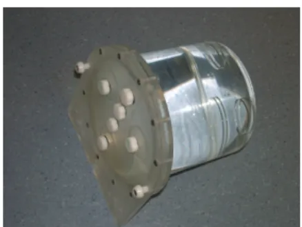
Fig. 1. Phantom (Uniform NU2-94 Phantom).
특성상 성인검사 보다는 소아환자에 있어서 많은 어려움을 가지고 있다. 특히, 수면 진정 상태, 금식, 검사 중 움직임 등 검사 시행 중 발생하는 문제점이 있을 수 있고, 영상 판독 시 소아 환자의 작은 체구로 인하여 병소의 위치 확인 및 정량분 석 평가에 어려움을 가지고 있다.3)
따라서 소아 환자의 영상 재구성 시 성인 기준인 DFOV (Display Field Of View)를 50 cm로 적용을 할 경우 해상력 저하와 표준화섭취계수(Standardized Uptake Value : SUV) 값이 다소 낮아지는 영향을 받을 수가 있다. 또한 작은 체구 로 인한 부분 체적 효과 (Partial Volume Effect : PVE)로 인 하여 수 mm의 작은 병변을 과소 평가하여 SUV 값에 다소 영향을 줄 수도 있다.4,5)
본 저자들은 소아환자를 대상으로 PET/CT에서의 영상재 구성 시 DFOV 변화에 따른 유용성을 평가하였다.
실험재료 및 방법
1. 환자검사
2007년 10월부터 2008년 1월까지 본원을 내원하여 소아 암 진단을 받거나 의심되는 환자 20명을 대상으로 하였으며, 연령 범위는 0 ~ 15세로 평균 연령은 6.7세, 몸무게는 50 kg 이하 등을 기준으로 소아과에서 의뢰한 것으로 국한하였다.
2. PET/CT 장비
Discovery LS8 PET/CT Scanner로 GE사 장비로 주요 제 원은 다음과 같다.
CT
- Scan Mode : Helical, Axial, ScoutTM
- Reconstruction Algorithms : Soft Tissue, Standard, Detail, Bone, Lung and Edge
- Reconstruction Matrix : 512
- CT Number scale : 1024 to 3071 HU - Helical Pitch: 5:1, 7:1, 10.8:1, 13.4:1 - Scan Technique - kVp : 80, 100, 120, 140
- mA : 10 to 440 mA, 10 mA increments
- Power : 0.8 to 53.2 kW
- Rotational speed : 0.5, 0.6, 0.7, 0.8, 0.9, 1.0, 2.0, 3.0 and 4.0 sec full scan (360° acquisition)
- Aperture : 70 cm - Maximum SFOV : 50 cm
PET
- Detector Ring : Bismuth Germanate (BGO) - Detector ring diameter : 92.7 cm
- Detector layout : 12.096 individual crystals arranged in 18 rings of 672 crystals each
- Transaxial FOV : 55 cm - Axial FOV : 15.2 cm
- Axial sampling interval : 4.25 mm - Number of image plane : 35 - Coincidence window : 12.5 nsec
- Transmission measurement rod source : 68Ge 10 mCi - Normalization rod source : 68Ge 1.5 mCi
- Resolution : Transaxial FWHM 4.8 mm 3. Phantom
Uniform NU2-94 Phantom
Specification : - Dimensions ; 8 “dia×8.98” l (20.3×22.8 cm) - Volume without inserts ; 5,640 mL
4. 방사성의약품
Phantom : 0.023 MBq/cc 소아 환자 : 5.29 MBq/kg 5. Phantom 영상
Uniform NU2-94 Phantom에 5,640 mL의 멸균증류수와
18F-FDG 0.023 M㏃/cc를 넣어 잘 혼합시킨다. 영상 획득은
Table 1. Post filter & Loop filter value of versus DFOV according to change
Unit : mm
50 cm 45 cm 40 cm 35 cm 30 cm 25 cm
Post Filter 2.73 3.27 3.82 4.36 4.91 5.45
Loop Filter 2.00 2.34 2.73 3.12 3.52 3.91
(A)
Pixel Size
(B)SUV
maxDFOV DFOV Fig. 2. (A) Pixel Size versus DFOV. (B) SUVmax versus DFOV.
1 frame에 CT 영상은 환자 검사 방법과 동일, PET 영상은 10 min 동안 얻었으며, 총 4회를 실시하여 평균값을 얻었다.
6. 환자 영상
소아 환자를 10시간 이상 금식을 시키고, 18F-FDG는 5.29 MBq/kg으로 정맥 및 IV Line을 통하여 주입한 후 방사성의 약품이 흡수될 때까지 최대한 잠을 자지 못하도록 하면서 40~50분 정도 대기하고, 검사 시작 10분 전에 sedation을 시 작하여 검사를 진행한다. 촬영 범위는 4~6 frame (57.8~86.7 cm), 영상획득 시간은 4 min/frame으로 하여 방출 촬영상을 획득하였다.
7. 영상 재구성
OSEM (Ordered Subset Expectation Maximization)과 반 복영상재구성(Iterative Reconstruction)으로 2회 반복(ite- ration)하고, 부분 집합(subsets)은 28로 정하였다. filter는 post filter와 loop filter를 적용하고, 영상재구성 시 matrix size의 크기는 128×128로 하였다.
감쇠보정방법으로는 측정된 감쇠보정법(Measured Attenu- ation Correction)을, 투과촬영은 CT를 이용한 감쇠지도를 적용하였다.
8. 영상 분석
- Phantom 영상 : 원형 관심영역(Region Of Interest)을 중 앙 부위에 지름 1 cm로 동일하게 설정하고, 평균과 최소, 최대 SUV 값을 측정하여 DFOV 50~25 cm까지 영상간 의 SUV 값의 차이와 백분율을 계산하였다.
- 환자 영상 : 최대표준화섭취계수(SUVmax) 값의 차이와 백 분율을 계산하였다.
결 과
- Phantom 영상 : DFOV가 50, 45, 40, 35, 30, 25 cm로 감 소함에 따라 pixel 크기는 3.906, 3.515, 3.125, 2.734, 2.343, 1.953 mm로 점차 감소하였다. 또한 SUVmax는 1.275, 1.323, 1.359, 1.418, 1.524, 1.685로 증가 양상을 보 였으며, 백분율은 DFOV 50 cm 기준으로 3.7, 6.5, 11.2, 19.5, 32.1%의 증가함을 알 수 있었다(Fig. 2).
- 환자 영상 : 그림 3-A에서 DFOV 50 cm 기준으로, SUVmax
는 4.629, 4.786, 4.995, 5.231, 5.373, 5.659의 변화가 있었 으며, 백분율은 3.3, 7.3, 12, 14, 18%로 증가하였다. 그림 3-B에서는 SUVmax는 11.9, 12.22, 12.43, 12.52, 12.80, 13.23으로 증가하였고, 백분율은 2.6, 4.3, 5.0, 7.0, 10.0%
의 증가를 나타냈다(Fig. 3).
(A) (B)
DFOV50 DFOV45 DFOV40
DFOV35 DFOV30 DFOV25
SUV
maxDFOV Fig. 3. (A) Pediatric imagesaccording to DFOV change. (B) SUV according to DFOV change in pediatric images.
(A) (B)
DFOV50 DFOV45 DFOV40
DFOV35 DFOV30 DFOV25
SUV
maxDFOV Fig. 4. (A) Pediatric imagesaccording to DFOV change. (B) SUV according to DFOV change in pediatric images.
따라서 DFOV 5 cm가 감소하면서 40 cm까지는 좋은 영상 을 얻을 수 있었지만, DFOV 35 cm 이하는 truncated artifact 가 나타나는 것을 알 수 있었다.
고찰 및 결론
영상 재구성에서 성인 방법으로 소아 암 환자에 DFOV 50 cm를 적용하였으나 소아 환자의 작은 체구로 인하여 병변 위치 파악에 어려움이 있었다. 그 이유는 병변 크기에 비하여 다소 큰 pixel 크기 때문에 부분체적 효과가 발생하게 되어 수 mm의 병변과 같은 작은 크기의 병변에 대한 정량평가에 서 결과가 과소평가(underestimated value)하게 되어 SUV 값 측정에 다소 영향을 줄 수가 있다.
phantom을 이용한 영상에서 SUVmax 값은 DFOV 50~25
cm 범위를 선정하여, 5 cm씩 감소함에 따라 DFOV 50 cm와 비교할 때 3.7, 6.5, 11.2, 19.5, 32.1%의 차이를 얻었다.
SUVmax값은 DFOV 50 cm와 비교할 때 그림 3-A에서 백분율 은 3.3, 7.3, 12, 14, 18%의 변화를 보였고, 그림 3-B에서의 백분율은 2.6, 4.3, 5.0, 7.0, 10.0%의 차이를 나타냈다. 그리고 병변에 따라 많은 SUVmax 값 차이를 보였기에 평균값을 구하 기에는 다소 무리가 있었다. 또한 시각적인 평가에 있어서는 DFOV 50~25 cm에서 전체적으로 큰 차이는 보이지 않았 으나, truncated artifact는 DFOV 35 cm 이하에서 발생함을 알 수 있었다. 그러나 phantom에서 데이터 결과상 영상의 질이 가장 좋았던 DFOV 25 cm로 적용할 경우 향후 동일한 소아환자의 추적검사 시 성장함에 따라 영상의 DFOV 범위 를 벗어나게 되어 truncated artifact가 발생할 수 있기 때문에 일정 크기 이하의 DFOV는 제한적으로 적용시켜야 한다. 따
라서 DFOV 40 cm 적용 시 안정적으로 결과를 얻는데 적합 하며, 성인보다 적은 DFOV의 적용은 재구성 시 matrix size 크기의 변화 없이 DFOV를 감소시키는 것만으로도 pixel 크 기가 줄어들기 때문에 영상의 질을 향상시킬 수 있다. 이는 소아 환자의 영상을 성인과 같은 크기의 DFOV를 적용한 영 상이 재구성 후 화면상에서 확대하여 보는 것보다 훨씬 효과 적이라 할 수 있다. 그러나 DFOV 35 cm 이하로 적용할 경우 truncated artifact가 발생 할 수 있으므로 제한적으로 적용 하여야 한다.6) 그러므로 소아 환자 검사 시 DFOV의 변화는 좋은 영상을 얻을 수 있지만, 영상 판독은 DFOV 변화에 따른 SUV값 변화를 고려하여 해야 할 것으로 사료된다.7)
요 약
목적 : 소아환자는 성인과는 다르게 작은 체구로 인하여 병소의 위치확인과 정량평가하기에 다소 어려움이 있다. 저 자들은 phantom 실험과 소아환자를 대상으로 PET/CT에서 의 영상재구성 시 DFOV 변화에 따른 SUV값의 유용성을 평 가하였다.
실험재료 및 방법 : Uniform NU2-94 Phantom에 0.023 MBq/cc의 18F-FDG를 채운 후 10 min 동안 얻었으며, 재구성 시 DFOV는 50, 45, 40, 35, 30, 25 cm로 변화를 주었다. 환자 영상은 2007년 10월부터 2008년 1월까지 소아 암 진단을 받았거나 의심되는 20명, 영상재구성 시 DFOV는 50~25 cm 까지 각 5 cm 변화를 주었다. phantom과 소아 환자의 재구성 된 영상에서 DFOV 변화에 따라 각각의 pixelsize와 SUVmax
값 변화를 비교⋅분석하였다.
결과 : phantom실험에서 DFOV가 50, 45, 40, 35, 30, 25 cm로 감소함에 따라 pixel size는 각각 3.906, 3.515, 3.125, 2.734, 2.343, 1.953 mm로 감소하였고, SUVmax는 각각 1.275, 1.323, 1.359, 1.418, 1.524, 1.685로 증가를 보였다. 환자 영상 에서는 DFOV 50 cm를 기준으로, SUVmax는 4.629, 4.786,
4.995, 5.231, 5.373, 5.659의 증가 변화와. 11.9, 12.22, 12.43, 12.52, 12.80, 13.23으로 증가를 나타냈다. 또한 DFOV 5 cm 가 감소하면서 40 cm 까지는 좋은 영상을 얻을 수 있었지만, DFOV 35 cm 부터는 truncated artifact가 나타나는 것을 알 수 있었다.
결론 : phantom을 이용한 SUVmax 값은 DFOV가 5 cm씩 감소함에 따라 평균 수치가 증가하였으며, DFOV 50 cm를 기준으로 각각 3.7, 6.5, 11.2, 19.5, 32.1%로 증가함을 알았다.
소아환자 영상에서도 DFOV가 감소함에 따라 phantom 실험 에서와 같이 증가하는 양상을 보였다. 영상 재구성 시 matrix size의 변화 없이 DFOV를 감소시키는 것만으로도 pixel size 가 줄어들기 때문에 영상의 질을 향상시킬 수 있으며, 이는 소아 환자의 영상을 성인에서와 같은 방법으로 재구성한 후 확대하여 보는 것 보다 효과적이라 할 수 있다. 그러나 DFOV 를 35 cm 이하로 적용할 경우 truncated artifact가 발생할 수 있으므로 제한적으로 적용해야 할 것이라 생각된다. 그러므로 DFOV의 변화는 소아 환자에 보다 좋은 영상을 얻을 수 있 지만, 영상 판독은 DFOV의 변화에 따른 SUV값의 변화를 고려해야 할 것으로 사료된다.
REFERENCES
1. 고창순.
핵의학
, 2판, 1997. 260-307.2. 최용, 이정림. 양전자방출단층촬영(Positron Emission Tomography) 의 원리와 응용.
대한핵의학기술학회지
1996. 1권. P26-34.3. Eric G. Roberts. Technical Issues in Performing PET Studies in Pediatric Patients. J Nucl Med 2004 32, (1) MARCH:5-9.
4. Olivier G. Rousset, Yilong Ma, and Alan C. Evans. Correction for Partial Volume Effects in PET Principle and Validation. J
Nucl Med 39:904-911.
5. Marine Soret, Stephen L. Bacharach, and Irène Buvat. Partial- Volume Effect in PET Tumor Imaging. J Nucl Med 48:932-945.
6. Thomas Beyer. Whole-Body 18F-FDG PET/CT in the Presence of Truncation. Artifacts J Nucl Med 2006; 47:91–99.
7. John W. Keyes, Jr. UV: Standard Uptake or Silly Useless Value?