ISSN 0378-6471 (Print)⋅ISSN 2092-9374 (Online)
http://dx.doi.org/10.3341/jkos.2015.56.5.771
Original Article
일산화질소가 섬유주단층세포층의 투과성에 미치는 영향
Effect of Nitric Oxide on the Permeability of Trabecular Meshwork Cell Monolayer
김현연⋅김재우
Hyun Yeon Kim, MD, Jae Woo Kim, MD, PhD
대구가톨릭대학교 의과대학 안과학교실
Department of Ophthalmology, Catholic University of Daegu School of Medicine, Daegu, Korea
Purpose: To investigate the effects of nitric oxide (NO) on the permeability of cultured human trabecular meshwork cell (HTMC) monolayer.
Methods: HTMCs were cultured until confluency in the Transwell inner chamber and then exposed to 0, 10 or 100 μm S-nitro- so-N-acetyl-DL-penicillamine (SNAP) and 0.5 mm L-NG-Nitroarginine methyl ester (L-NAME) for 24 hours. Permeabilities of car- boxyfluorescein through the HTMC monolayer were measured using a spectrofluorometer after 2 hours in the outer chamber.
Cellular viabilities and production of NO were assessed using 3-(4, 5 –dimethylthiazol-2-yl)-2, 5-diphenyltetrazolium bromide (MTT) and Griess assay, respectively.
Results: The cellular survival was not affected by 10 or 100 μm SNAP (p > 0.05) but NO production increased in a dose-depend- ent manner (p < 0.05). SNAP significantly increased the permeability of carboxyfluorescein through the HTMC monolayer in a dose-dependent manner compared with non-exposed control (p < 0.05). The endothelial NO synthase inhibitor L-NAME abol- ished SNAP-induced increase of the carboxyfluorescein permeability (p > 0.05).
Conclusions: NO increased the permeability of carboxyfluorescein through the HTMC monolayer in a dose-dependent manner.
Thus, NO could increase trabecular outflow by increasing the permeability of trabecular cell layer in addition to trabeular mess- work (TM) relaxation.
J Korean Ophthalmol Soc 2015;56(5):771-775
Key Words: Carboxyfluorescein, Nitric oxide, Permeability, Trabecular meshwork cells
■Received: 2014. 12. 27. ■ Revised: 2015. 1. 6.
■Accepted: 2015. 4. 20.
■Address reprint requests to Jae Woo Kim, MD, PhD Department of Ophthalmology, Daegu Catholic University Medical Center, #33 Duryugongwon-ro 17-gil, Nam-gu, Daegu 705-718, Korea
Tel: 82-53-650-4728, Fax: 82-53-627-0133 E-mail: [email protected]
ⓒ2015 The Korean Ophthalmological Society
This is an Open Access article distributed under the terms of the Creative Commons Attribution Non-Commercial License (http://creativecommons.org/licenses/by-nc/3.0/) which permits unrestricted non-commercial use, distribution, and reproduction in any medium, provided the original work is properly cited.
섬유주세포는 녹내장에서 방수유출로의 조절에 중요한 역할을 하는데, 섬유주의 변성 또는 손상으로 인해 방수유 출로의 저항이 증가되면 개방각녹내장을 유발할 수 있는 기전이 된다.1,2
섬유주를 통한 방수유출을 조절함에 있어서 일산화질소
(Nitric oxide, NO)가 중요한 역할을 하는데 섬유주세포에 서도 NO합성효소(endothelial NO synthase, eNOS)가 발현 될 뿐만 아니라3-5 NO는 섬유주를 이완시켜 방수유출을 촉 진하는 것으로 알려졌으며6,7 녹내장이 있는 경우에는 eNOS 의 활성이 감소되어 있는 것으로 알려졌다.8
최근 플루오레신 제제를 변형한 carboxyfluorescein을 이용하여 배양한 단일세포층의 투과도를 측정하여 약제 가 세포에 미치는 독성 또는 스트레스를 측정하는 민감한 방법으로 보고되었는데9-13 이 방법을 이용하여 원하는 세 포를 단일세포층으로 충만하게 배양한 후 약제에 장기간 노출시킬 경우 약제가 세포의 투과성에 미치는 영향을 알 수 있다.14-16
NO가 섬유주를 이완시켜 방수유출을 증가시키는 것으로 알려졌으나 섬유주세포층에 미치는 영향은 아직 알려져 있 지 않다. 따라서 본 연구에서는 carboxyfluorescein을 이용 하여 NO가 배양된 인체의 섬유주단층세포층의 투과성에 미치는 영향을 알아보고자 하였다.
대상과 방법
세포배양
안구은행에서 얻은 사후 6시간 이내에 적출한 안구의 전방각에서 섬유주를 벗겨내어 폴리라이신(Sigma-Aldrich, MO, USA)으로 처리한 배양접시에 옮긴 후 항생제(50 μg/mL gentamicin and 2.5 mg/mL Fungizone, Gibco, Invitrogen, Carlsbad, CA, USA)와 15% 우태아혈청(Hyclone, Thermoscientific, Carlsbad, CA, USA)이 포함된 Dulbecco’s modified Eagle’s medium 배지(DMEM, Gibco, Invitrogen, Carlsbad, CA, USA)를 사용하여 5% CO2 배양기에서 초대배양하였다. 섬 유주세포가 이식된 조직편 주위로 자라나온 것을 확인한 후 섬유주조직의 이식편을 제거하고 배양을 계속하였으며 세포가 배양접시에 충만해지면 10% 우태아혈청(Gibco, Invitrogen, Carlsbad, CA, USA)을 포함한 배지로 1:3의 비 율로 트립신 처리하여 계대배양하였다.
약물처리
일차배양한 인체의 섬유주세포를 트립신 처리한 후 12-well의 Transwell (Corning, No.3460, Costar, Acton, MA, USA)의 내측 chamber (insert diameter 12 mm, pore size 0.4 mm)에 2x10⁴ cells/mL의 농도로 각 well에 고르게 세포 를 분주하여 10% 우태아혈청을 포함한 배지로 배양하였다.
역위상차현미경으로 섬유주세포가 단층으로 충만하게 자 란 것을 확인한 후 혈청에 포함된 단백질 등의 영향을 배제 하기 위하여 1% 우태아혈청을 포함한 배지로 교환한 후 NO 공여자인 S-Nitroso-N-acetyl-DL-penicillamine (SNAP, Sigma- Aldrich, St. Louis, MO, USA)에 10, 100 μm의 농도로 24시간 노출시켰으며, eNOS 저해제인 0.5 mm L-NG-Nitroarginine methyl ester (L-NAME, Sigma-Aldrich, St. Louis, MO, USA)에도 각각 노출시켜 비교하였다.
MTT assay
세포의 생존에 대한 효과는 세포증식과 세포독성의 screening test로 흔히 이용되고 있는 colorimetric test의 일 종인 MTT (3-[4, 5 –dimethylthiazol-2-yl]-2, 5-diphenylte- trazolium bromide, Sigma-Aldrich, St. Louis, MO, USA) as- say를 이용하였다.17,18 인체의 섬유주세포를 단층으로 충만
하게 배양한 후 24시간 각 농도의 약제에 노출시킨 세포의 배지에 MTT를 각 well당 100 μL씩 투여한 후 4시간 동안 정치배양하였다. 그 다음 phosphate buffered saline (PBS)으 로 씻어낸 후 dimethylsulfoxide (Sigma, St. Louis, MO, USA)를 각 well당 0.5 mL씩 넣어 10분 이상 흔든 다음 96-well plate에 200 μL씩 옮겨 spectrophotometer (Fluostar Optima, BMG Labtech, Offenburg, Germany)로 570 nm에서 흡광도를 측정하였다. 이때 세포의 증식 정도는 실험군의 값을 약물처리를 하지 않은 대조군의 비로 나누어 백분율 로 나타내었다.
Griess assay
섬유주세포에서 NO의 생성은 Griess assay를 이용하여 측정하였다.19 24시간 각 농도의 약제에 노출시킨 다음 배 지에 동량의 Griess reagent (Sigma, St Louis, MO, USA)를 섞은 후 96-well plate에 옮겨 spectrophotometer (FLUOstar Optima, BMG Labtech, Offenburg, Germany)로 540 nm에 서 흡광도를 측정하였다. 이때 표준치를 구하기 위해 so- dium nitrite (Sigma, St Louis, MO, USA)를 단계적으로 희 석하여 사용하였다.
Carboxyfluorescein permeability assay
Transwell의 내측 chamber에 세포가 단일세포층으로 충 만하게 자란 것을 확인한 후 24시간 동안 각 약제에 노출시 킨 후 투과도 검사를 시행하였다. 내측 chamber에 자라고 있는 세포를 PBS로 3회 세척한 다음 50 mm carboxy- fluorescein (Sigma-Aldrich, St. Louis, MO, USA)을 노출시 켰다. 노출 2시간 후 transwell을 통하여 외측 chamber로 투 과된 carboxyfluorescein의 농도를 532 nm에서 spectro- fluorometerer (Fluostar Optima, BMG Labtech, Offenburg, Germany)로 측정하여 백분율로 나타내었다.
통계적 처리
모든 실험은 3계대에서 5계대 사이의 세포를 이용하였 다. 실험군과 대조군의 비교는 외측 chamber에서 측정한 형광치를 평균 ± 표준오차로 나타내어 unpaired t-test를 사 용하여 비교하였으며 유의수준은 p<0.05로 정하였다. 대조 군의 carboxyfluorescein 투과도를 100%로 하여 백분율로 나타내었다.
결 과
NO 공여자가 섬유주세포의 생존에 미치는 영향 Transwell의 내측 chamber에 섬유주가 단층으로 충만하
Figure 1. Effect of nitric oxide donor on the survival of con-
fluently cultured trabecular meshwork cells in monolayer. 10, 100 μm SNAP and co-exposed 0.5 mm L did not affect on the survival significantly compared to non-exposed control (p > 0.05).SNAP = S-Nitroso-N-acetyl-DL-penicillamine; L = L-NG- Nitroarginine methyl ester.
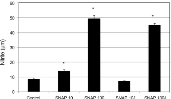
Figure 2. Effect of nitric oxide donor on the production of ni-
trite in confluently cultured trabecular meshwork cells. 10, 100 μm SNAP, and 100 μm SNAP with 0.5 mm L increased nitrite production significantly compared to non-exposed control. SNAP = S-Nitroso-N-acetyl-DL-penicillamine; L = L-NG-Nitroarginine methyl ester. *p < 0.05.Figure 3. Effects of nitric oxide donor on the permeability of car-
boxyfluorescin through the trabecular meshwork cell monolayer.10, 100 μm SNAP increased the permeability of carboxyfluorescin significantly compared to non-exposed control and abolished by co-exposed 0.5 mm L. Carboxyfluorescein intensity of outer chamber normalized to the mean value obtained using non-ex- posed control (permeability 100%). SNAP = S-Nitroso-N- ace- tyl-DL-penicillamine; L = L-NG-Nitroarginine methyl ester.
*p < 0.05.
게 자란 것을 확인한 후 SNAP에 24시간 노출시킨 결과 SNAP은 10, 100 μm의 농도에서 단층으로 충만하게 자란 섬유주세포의 생존에 영향을 미치지 않았다(p>0.05) (Fig. 1).
또한 0.5 mm L-NAME에 동시에 노출시킨 경우에도 세 포의 생존에 유의한 영향을 미치지 않았다(p>0.05). 따 라서 섬유주세포층의 투과도를 측정하기 위한 본 실험 에서 사용한 농도의 각 약제는 세포의 생존 변화에 의한 섬유주단층세포층의 투과도에는 영향을 미치지 않음을 알 수 있었다.
NO 공여자가 NO의 생성에 미치는 영향
SNAP은 배지에서 nitrite의 생성을 농도에 비례하여 유 의하게 증가시켰다(p<0.05) (Fig. 2). 10 μm의 농도에서는 대조군에 비해 5.48 μm, 그리고 100 μm의 농도에서는
40.71 μm의 nitrite를 더 많이 생성하였다(p=0.002, 0.001).
eNOS 저해제인 0.5 mm L-NAME에 동시에 노출시킨 경우, 각각 6.67 μm, 4.09 μm의 nitrite 생성을 감소시켰다 (p=0.001, 0.130). 따라서 SNAP은 섬유주단층세포층에서 NO의 생성을 유의하게 증가시켰으며, 이는 L-NAME에 의 해 NO의 생성이 억제됨으로써 확인할 수 있었다.
NO 공여자가 섬유주단층세포층의 투과도에 미치는 영향 약제에 노출되지 않은 대조군에 비하여 SNAP은 carbox- yfluorescein의 섬유주단층세포층의 투과도를 10 μm 농도 에서 5.9%, 100 μm 10.75% 증가시켰다(p=0.02, 0.001) (Fig. 3).
eNOS 저해제인 0.5 mm L-NAME에 동시에 노출시킨 경우, 약제에 노출되지 않은 대조군에 비하여 10 μm SNAP은 투 과도가 감소하였고(p=0.001), 100 μm SNAP의 경우 대조군 과 차이를 나타내지 않았다(p=0.064). 또한 10 μm SNAP에 노출시킨 경우에 비해 12.4%, 100 μm SNAP에 노출시킨 경우에 비해 13.2%씩 각각 투과도가 감소하였다(p=0.001, 0.001). 이는 0.5 mm 농도의 L-NAME는 100 μm SNAP에 의한 NO의 생성을 억제하기에는 충분하지 않은 것을 보여 주고 있다.
고 찰
본 연구의 결과는 섬유주단층세포층을 이용한 연구모델 에서 NO가 섬유주단층세포층의 투과도를 증가시킴을 보여 주고 있다.
실제 섬유주를 통한 방수유출의 정도를 정확하게 측정하 기는 매우 어려운데 주된 이유는 섬유주만을 통한 방수유 출의 측정이 어렵고, 각각의 연구마다 실험에 사용한 측정 방법이 다르기 때문이다. Carboxyfluorescein은 플푸오레신 과 분자의 크기와 형광도는 유사하지만 플루오레신에 비해 약 1,000배 정도 더 친수성이 있기 때문에 세포층의 투과도 를 측정하는 보다 민감한 방법이다.9-16 이 방법은 세포 스트 레스를 유발하지 않는 저농도로 장기간 노출시킨 다음 투 과도를 측정하면 혈액-안구장벽의 기능을 측정할 수도 있 을 뿐만 아니라14 이를 이용하면 약제가 섬유주의 투과도에 미치는 영향, 즉 섬유주를 통한 방수유출의 정도도 측정할 수 있다.15,16 이에 본 연구에서는 기존에 사용되지 않았던 carboxyfluorescein을 이용하여 NO가 섬유주세포층의 투과 도에 미치는 영향에 대한 연구를 시행하게 되었다.
NO는 섬유주를 이완시켜 섬유주를 통한 방수유출을 증 가시키는 것으로 알려졌으나3-8 섬유주세포층에 미치는 영 향은 알려져 있지 않다. 본 연구에서는 섬유주단층세포층 의 경우에도 NO에 의해 투과도가 증가되는지 확인하기 위 하여 섬유주세포를 단층으로 배양한 다음 NO 공여자에 24 시간 노출시켜 섬유주단층세포층의 투과도에 미치는 영향 을 측정하였다. 만약 노출시킨 약제가 세포의 생존에 영향 을 미친다면 죽은 세포의 탈락에 의해 투과도 측정에 오류 가 유발될 가능성을 배제할 수 없기 때문에 10, 100 μm의 농도에서 세포의 생존에 영향을 미치지 않는 것을 확인한 다음 투과성에 대한 실험을 시행하였다. 본 연구에서 사용 한 NO 공여자는 NO의 생성을 농도에 비례하여 유의하게 증가시켰으며 이는 eNOS 저해제인 L-NAME에 의해 NO의 생성이 억제됨으로써 확인할 수 있었다. 100 μm SNAP에 노출시킨 경우 10 μm에 노출시킨 경우에 비해 nitrite의 생 성 억제가 크게 차이가 나지 않는 것으로 보아 0.5 mm 농 도의 L-NAME는 100 μm SNAP에 의한 NO의 생성을 억제 하기에는 충분하지 않은 것으로 생각된다. 고농도의 NO 공 여자와 L-NAME에 노출시킨 경우, NO의 생성이 증가하였 음에도 불구하고 투과도가 떨어진 이유는 세포의 생존 자체 에는 영향을 미치지 않았으나 과량의 NO가 세포독성을 유발 하여 세포의 기능에 영향을 주었을 가능성이 있고, L-NAME 는 비특이적 NO 합성 저해제로서 다양한 작용을 나타낼 수 있기 때문으로 생각되며 이에 대해서는 L-NAME를 비롯한 여러 NO 합성 저해제의 작용기전에 대한 보다 자세한 연구 가 필요할 것으로 생각된다.
본 연구의 결과 NO는 섬유주단층세포층의 투과도를 농 도에 비례하여 증가시켰으며, eNOS 저해제에 동시에 노출 시킨 경우에는 섬유주단층세포층의 투과도를 감소시켰다.
따라서 NO가 섬유주단층세포층의 투과도를 증가시킴을 확
인할 수 있었다. 본 실험에서 사용한 carboxyfluorescein을 이용하여 섬유주단층세포층의 투과도를 측정하는 연구방 법은 기존에 보고된 fluorescein isothiocyanate-dextran을 이 용한 방법보다15,16 좀 더 민감하게 단층세포층의 투과도를 측정할 수 있는 방법으로 생각되며, NO뿐만 아니라 약제가 섬유주를 통한 방수유출에 미치는 영향을 측정하는 또 다 른 유용한 실험방법이 될 것으로 생각된다.
NO가 안압을 감소시키는 원인은 명확하지 않으나 섬유 주의 이완을 유발하여 방수를 유출시키는 것이 주된 기전 으로 여겨지고 있다.6,20 그런데 안구 내 생리적 조건과는 달 리 본 연구와 같이 섬유주 자체의 이완이 일어날 수 없는 조건인 단층세포층에서도 투과도를 증가시키는 것으로 나 타났다. 그 기전으로 생각해 볼 수 있는 것은 NO가 섬유주 세포 사이의 세포결합에 영향을 미쳐서 투과도를 증가시켰 을 가능성이 있으며,21 또한 NO가 기질금속단백분해효소 (matrix metalloproteinase)에 작용하여 세포외기질을 감소 시켜 투과도를 증가시켰을 가능성도 있고22 섬유주세포의 부피를 감소시켰을 수도 있으나23 이에 관해서는 향후 좀 더 자세한 연구를 요한다.
그러나 섬유주단층세포층을 이용한 실험실 내 결과가 실 제 안구 내 생리적 환경과는 많은 차이가 있으며 NO가 쉴 렘관과 공막을 비롯한 섬유주 주위의 여러 조직에도 영향 을 줄 수 있기 때문에 섬유주단층세포층을 이용한 실험의 결과에 의미를 부여하는 데 주의해야 할 것이다.
결론적으로 섬유주의 이완을 통해 방수유출을 증가시키 는 것으로 알려진 NO는 섬유주세포층의 방수유출을 증가 시키는 것을 알 수 있었으며, 이러한 방법은 향후 다른 약 제들이 섬유주를 통한 방수유출에 미치는 영향을 알아볼 수 있는 또 다른 연구방법이 될 수 있을 것으로 생각된다.
REFERENCES
1) Alvarado J, Murphy C, Juster R. Trabecular meshwork cellularity in primary open-angle glaucoma and nonglaucomatous normals.
Ophthalmology 1984;91:564-79.
2) Rohen JW, Lütjen-Drecoll E, Flügel C, et al. Ultrastructure of the trabecular meshwork in untreated cases of primary open-angle glaucoma (POAG). Exp Eye Res 1993;56:683-92.
3) Nathanson JA, McKee M. Identification of an extensive system of nitric oxide-producing cells in the ciliary muscle and outflow path- way of the human eye. Invest Ophthalmol Vis Sci 1995;36:
1765-73.
4) Geyer O, Podos SM, Mittag T. Nitric oxide synthase activity in tis- sues of the bovine eye. Graefes Arch Clin Exp Ophthalmol 1997;235:786-93.
5) Meyer P, Champion C, Schlötzer-Schrehardt U, et al. Localization of nitric oxide synthase isoforms in porcine ocular tissues. Curr
= 국문초록 =
일산화질소가 섬유주단층세포층의 투과성에 미치는 영향
목적: Carboxyfluorescein을 이용하여 일산화질소가 섬유주단층세포층의 투과성에 미치는 영향을 알아보고자 하였다.
대상과 방법: Transwell의 내측 chamber에 인체의 섬유주세포를 단층으로 충만하게 배양한 후 일산화질소 공여자인 0, 10, 100 μm S-Nitroso-N-acetyl-DL-penicillamine (SNAP)과 일산화질소 합성효소 저해제인 0.5 mm L-NAME에 24시간 노출시킨 다음 내측 chamber에 50 mm carboxyfluorescein을 2시간 동안 노출시킨 후 외측 chamber에서 투과된 carboxyfluorescein의 농도를 측정하 였다. 이때 세포의 생존은 MTT assay로, 일산화질소의 생성은 Griess assay로 각각 측정하였다.
결과: SNAP은 10, 100 μm의 농도에서 세포의 생존에 유의한 영향을 미치지 않았으며 일산화질소의 생성을 농도에 비례하여 유의하 게 증가시켰다. SNAP은 대조군에 비해 농도에 비례하여 섬유주단층세포층의 투과도를 유의하게 증가시켰다(p<0.05). 또한 L-NAME는 각 농도의 SNAP에 대해 유의하게 투과도를 감소시켰다(p<0.05).
결론: 일산화질소는 농도에 비례하여 섬유주단층세포층에서 carboxyfluorescein의 투과도를 증가시켰다. 따라서 섬유주를 이완시켜 안압을 하강시키는 것으로 알려진 일산화질소는 섬유주세포층을 통해서도 방수유출을 증가시킬 가능성이 있을 것으로 생각된다.
<대한안과학회지 2015;56(5):771-775>
Eye Res 1999;18:375-80.
6) Wiederholt M, Sturm A, Lepple-Wienhues A. Relaxation of tra- becular meshwork and ciliary muscle by release of nitric oxide.
Invest Ophthalmol Vis Sci 1994;35:2515-20.
7) Behar-Cohen FF, Goureau O, D'Hermies F, Courtois Y. Decreased intraocular pressure induced by nitric oxide donors is correlated to nitrite production in the rabbit eye. Invest Ophthalmol Vis Sci 1996;37:1711-5.
8) Nathanson JA, McKee M. Alterations of ocular nitric oxide syn- thase in human glaucoma. Invest Ophthalmol Vis Sci 1995;36:1774-84.
9) Araie M. Carboxyfluorescein. A dye for evaluating the corneal en- dothelial barrier function in vivo. Exp Eye Res 1986;42:141-50.
10) Araie M. Barrier function of corneal endothelium and the intra- ocular irrigating solutions. Arch Ophthalmol 1986;104:435-8.
11) Tsuboi S, Pederson JE. Permeability of the isolated dog retinal pig- ment epithelium to carboxyfluorescein. Invest Ophthalmol Vis Sci 1986;27:1767-70.
12) Blair NP, Rusin MM. Blood-retinal barrier permeability to carbox- yfluorescein and fluorescein in monkeys. Graefes Arch Clin Exp Ophthalmol 1986;224:419-22.
13) Grimes PA. Carboxyfluorescein transfer across the blood-retinal barrier evaluated by quantitative fluorescence microscopy: com- parison with fluorescein. Exp Eye Res 1988;46:769-83.
14) Nakagawa S, Usui T, Yokoo S, et al. Toxicity evaluation of anti- glaucoma drugs using stratified human cultivated corneal epi- thelial sheets. Invest Ophthalmol Vis Sci 2012;53:5154-60.
15) Kameda T, Inoue T, Inatani M, et al. The effect of Rho-associated protein kinase inhibitor on monkey Schlemm's canal endothelial
cells. Invest Ophthalmol Vis Sci 2012;53:3092-103.
16) Rao PV, Deng PF, Kumar J, Epstein DL. Modulation of aqueous humor outflow facility by the Rho kinase-specific inhibitor Y-27632. Invest Ophthalmol Vis Sci 2001;42:1029-37.
17) Mosmann T. Rapid colorimetric assay for cellular growth and sur- vival: application to proliferation and cytotoxicity assays. J Immunol Methods 1983;65:55-63.
18) Freimoser FM, Jakob CA, Aebi M, Tuor U. The MTT [3-(4,5-di- methylthiazol-2-yl)-2,5-diphenyltetrazolium bromide] assay is a fast and reliable method for colorimetric determination of fungal cell densities. Appl Environ Microbiol 1999;65:3727-9.
19) Green LC, Wagner DA, Glogowski J, et al. Analysis of nitrate, ni- trite, and [15N]nitrate in biological fluids. Anal Biochem 1982;
126:131-8.
20) Stamer WD, Lei Y, Boussommier-Calleja A, et al. eNOS, a pres- sure-dependent regulator of intraocular pressure. Invest Ophthalmol Vis Sci 2011;52:9438-44.
21) Predescu D, Predescu S, Shimizu J, et al. Constitutive eNOS-de- rived nitric oxide is a determinant of endothelial junctional integrity. Am J Physiol Lung Cell Mol Physiol 2005;289:L371-81.
22) Pfeilschifter J, Eberhardt W, Huwiler A. Nitric oxide and mecha- nisms of redox signalling: matrix and matrix-metabolizing en- zymes as prime nitric oxide targets. Eur J Pharmacol 2001;429:
279-86.
23) Dismuke WM, Mbadugha CC, Ellis DZ. NO-induced regulation of human trabecular meshwork cell volume and aqueous humor out- flow facility involve the BKCa ion channel. Am J Physiol Cell Physiol 2008;294:C1378-86.