DOI : 10.3341/jkos.2008.49.10.1665
과산화수소에 의한 산화스트레스가 섬유주세포의 노화에 미치는 영향
김재우․김신후․이재형 대구가톨릭대학교 의과대학 안과학교실
목적: 산화스트레스가 섬유주세포의 노화에 미치는 영향을 알아보고 아스코르빈산이 노화억제에 미치는 영향을 알아보 고자 하였다.
대상과 방법: 사람의 섬유주세포를 일차배양한 후 0, 0.05, 0.1 mM의 과산화수소에 30분간 노출시킨 후 일주일간 정치배양한 다음 일산화질소의 생성과 세포의 생존, 세포노화의 정도를 각각 Griess assay, MTT assay, SA-β -gal assay로 조사하였다. 이때 항산화제의 효과를 알아보기 위하여 0.1 mM의 아스코르빈산을 함께 투여하였다.
결과: 과산화수소는 일산화질소의 생성과 세포의 생존을 저하시켰으며, 섬유주세포의 노화를 유발하였으나 아스코르빈 산은 유의한 영향을 미치지 않았다.
결론: 과산화수소에 의한 산화스트레스는 섬유주세포의 노화를 유발하였다. 따라서 산화스트레스는 섬유주세포를 노화 시켜 섬유주의 기능을 저하시킬 수 있을 것으로 생각된다.
<대한안과학회지 2008;49(10):1665-1670>
<접수일 : 2008년 5월 9일, 심사통과일 : 2008년 7월 10일>
통신저자 : 김 재 우
대구시 남구 대명4동 3056-6 대구가톨릭대학병원 안과
Tel: 053-650-4728, Fax: 053-627-0133 E-mail: [email protected]
* 이 연구는 2006년도 대구가톨릭대학교 의과학연구소 연구비의 지원으로 이루어졌음
안구 내에서는 항상 활성산소가 방출되며 방수 내에 도 다량의 활성산소가 존재하고 이에 따른 조직의 손상 을 막기 위한 다양한 형태의 항산화 방어체계가 존재한 다.1 방수 내에는 활성산소가 지속적으로 생성되어 방 수유출경로인 섬유주는 항상 활성산소에 노출되어 있으 므로 섬유주는 산화스트레스에 의한 손상을 받아 방수 유출로의 저항을 증가시켜 녹내장을 유발할 수 있는 것 으로 알려져 있다.2,3 또한 원발개방각녹내장에서 섬유 주의 조직소견이 섬유주세포가 노화된 양상으로 나타나 는 것으로 보고되었다.4,5
그러나 섬유주세포에서 산화스트레스가 유발되었을 때 세포의 노화가 유발되는 지에 대해서는 아직 자세히 밝혀져 있지 않았으며 항산화 작용을 가진 아스코르빈 산이 섬유주세포의 노화에 미치는 영향도 아직 알려져
있지 않다. 따라서 본 연구에서는 섬유주세포를 과산화 수소에 노출시켜 산화스트레스를 유발한 후 산화스트레 스에 의해 섬유주세포의 노화가 유발되는 지 알아보고 아스코르빈산이 산화스트레스에 의한 섬유주세포의 노 화에 미치는 영향을 알아보고자 하였다.
대상과 방법
섬유주세포의 일차배양과 약물처리
안구은행에서 사후 6시간 이내에 적출한 특이한 병력 이 없는 인체의 안구를 이용하여 섬유주세포를 일차배 양한 후 배지에 포함된 혈청단백이 과산화수소의 작용 에 미치는 영향을 피하기 위하여 무혈청배지를 이용하 여 0, 0.05, 0.1 mM의 과산화수소에 30분간 노출시 켜 산화스트레스를 유발한 다음 정상배지로 교환하여 1 주일간 정치배양하였다. 이때 항산화제가 세포 노화에 미치는 영향을 알아보기 위하여 0.1 mM의 아스코르빈 산(L-ascorbic acid, Sigma, USA)에도 동시에 노 출시켰다.
일산화질소의 생성과 세포의 생존
일산화질소(NO, Nitric oxide)의 생성은 배지에서
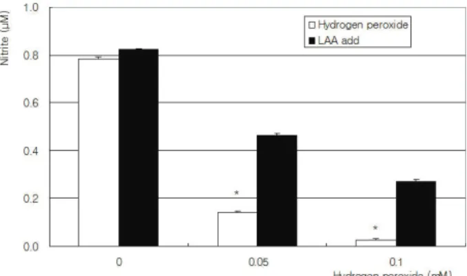
Figure 1. Effect of hydrogen-peroxide-induced oxidative stress on the production of NO in cultured trabecular meshwork cells. Hydrogen peroxide inhibited NO production significnatly, which was abolished by co-exposed L-ascorbic acid (LAA) (* p<0.05).
Figure 2. Effect of hydrogen peroxide on the survival of trabecular meshwork cells. Hydrogen peroxide decreased cellular survival significantly in a dose dependent manner.
Co-exposed 0.1 mM L-ascorbic acid did not affect on the survival (* p<0.05).
생성된 아질산염의 농도를 Griess assay로 측정하였 으며 세포의 생존율은 MTT assay로 조사하였다.6 MTT assay는 약물처리한 세포의 배지에 methyl thiazolyldiphenyl-tetrazolium bromide (MTT, Sigma, USA)를 각 well 당 100 µl씩 투여한 후 4시 간 동안 정치배양한 다음 염류용액으로 씻어낸 후 dimethylsulfoxide (Sigma, USA)를 각 well당 0.5 ml씩 넣어 10분 이상 흔든 다음 96-well plate 에 200 µl씩 옮겨 570 nm에서 흡광도를 측정하였다.
이때 세포의 증식정도는 실험군의 값을 약물처리를 하 지않은 대조군의 비로 나누어 백분율로 나타내었다.
NO의 생성은 NO의 대사물인 아질산염의 양을 측정하 는 Griess assay를16 이용하였는데 약물처리한 세포 의 배지에 동량의 Griess 반응액(Sigma, USA)를 섞 은 후 96-well plate에 옮겨 540 nm에서 흡광도를 측정하였다. 표준곡선은 sodium nitrite (Sigma, USA)를 단계적으로 희석하여 구하였다.
섬유주세포의 노화
노화된 세포에서 특징적으로 발현되는
β
-galacto sidase의 활성을 측정하기 위해 상용의 senescence -associatedβ
-galactosidase kit (SA-β
-gal, Sigma, St Louis, MO, USA)을 이용하였다.7,8 배 지를 흡입제거한 후 염류용액으로 세척한 후 고정한 다 음 염색액을 넣어 2시간 동안 배양하였다. 이때 노화된 세포는β
-galactosidase를 발현하여 푸른 색을 나타 내므로 광학현미경으로β
-galactosidase 양성인 푸 르게 염색된 노화된 세포수와 전체 세포수를 세어서 노 화된 세포의 정도를 백분율로 표시하였다.통계적 처리
모든 실험은 3계에서 5계대 사이의 세포를 이용하였 고 3회 이상 반복하여 시행하였다. 모든 실험에서 대조 군은 약물처리를 하지 않은 군으로 하였고 실험군과 대 조군의 비교는 unpaired t-test를 이용하였으며 유의 수준은 0.05% 이하로 정하였다.
결 과
세포배양
초대배양 2주째부터 섬유주조직의 이식편 주위로 섬 유주세포가 자라 나오기 시작하였으며 섬유주세포의 확 인은 특징적인 형태학적인 양상과 섬유주 조직의 이식 편 주위에서 위성양상으로 자라나는 섬유주세포의 특징 적인 성장양상으로 확인하였다.9,10
NO의 생성과 세포의 생존
과산화수소는 농도에 비례하여 NO의 생성을 유의하 게 감소시켰다(p<0.05). 산화스트레스가 유발된 경우 아스코르빈산이 NO의 생성에 미치는 영향을 알아보기 위하여 과산화수소와 아스코르빈산을 동시에 노출시킨 경우에는 아스코르빈산은 각 농도에서 과산화수소의 NO 생성억제작용을 상쇄시켰다(p>0.05)(Fig. 1). 또 한 과산화수소는 세포의 생존을 유의하게 저하시켰으나 (p<0.05) 동시에 투여한 아스코르빈산은 과산화수소에 의한 섬유주세포의 생존저하에 유의한 영향을 미치지 않았다(p>0.05)(Fig. 2).
Figure 4. Effect of hydrogen peroxide on the induction of cellular senescence in cultured trabecular meshwork cells.
Hydrogen peroxide increased cellular senescence significantly, which was not abolished by co-exposed 0.1 mM L-ascorbic acid (* p<0.05).
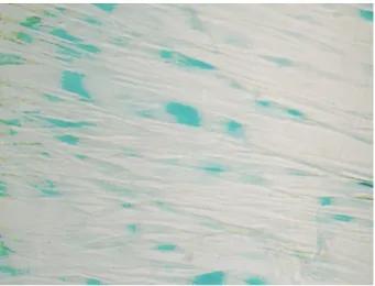
Figure 3. Photograph of SA-β-gal positive senescent trabecular meshwork cells (blue color) after exposed to 0.05 mM of hydrogen peroxide. Original magnification ×100.
산화스트레스에 의한 섬유주세포의 노화
노화된 섬유주세포는 SA-
β
-gal에 의해 푸르게 염색 되었으며(Fig. 3) 과산화질소는 농도에 비례하여 유의 하게 섬유주세포의 노화를 촉진하였다(p<0.05). 동시 에 투여한 0.1 mM의 아스코르빈산은 과산화수소에 의 한 섬유주세포의 노화촉진을 저하시키는 경향을 보였으나 통계학적으로 유의하지는 않았다(p>0.05)(Fig. 4).고 찰
섬유주세포는 섬유주를 통한 방수유출을 정상적으로 유지하는 중요한 역할을 하며 섬유주세포가 손상을 받 으면 섬유주의 방수유출기능이 저하되어 안압이 상승하 여 녹내장이 유발되거나 악화될 수 있다. 인체 내에서 산화스트레스는 다양한 병적 상태를 유발하며 녹내장의 경우에도 산화스트레스에 의해 망막신경절 세포의 손상 을 유발하는 것은 이미 잘 알려져 있다.11,12 산화스트레 스는 녹내장성 시신경손상을 유발할 뿐만 아니라 방수 내에는 다량의 활성산소가 발생하면 이에 따른 산화스 트레스에 의해 섬유주세포가 병적인 손상을 받아 안압 이 상승되는 기전도 될 수 있다.13,14 녹내장 환자의 방 수에서 산화스트레스를 받고 있는 상태를 나타내는 다 양한 결과가 보고되었으며3 인체 내에는 이러한 산화스 트레스에 대해 다양한 방어기전이 존재하는데15 아스코 르빈산은 혈액에 비해 방수 내에서 매우 고농도로 존재 하며 방수 내에서 중요한 항산화물질로 작용한다.1,16 혈관내피세포의 기능장애는 적절한 양의 NO의 생성장 애에 의해 유발되는데17 아스코르빈산은 잘 알려진 항 산화작용 뿐만 아니라 혈관내피세포의 NO합성을 촉진
하여 내피세포의 기능장애를 방지하는 역할을 하는 것 으로도 알려져 있고18,19 그 기전은 혈관내피세포의 경 우 NO합성의 필수적인 요소인 tetrahydrobippte rin를 화학적으로 안정화시켜 활성산소에 의한 NO의 산화적 변성을 방지하여 NO를 유지하는 것으로 보고 되었다.20,21
녹내장에서 섬유주의 기능이 저하되는 병적 소견으 로 섬유주의 노화가 제시되었는데4 산화스트레스는 세 포를 노화시켜 세포의 복제를 억제하여 그 기능을 상실 하게 되며 이러한 노화에 의한 세포복제의 억제에 NO 가 관여하는 것으로 알려져 있다.22 뿐만 아니라 산화스 트레스는 섬유주세포의 구조적 변성을 유발하여 세포외 기질에의 부착성을 감소시켜 세포의 소실을 유발하는 것으로 알려져 있다.23,24 섬유주의 변성 또는 노화에 따 른 기능의 저하는 개방각녹내장의 중요한 발생기전으로
생각되며25-27 활성산소의 발생이 세포노화의 중요한 원
인으로 받아들여 지고 있다.28-30 NO는 세포복제에 작 용하는 telomerase를 활성화시켜 세포노화를 방지하 는 역할을 하는 것으로 알려져 있는데22,28 활성산소에 지속적을 노출되어 있는 섬유주세포의 경우 산화스트레 스에 대한 방어체계는 섬유주의 노화를 방지하여 그 기 능을 정상적으로 유지하는 데 매우 중요할 것으로 생각 된다. 따라서 본 연구에서 산화스트레스가 섬유주세포 의 NO생성에 미치는 영향과 세포노화를 유발하는 지 알아보기 위해 과산화수소를 이용하여 섬유주세포에 산 화스트레스를 유발한 결과 농도에 비례하여 세포의 생 존이 감소하면서 동시에 NO의 생성도 감소하였다.
또한 본 연구에서 과산화수소는 농도에 비례하여 NO의 생성 감소와 함께 섬유주세포의 노화를 유발하 였으므로 기존의 여러 결과와 같이 NO의 생성 저하가
세포 노화의 기전으로 생각된다. 이에 대해 항산화제인 아스코르빈산은 과산화수소에 의해 유도된 산화스트레 스에 대해 NO의 생성 저하를 억제하고 섬유주세포의 노화를 억제하는 경향을 보이기는 하였으나 유의하게 산화스트레스에 의한 세포노화를 억제하지는 못하였 다.31 따라서 본 연구의 결과에서 아스크르빈산만으로 는 산화스트레스에 의한 섬유주세포의 노화를 방지할 수는 없다는 것을 알 수 있었으나 이에 관해서는 보다 자세한 연구가 필요할 것이며32,33 녹내장에 대한 새로 운 치료방법의 하나로 항산화제를 이용하여 NO의 생 성을 유지시키고 세포의 노화를 방지하여 정상적인 기 능을 회복하려는 시도와 연구가 활발히 행해지고 있으 며, 활성산소와 관련하여 섬유주세포의 기능을 유지하 고 노화를 방지하기 위한 연구가 앞으로 많이 이루어 질 것으로 예상된다.11
결론적으로 과산화수소에 의한 산화스트레스는 섬유 주세포에서 NO의 생성을 감소시켰으며 섬유주세포의 생존을 감소시키고 세포의 노화를 유발하였으나 항산화 제인 아스코르빈산은 산화스트레스에 의한 섬유주세포 의 노화에 유의한 영향을 미치지 않았다. 과다한 산화 스트레스는 섬유주세포의 노화를 유발함으로써 그 기능 을 저하시켜 방수유출을 감소시켜 안압상승을 유발할 수 있는 원인이 될 수 있을 것으로 생각된다.
참고문헌
1) Becker B. Chemical composition of human aqueous humor.
Effects of acetazolamide. Arch Ophthalmol 1957;57:793-800.
2) Zhou L, Li Y, Yue BY. Oxidative stress affects cytoskeletal structure and cell-matrix interactions in cells from an ocular tissue: The trabecular meshwork. J Cell Physiol 1999;180:182-9.
3) Sacca SC, Pascotto A, Camicione P, et al. Oxidative DNA damage in the human trabecular meshwork. Arch Ophthalmol 2005;123:458-63.
4) Liton PB, Challa P, Stinnett S, et al. Cellular senescence in the glaucomatous outflow pathway. Exp Gerontol 2005;40:745-8.
5) Yamazaki Y, Matsunaga H, Nishikawa M, et al. Senescence in cultured trabecular meshwork cells. Br J Ophthalmol 2007;91:
808-11.
6) Green LC, Wagner DA, Glogoski J, et al. Analysis of nitrate, nitrite and [15N] nitrate in biologic fluids. Anal Biochem 1982;126:131-8.
7) Matsunaga H, Handa JT, Aotaki-Keen A, et al. β-galactosi dase histochemistry and telomere loss in senescent retinal pigment epithelial cells. Invest Ophthalmol Vis Sci 1999;40:
197-202.
8) Dimri GP, Lee X, Basilr G, et al. A biomarker that identifies senescent human cells in culture and in aging skin in vitro.
Proc Natl Acad Soc U S A 1995;92:9363-7.
9) Polansky JR, Weinreb RN, Baxter JD, Alvarado J. Human trabecular cells. I. Establishment in tissue culture and growth characteristics. Invest Ophthalmol Vis Sci 1979;18:1043-9.
10) Alvarado JA, Wood I, Polansky JR. Human trabecular cells. II.
Growth pattern and ultrastructural characteristics. Invest Ophthalmol Vis Sci 1982;23:464-78.
11) Chen JZ, kadlubar FF. A new clue to glaucoma pathogenesis.
Am J Med 2003;114:697-8.
12) Ko ML, Hu DN, Ritch R, Sharma SC. The combined effect of brain-derived neurotrophic factor factor and free radical scavenger in experimental glaucoma. Invest Ophthalmol Vis Sci 2000;41:2967-71.
13) Izzotti A, Sacca SC, Cartiglia C, De Flora S. Oxidative deoxyribonucleic acid damage in the eyes of glaucoma patients. Am J Med 2003;114:638-46.
14) Ferreira SM, Lerner SF, Brunzini R, et al. Oxidative stress markers in aqueous humor of glaucoma patients. Am J Ophthalmol 2004;137:62-9.
15) Padgaonkar V, Giblin FJ, Leverenz V, et al. Studies of H2O2-induced effects on cultured bovine trabecular meshwork cells. J Glaucoma 1994;3:123-31.
16) Erb C, Nau-Staudt K, Flammer J, Nau W. Ascorbic acid as a free radical scavenger in porcine and bovine aqueous humor.
Ophthalmic Res 2004;36:38-42.
17) Ross R. Atherosclerosis-an inflammatory disease. N Eng J Med 1999;340:115-26.
18) Heller R, Münscher-Paulig F, Gräbner R, Till U. L-ascorbic acid potentiates nitric oxide synthesis in endothelial cells. J Biol Chem 1999;274:8254-60.
19) May JM. How does ascorbic acid prevent endothelial dysfunction? Free Radic Biol Med 2000;28:1421-9.
20) Huang A, Vita JA, Venema RC, Keaney Jr. JF. Ascorbic acid enhances endothelial nitric-oxide synthase activity by increasing intracellular tetrahydrobiopterin. J Biol Chem 2000;275:17399-406.
21) Heller R, Unbehaun A, Schnellenberg B, et al. L-ascorbic acid potentiates endothelial nitric oxide synthesis via a chemical stabilization of tetrahydrobiopterin. J Biol Chem 2001;276:40-7.
22) Vasa M, Breitschopf K, Zeiher AM, Dimmeler S. Nitric oxide activates telomerase and delays endothelial cell senescence.
Circ Res 2000;87:540-2.
23) Zhou L, Li Y, Yue BY. Oxidative stress affects cytoskeletal structure and cell-matrix interactions in cells from ocular tissue: the trabecular meshwork. J Cell Physiol 1999;180:182-9.
24) Alvarado J, Murphy C, Juster R. Trabecular meshwork cellularity in primary open-angle glaucoma and nonglauco matous normals. Ophthalmology 1984;91:564-79.
25) Millard CB, Tripathi BJ, Tripathi RC. Age-related changes in protein profiles of the normal human trabecular meshwork.
Exp Eye Res 1987;45:623-31.
26) Schchschabel DO, Binninger E. Aging of trabecular meshwork cells of the human eye in vitro. Z Gerontol 1990;23:133-5.
27) Horstmann HJ, Rohen JW, Sames K. Age-related changes in
the composition of proteins in the trabecular meshwork of the human eye. Mech Ageing Dev 1983;21:121-36.
28) Kurz DJ, Decary S, Hong Y, et al. Chronic oxidative stress compromises telomere integrity and accelerates the onset of senescence in human endothelial cells. J Cell Sci 2004;117:
2417-26.
29) von Zglinicki T. Role of oxidative stress in telomere length regulation and replicative senescence. Ann N Y Acad Sci 2000;908:99-110.
30) Wei YH. Oxidative stress and mitochondrial DNA mutations in human aging. Proc Soc Exp Biol Med 1998;217:53-63.
31) Kim JW. Ascorbic acid enhances nitric oxide production in cultured trabecular meshwork cell. Korean J Ophthalmol 2005;19:297-31.
32) Furumoto K, Inoue E, Nagao N, et al. Age-dependent telomere shortening is slowed down by enrichment of intracellular vitamin C via suppression of oxidative stress. Life Sci 1998;63:935-48.
33) Roques SC, Landrault N, Teisse’dre P, et al. Hydrogen peroxide generation in Caco-2 cell culture medium by addition of phenolic compounds: Effect of ascorbic acid. Free Radic Res 2002;36:593-9.
=ABSTRACT=
Effect of Hydrogen Peroxide-induced Oxidative Stress on the Senescence of Trabecular Meshwork Cells
Jae Woo Kim, M.D., Sin Hoo Kim, M.D., Jae Hyung Lee, M.D.
Department of Ophthalmology, Catholic University of Daegu School of Medicine, Daegu, Koera
Purpose: To investigate the effects of oxidative stress on the senescence of trabecular meshwork (TM) cells and the effect of L-ascorbic acid (LAA) against oxidative stress-induced senescence.
Methods: Primary cultured human TM cells were exposed to 0.05 or 0.1 mM hydrogen peroxide for 30 minutes and incubated for 1 week with or without co-exposure of LAA. Cellular survival, nitrite production, and senescence were assessed with MTT, Griess, and SA-β-gal assays, respectively.
Results: Hydrogen peroxide decreased cellular survival and NO production accompanied increased cellular senescence. LAA did not prevent hydrogen peroxide-induced senescence.
Conclusions: Oxidative stress-induced senescence of TM cells may be related to the dysfunction of trabecular meshwork in glaucoma.
J Korean Ophthalmol Soc 2008;49(10):1665-1670
Key Words: Ascorbic acid, Hydrogen peroxide, Oxidative stress, Senescence, Trabecular meshwork cells
Address reprint requests to Jae Woo Kim, M.D.
Department of Ophthalmology, Catholic University of Daegu College of Medicine
#3056-6 Daemyeung-4-dong, Nam-gu, Daegu 705-718, Korea Tel: 82-53-650-4728, Fax: 82-53-627-0133, E-mail: [email protected]