서 론
섬유주세포는 녹내장에서 방수 유출의 조절에 중요한 역할을 하는데, 섬유주의 변성으로 방수 유출로의 저항이 증가되면 개 방각녹내장을 유발할 수 있는 기전이 된다.1,2 기존의 녹내장 치료 약제의 대부분은 방수의 생성을 줄여 안압하강 효과를 나타내지 만 최근 섬유주를 통한 방수 유출을 직접 증가시키는 약제가 많 이 연구되고 있으며3 이러한 약제의 하나로 Rho-kinase (ROCK) 저해제가 있다.4
ROCK 저해제는 섬유주를 이완시켜 방수 유출을 증가시키는 데, 주된 기전은 F-액틴의 중합을 저해하고 세포내 칼슘을 조절함 으로써 세포 간 밀착연접(tight junction)을 해체시켜 섬유주를 통 한 방수 유출을 증가시키는 것으로 알려져 있다.5 ROCK 저해제 의 일종인 Y-27632는 여러 연구에서 방수 유출을 증가시키는 작 용을 나타내는 것으로 보고되어 있다.6-9
자유유리기인 일산화질소(nitric oxide, NO)는 세포의 종류에 따라 다양한 역할을 나타낼 수 있으며, 평활근 이완 효과도 나타 내는 것으로 알려져 있다. 섬유주세포는 형태학적 연구와 전기생 리학적 연구에서 평활근과 유사한 성질을 가진 것으로 알려져 있
으며10,11 endothelial nitric oxide synthase (eNOS)에서 생산하는
NO는 섬유주를 이완시켜 방수 유출을 촉진하는 것으로 알려져 있다.12-14
ROCK 저해제의 작용기전은 섬유주 주변 세포의 형태학적 변 화와 액틴스트레스 섬유에 영향을 미쳐 섬유주를 이완시키고 쉴
저농도의 Rho-kinase 저해제가 섬유주 투과성과 일산화질소 생성에 미치는 영향
Effect of Low Concentration Rho-kinase Inhibitor on the Permeability and Nitric Oxide Production in Trabecular Meshwork
김재우
Jae Woo Kim, MD, PhD
대구가톨릭대학교 의과대학 안과학교실
Department of Ophthalmology, Daegu Catholic University School of Medicine, Daegu, Korea
Purpose: To investigate the effects of the lower concentration of Rho-kinase (ROCK) inhibitor on the permeability of trabecular cell monolayer and its relationship with nitric oxide (NO) production in cultured human trabecular meshwork cells (HTMC).
Methods: Primarily cultured HTMC were exposed to 0, 5, 10, 20 mM Y-27632 for 24 hours. NO production and expression of endothelial nitric oxide synthase (eNOS) mRNA were measured with Griess assay and Reverse Transcription-Polymerase Chain Reaction (RT-PCR). The permeability of the HTMC monolayer was determined using Transwell and carboxyfluorescein. After inducing oxidative stress with 200 mM hydrogen peroxide, above experiments were done simultaneously.
Results: In HTMC, 5, 10, and 20 mM Y-27632 significantly increased the permeability of the HTMC monolayer (all p < 0.05) in a dose-dependent manner. In contrast, 5, 10 mM Y-27632 did not increase NO production and expression of eNOS mRNA significantly (p > 0.05). Experiments under the oxidative stress showed similar results.
Conclusion: ROCK inhibitor increased trabecular permeability in a dose-dependent manner but this increase was not accompanied with increased NO production. At low concentration, ROCK inhibitor-induced increased trabecular outflow may be not associated with NO production.
Key words: Nitric oxide, Oxidative stress, Permeability, ROCK inhibitor, Trabecular meshwork
Received: 2019. 11. 22. Revised: 2019. 12. 13.
Accepted: 2019. 12. 15.
Corresponding Author: Jae Woo Kim, MD, PhD
Department of Ophthalmology, Daegu Catholic University Hospital,
#33 Duryugongwon-ro 17-gil, Nam-gu, Daegu 42472, Korea Tel: +82-53-650-4728, Fax: +82-53-627-0133
E-mail: [email protected]
렘관세포의 투과성을 증가시키는 것인데7,15 혈관내피세포와 섬유 주세포를 이용한 여러 연구에서 ROCK 저해제가 NO의 생성을 증가시킨다는 보고가 있다.16-21 이와 대조적으로 ROCK 저해제의 농도에 따라 NO의 생성에 효과가 다르게 나타난다는 보고가 있
는데22,23 고농도에서는 ROCK 저해제가 NO의 생성을 증가시키지
만 저농도에서는 오히려 NO의 생성을 감소시켜 원치 않는 해로 운 효과를 나타낼 수 있다고 하였다.23
섬유주세포에서 ROCK 저해제는 고농도에서는 eNOS의 발현 과 NO의 생성을 증가시키는 것으로 이미 잘 알려져 있지만, 저농 도에서 ROCK 저해제가 섬유주의 투과도에 미치는 영향과 NO 의 생성에 미치는 영향에 대해서는 아직 자세히 연구되어 있지 않 다. 이에 본 연구에서는 사람의 섬유주세포를 일차배양하여 저농 도의 ROCK 저해제가 NO의 생성에 미치는 영향과 섬유주단층 세포층의 투과도에 미치는 영향을 알아보고자 하였다.
대상과 방법
세포배양과 약물처리
본 연구는 대구가톨릭대학교병원의 의학윤리심의위원회(IRB) 승인을 받았고(승인 번호: CR-16-117), 헬싱키선언을 따라 시행되 었다. 안구은행에서 얻은 사후 6시간 이내에 적출한 안구의 앞방 각에서 섬유주를 벗겨내어 배양접시에 옮긴 후 항생제(penicillin 10,000 units/mL, streptomycin 10,000 µg/mL, amphotericin B 25 µg/mL; Gibco, Invitrogen, Carlsbad, CA, USA)와 15% 우 태아혈청이 포함된 Dulbecco’s modified Eagle’s medium 배지 (DMEM, Gibco, Invitrogen)를 사용하여 5% CO2 배양기에서 초대배양하였다. 섬유주세포가 이식된 조직편 주위로 자라나온 것을 확인한 후 섬유주조직의 이식편을 제거하고 배양을 계속하 였으며, 세포가 배양접시에 충만해지면 10% 우태아혈청(Gibco, Invitrogen)을 포함한 배지로 1:3의 비율로 트립신 처리하여 계대 배양하였다. 일차배양한 섬유주세포를 배양접시에 부착시킨 다음 ROCK 저해제인 Y-26732 (Sigma, St. Louis, MO, USA)를 0, 5, 10, 20 µM의 농도로 24시간 노출시켰다. 이때 Y-26732 처리 전에 200 µM 과산화수소에 먼저 노출시켜 산화스트레스를 유도 한 다음 동일한 실험을 시행하였다.
MTT assay
세포의 생존에 대한 효과는 3-[4, 5 –dimethylthiazol-2-yl]-2, 5-diphenyltetrazolium bromide (MTT, Sigma) assay를 이용
하였다.24,25 24 시간 약물처리한 세포의 배지에 MTT를 각 well
당 100 µL씩 투여한 후 4시간 동안 정치배양한 다음 염류용액
(phosphated buffered saline [PBS], Gibco, Invitrogen)으로 씻 어낸 후 dimethylsulfoxide (Sigma)를 각 well당 0.5 mL씩 넣 어 96-well plate에 옮겨 분광광도계(Fluostar Optima, BMG labtech, Offenberg, Germany)로 570 nm에서 흡광도를 측정하 였다. 이때 세포의 생존 정도는 실험군의 값을 약물처리를 하지 않은 대조군의 비로 나누어 백분율로 나타내었다.
Griess assay
섬유주세포에서 NO의 생성은 Griess assay를 이용하여 배지 에서의 nitrite 농도를 측정하였다.26 24시간 각 농도의 Y-26732에 노출시킨 다음 세포배양액 100 µL를 후 96-well plate에 옮겨 동 량의 Griess reagent (Sigma)를 배지에 섞어 10분간 정치배양한 다음 spectrophotometer (Fluostar Optima, BMG Labtech)로 540 nm에서 흡광도를 측정하였다. 이때 측정된 nitrite를 정량화 하기 위해 표준치를 구하기 위해 sodium nitrite (Sigma)를 단계 적으로 희석하여 표준치를 구하여 비교하였다.
eNOS mRNA 발현을 측정하기 위한 RT-PCR
섬유주세포에서 Trizol (Invitrogen)을 이용하여 RNA를 분리 한 후 분리한 RNA와 Oligo dT primer, Nuclease-free water를 혼합하여 만든 RNA denaturation mix를 70℃에 5분간 변성시키 고 Prime RT premix (Genet bio, Seoul, Korea)와 혼합하여 반 응시켜 cDNA로 합성하였다. 합성한 cDNA에 2XGoTaq Green Master Mix (Promega, Fitchburg, WI, USA)와 forward primer (ctg gct ttc cct tcc agt tc, 225 bp), reverse primer (cct tcc aga tta agg cgg ac, 225 bp)를 각각 혼합하여 DNAEngine cycler (Bio- Rad, Hercules, CA, USA)를 사용하여 총 30 cycles를 시행하여 반응시켰다. 증폭된 PCR product를 전기 영동하여 DNA band를 multi Gauge v2.02 (Fujifilm, Tokyo, Japan)을 이용하여 분석하 였다. 이때 β-action을 internal standard로 사용하였다.
Carboxyfluorescein permeability assay
Transwell (No. 3460, Corning, Tewksbury, MA, USA)의 내 측 chamber에 세포가 단일세포층으로 충만하게 자란 것을 확인 한 후 6 시간 동안 각 약제에 노출시킨 후 투과도 검사를 시행하
였다.27-29 내측 chamber에 자라고 있는 세포를 PBS로 3회 세척
한 다음 50 µM carboxyfluorescein (Sigma)을 노출시켰다. 노출 2시간 후 외측 chamber로 투과된 carboxyfluorescein의 농도를 532 nm에서 분광형광계(Fluostar Optima, BMG Labtech)로 측 정하여 백분율로 나타내었다.
통계적 처리
모든 실험은 3계대에서 5계대 사이의 세포를 이용하였다. 세포 의 생존과 NO의 생성은 평균±표준오차로 나타내었으며, eNOS mRNA의 발현과 투과도의 변화는 대조군에 대한 백분율로 나타 내었다. Unpaired t-test를 사용하여 유의성을 비교하였으며 유의 수준은 5%로 정하였다.
결 과
Y-26732가 섬유주세포의 생존에 미치는 영향
0, 5, 10, 20 µM 농도의 Y-26732에 24시간 노출시킨 후 시행 한 MTT assay에서 모든 농도에서 약물에 노출되지 않은 대조군 에 비해 세포의 생존율에 유의한 차이를 나타내지 않았으며, 산 화스트레스를 유발한 경우에도 과산화수소만 처치한 군에 비해 세포의 생존율에 있어서 유의한 차이를 나타내지 않았다(모두 p>0.05).
Y-26732가 NO의 생성에 미치는 영향
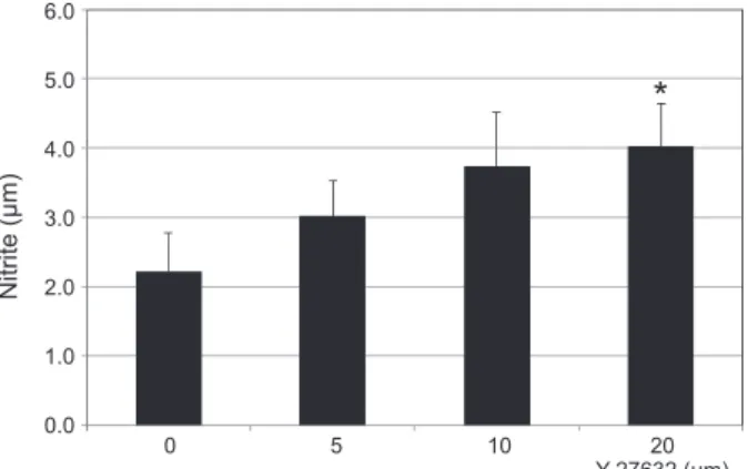
섬유주세포에서 Y-26732는 약물 처리 후 1일째에 20 µM 농 도에서만 약물처리를 하지 않은 대조군에 비하여 배지에서의 nitrite 생성량이 유의하게 증가하였다(p=0.028) (Fig. 1). 또한 산화 스트레스를 유발한 경우에도 과산화수소만 처치한 군에 비해 20 µM 농도에서만 nitrite 생성량이 유의하게 증가하였다(p=0.044) (Fig. 2).
Y-26732가 eNOS mRNA의 발현에 미치는 영향
약물처리를 하지 않은 대조군에 비하여 0, 5, 10, 20 µM 농도
의 Y-26732는 농도에 비례하여 eNOS mRNA의 발현을 증가시 키는 경향을 나타내었으나 통계적으로 유의하지는 않았다(모두 p>0.05) (Fig. 3). 산화스트레스를 유발한 경우에는 과산화수소 만 처치한 군에 비해 20 µM 농도에서만 유의하게 eNOS mRNA 의 발현을 증가시켰다(p=0.021) (Fig. 4).
Y-26732가 섬유주단층세포층의 투과성에 미치는 영향
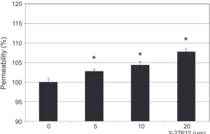
약물처리를 하지 않은 대조군에 비하여 5, 10, 20 µM의 Y-26732는 5 µM의 저농도에서부터 농도에 비례하여 섬유주단 층세포층의 투과도를 유의하게 증가시켰다(모두 p<0.05) (Fig. 5).산화스트레스를 유발한 경우에는 과산화수소만 처치한 군에 비 해 10 µM과 20 µM 농도에서 각각 유의하게 투과도를 증가시켰 다(p=0.001, p=0.001) (Fig. 6).
고 찰
본 연구의 결과는 사람의 섬유주세포에서 저농도의 ROCK 저 해제인 Y-26732가 농도에 비례하여 섬유주의 투과도를 증가시키 지만 이러한 투과도의 증가는 eNOS의 발현이나 NO의 생성과는 연관성이 적음을 보여주고 있다.
ROCK 저해제가 섬유주를 통한 방수 유출을 촉진하는 주된 기전은 섬유주를 이완시키며 액틴스트레스 섬유와 초점접착역 (focal adhesion)에 작용하여 세포 주변을 통해 방수 유출을 촉진 하는 것이며,5,7,15 ROCK 저해제의 또 다른 작용 기전으로 eNOS mRNA의 발현과 NO의 생성을 증가시키는 것이 있다.16,17 NO 역 시 섬유주를 이완시켜 방수 유출을 촉진하므로12-14 ROCK 저
Nitrite (µm)
0 5 10 20
6.0 5.0 4.0 3.0 2.0 1.0
0.0
Y-27632 (µm)
Figure 1. Effects of Y-27632 (0, 5, 10, 20 µM) on the produc- tion of nitric oxide in cultured human trabecular meshwork cells.
Twenty µM Y-27632 increased the production of nitric oxide sig- nificantly compared to non-exposed controls (*p = 0.028).
*
Nitrite (µm)
0 5 10 20
6.0 5.0 4.0 3.0 2.0 1.0
0.0
Y-27632 (µm)
Figure 2. Effects of Y-27632 (0, 5, 10, 20 µM) on the production of nitric oxide in cultured human trabecular meshwork cells under oxidative stress. Twenty µM Y-27632 increased the production of nitric oxide significantly compared to hydrogen peroxide-exposed controls (*p = 0.044).
*
해제가 혈관내피세포에서처럼 NO의 생성에 영향을 미친다면 ROCK 저해제에 의한 NO 생성 증가가 ROCK 저해제의 또 다른 작용기전이 될 수 있을 것이다.
ROCK 저해제인 Y-26732의 작용 기전을 연구한 결과들은 대 부분 10 µM 이상의 고농도로 시행되었는데30,31 실험실 내 연구에 서도 10, 100 µM의 농도에서 Y-26732가 쉴렘관세포의 투과도와 형태학적 변화를 유발하는 것으로 알려졌다.6-8 그러나 Y-26732
의 농도에 따라 상반된 결과를 나타낸다는 보고들이 있다.32,33 5 µM 이하의 저농도 Y-26732는 독성 작용을 나타낼 수 있으며 NO의 생성을 억제하여 오히려 유해한 작용을 나타낸다는 보고도
있어23,34 ROCK 저해제는 농도에 따라 효과가 다르게 나타날 수
있다. 녹내장 치료제로 ROCK 저해제가 많이 연구되고 있는데4,5 저농도에서 NO의 생성을 억제한다면 ROCK 저해제는 저농도에 서 오히려 방수 유출을 저하시킬 가능성이 있다. 이를 규명하기
Relative activity (% of control)
0 5 10 20
0 5 10 20
250 200 150 100 50
0
Y-27632 (µm)
Figure 4. Effects of Y-27632 (0, 5, 10, 20 µM) on the expression of eNOS mRNA in cultured human trabecular meshwork cells under oxidative stress. 20 µM Y-27632 increased the expression of eNOS mRNA significantly compared to hydrogen peroxide- exposed controls (*p = 0.021).
*
Permeability (%)
0 5 10 20
120 115 110 105 100 95
90
Y-27632 (µm)
Figure 5. Effects of Y-27632 on the permeability of carboxyfluo- rescin through the trabecular meshwork cell monolayer. Expo- sure to 5, 10 and 20 µM Y-27632 increased the permeability of carboxyfluorescein significantly in a dose-dependent manner compared with non-exposed controls (*p < 0.05). Carboxyfluo- rescein intensity of outer chamber normalized to the mean value obtained using non-exposed control (permeability 100%).
* *
*
Permeability (%)
0 5 10 20
120 115 110 105 100 95 90
Y-27632 (µm)
Figure 6. Effects of Y-27632 on the permeability of carboxyfluo- rescin through the trabecular meshwork cell monolayer under oxidative stress. Exposure to 10 and 20 µM Y-27632 increased the permeability of carboxyfluorescein significantly in a dose- dependent manner compared to hydrogen peroxide-exposed con- trols (*p < 0.05). Carboxyfluorescein intensity of outer chamber normalized to the mean value obtained using hydrogen peroxide- exposed controls (permeability 100%).
* *
Relative activity (% of control)
0 5 10 20
0 5 10 20
250 200 150 100 50
0
Y-27632 (µm)
Figure 3. Effects of Y-27632 (0, 5, 10, 20 µM) on the expression of eNOS mRNA in cultured human trabecular meshwork cells.
Y-27632 did not affect the expression of eNOS mRNA compared to non-exposed controls (all p > 0.05).
위해 시행한 본 연구에서는 세포의 생존에는 유의한 영향을 미치 지 않아 독성 작용이 나타나지 않음을 알 수 있었다. 또한 5 µM 의 저농도에서 Y-26732는 NO의 생성과 eNOS의 발현에 유의한 영향을 미치지 않아 섬유주에서 저농도의 ROCK 저해제는 NO 의 생성 억제에 의한 방수 유출의 저하는 나타내지 않을 것으로 생각된다. 저농도에서 Y-26732는 NO의 생성에는 유의한 영향을 미치지 않았으나 이와 대조적으로 Y-26732는 5 M의 저농도에서 부터 섬유주단층세포츠의 투과도를 농도에 비례하여 증가시켰다.
따라서 저농도의 ROCK 저해제는 방수 유출을 증가시키기는 하 였으나 NO의 생성과는 상관없으므로, 저농도에서 ROCK 저해 제에 의한 투과도의 증가는 ROCK 저해제의 주된 기존의 작용기 전인 섬유주의 이완과 쉴렘관연접부세포의 형태학적 변화에 의 한 것으로 생각된다.7,15 한편 고농도에서는 이에 더하여 섬유주세 포에서 eNOS mRNA의 발현과 NO 생성을 증가시킴으로써 섬유 주를 통한 방수 유출을 촉진시킬 것으로 생각된다.18
안구 내 방수에는 활성산소가 지속적으로 생성되어 방수의 주 된 유출 경로인 섬유주는 항상 활성산소에 노출되어 있으므로 섬 유주는 산화스트레스에 의한 손상을 받아 방수 유출로의 저항을 증가시켜 녹내장을 유발할 수 있다.35,36 혈관내피세포에서 전단응 력을 유발하여 시행한 기존의 연구에서24 저농도의 Y-26732는 과 산화수소의 생성량을 증가시켜 산화스트레스를 유발하며 NO의 생성을 억제한다고 보고하였으므로 본 연구에서는 과산화수소로 산화스트레스를 유발하여 그 변화를 살펴보았다. 그 결과 20 µM 의 고농도에서는 Y-26732는 NO의 생성과 eNOS의 발현이 증가 하였으나 5 µM의 저농도에서는 NO의 생성과 eNOS의 발현에 유 의한 영향을 미치지 않았으며 농도의 증가에 따라 섬유주단층세 포층의 투과도를 증가시켰다. 따라서 섬유주가 지속적으로 산화 스트레스에 노출된 녹내장의 경우에도 ROCK 저해제는 저농도 에서도 방수 유출을 증가시키는 작용을 나타낼 수 있을 것이다.
결론적으로 ROCK 저해제는 저농도에서 NO의 생성에는 유의 한 영향을 미치지 않으면서 방수 유출을 촉진할 수 있을 것으로 생각된다. 그러나 본 연구는 섬유주세포를 배양하여 시행한 실험 실 내 연구이므로 실제 ROCK 저해제의 농도에 따른 작용은 향 후 동물 실험 등을 통한 보다 자세한 연구가 필요할 것이다.
References
1. Alvarado J, Murphy C, Juster R. Trabecular meshwork cellularity in primary open-angle glaucoma and nonglau- comatous normals. Ophthalmology 1984;91:564-79.
2. Rohen JW, LÜtjen-Drecoll E, FlÜgel C, et al. Ultrastruc- ture of the trabecular meshwork in untreated cases of pri-
mary open-angle glaucoma. Exp Eye Res 1993;56:683-92.
3. Schmidl D, Schmetterer D, Garhöfer G, Popa-Chereche- anu A. Pharmacotherapy of glaucoma. J Ocul Pharmacol Ther 2015;31:63-77.
4. Kopczynski CC, Epstein DL. Emerging trabecular outflow drugs. J Ocul Pharmacol Ther 2014;30:85-7.
5. Inoue T, Tanihara H. Rho-associated kinase inhibitors: a novel glaucoma therapy. Prog Retin Eye Res 2013;37:1- 12.
6. Thieme H, Nuskovski M, Nass JU, et al. Mediation of calcium-independent contraction in trabecular meshwork through protein kinase C and rho-A. Invest Ophthalmol Vis Sci 2000;41:4240-6.
7. Rao PV, Deng PF, Kumar J, Epstein DL. Modulation of aqueous humor outflow facility by the Rho kinas- especific inhibitor Y-27632. Invest Ophthalmol Vis Sci 2001;42:1029-37.
8. Honjo M, Tanihara H, Inatani M, et al. Effects of rho- associated protein kinase inhibitor Y-27632 on intraocular pressure and outflow facility. Invest Ophthalmol Vis Sci 2001;42:137-44.
9. Novack GD. Rho kinase inhibitors for the treatment of glaucoma. Drugs Future 2013;38:107-13.
10. Wiederholt M, Dörschner N, Groth J. Effect of diuret- ics, channel modulators, and signal interceptors on con- tractility of the trabecular meshwork. Ophthalmologica 1997;211:153-60.
11. Wiederholt M, Stumpff F. The trabecular meshwork and aqueous humor reabsorption. In: Civan MM, ed. Current topics in membranes. The eye’s aqueous humor: from se- cretion to glaucoma. San Diego: Academic Press, 1998; v.
45. chap. 7.
12. Wiederholt M, Sturm A, Lepple-Wienhues A. Relaxation of trabecular meshwork and ciliary muscle by release of nitric oxide. Invest Ophthalmol Vis Sci 1994;35:2515-20.
13. Behar-Cohen FF, Goureau O, D’Hermies F, Courtois Y.
Decreased intraocular pressure induced by nitric oxide donors is correlated to nitrite production in the rabbit eye.
Invest Ophthalmol Vis Sci 1996;37:1711-5.
14. Dismuke WM, Mbadugha CC, Ellis DZ. NO-induced regulation of human trabecular meshwork cell volume and aqueous humor outflow facility involve the BKCa ion channel. Am J Physiol Cell Physiol 2008;294:C1378-86.
15. Sanka K, Maddala R, Epstein DL, Rao PV. Influence of actin cytoskeletal integrity on matrix metalloproteinase-2 activation in cultured human trabecular meshwork cells.
Invest Ophthalmol Vis Sci 2007;48:2105-14.
16. Ming XF, Viswambharan H, Barandier C, et al. Rho GT- Pase/Rho kinase negatively regulates endothelial nitric
oxide synthase phosphorylation through the inhibition of protein kinase B/Akt in human endothelial cells. Mol Cell Biol 2002;22:8467-77.
17. Takemoto M, Sun J, Hiroki J, et al. Rho-kinase mediates hypoxia-induced downregulation of endothelial nitric ox- ide synthase. Circulation 2002;106:57-62.
18. Kim JW, Kim KH, Hwang SJ. Effect of Rho kinase on the production of nitric oxide in trabecular meshwork cells. J Korean Ophthalmol Soc 2016;57;650-6.
19. Hein TW, Rosa RH Jr, Yuan Z, et al. Divergent roles of nitric oxide and rho kinase in vasomotor regulation of human retinal arterioles. Invest Ophthalmol Vis Sci 2010;
51:1583-90.
20. Guan R, Xu X, Chen M, et al. Advances in the studies of roles of Rho/Rho-kinase in diseases and the development of its inhibitors. Eur J Med Chem 2013;70:613-22.
21. Eto M, Barandiér C, Rathgeb L, et al. Thrombin sup- presses endothelial nitric oxide synthase and upregulates endothelin-converting enzyme-1 expression by distinct pathways: role of Rho/ROCK and mitogen-activated pro- tein kinase. Circ Res 2001;89:583-90.
22. Malek AM, Izumo S. Mechanism of endothelial cell shape change and cytoskeletal remodeling in response to fluid shear stress. J Cell Sci 1996;109(Pt 4):713-26.
23. Kolluru GP, Majumder S, Chatterjee S. Rho-kinase as a therapeutic target in vascular diseases: striking nitric oxide signaling. Nitric Oxide 2014:43:45-54.
24. Mosmann T. Rapid colorimetric assay for cellular growth and survival: application to proliferation and cytotoxicity assays. J Immunol Methods 1983;65:55-63.
25. Freimoser FM, Jakob CA, Aebi M, Tuor U. The MTT [3-(4,5-dimethylthiazol-2-yl)-2,5-diphenyltetrazolium bro- mide] assay is a fast and reliable method for colorimetric determination of fungal cell densities. Appl Environ Mi- crobio 1999;65:3727-9.
26. Green, LC, Wagner DA, Glogowski J, et al. Analysis of nitrate, nitrite and [15N] nitrate in biologic fluids. Analyti-
cal Biochem 1982;126:131-8.
27. Grimes PA, Stone RA, Laties AM, Li W. Carboxyfluo- rescein. A probe of the blood-ocular barriers with lower membrane permeability than fluorescein. Arch Ophthalmol 1982;100:635-9.
28. Nakagawa S, Usui T, Yokoo S, et al. Toxicity evaluation of antiglaucoma drugs using stratified human cultivated corneal epithelial sheets. Invest Ophthalmol Vis Sci 2012;53:5154-60.
29. Lei Y, Stamer WD, Wu J, Sun X. Oxidative stress impact on barrier function of porcine angular aqueous plexus cell monolayers. Invest Ophthalmol Vis Sci 2013;54:4827-35.
30. Seasholtz TM, Brown JH. RHO signaling in vascular dis- eases. Mol Interv 2004;4:348-57.
31. Versteilen AM, Korstjens IJ, Musters RJ, et al. Rho-kinase regulates renal blood flow by modulating eNOS activity in ischemia-reperfusion of the rat kidney. Am J Physiol Re- nal Physiol 2006;291:F606-11.
32. Uchida S, Watanabe G, Shimada Y, at al. The suppression of small GTPase rho signal transduction pathway inhibits angiogenesis in vitro and in vivo. Biochem Biophys Res Commun 2000;269:633-40.
33. Noma K, Oyama N, Liao JK. Physiological role of ROCKs in the cardiovascular system. Am J Physiol Cell Physiol 2006;290:C661-8.
34. Zhao Z, Rivkees SA. Rho-associated kinases play a role in endocardial cell differentiation and migration. Dev Biol 2004;275:183-91.
35. Zhou L, Li Y, Yue BY. Oxidative stress affects cytoskel- etal structure and cell-matrix interactions in cells from an ocular tissue: the trabecular meshwork. J Cell Physiol 1999;180:182-9.
36. Saccà SC, Pascotto A, Camicione P, et al. Oxidative DNA damage in the human trabecular meshwork: clinical corre- lation in patients with primary open-angle glaucoma. Arch Ophthalmol 2005;123:458-63.
국문초록
저농도의 Rho-kinase 저해제가 섬유주 투과성과 일산화질소 생성에 미치는 영향
목적: 저농도 Rho-kinase (ROCK) 저해제가 섬유주의 투과성에 미치는 영향과 일산화질소 생성과의 연관성을 알아보고자 하였다.
대상과 방법: 사람의 섬유주세포를 일차배양한 후 ROCK 저해제인 Y-27632를 0, 5, 10, 20 μM의 농도로 24시간 노출시킨 다음 일산화질소의 생성량과 endothelial nitric oxide synthase (eNOS) mRNA의 발현 정도를 Griess assay와 Reverse Transcription-Polymerase Chain Reaction (RT-PCR)을 이용하여 측정하였다. 또한 Transwell과 carboxyfluorescein을 이용하여 섬유주단층세포층의 투과도를 측정하였다. 이때 200 μM의 과산화수소로 산화스트레스를 유발한 다음 위의 실험을 함께 시행하였다.
결과: Y-27632는 5, 10, 20 μM에서 농도에 비례하여 섬유주단층세포층의 투과도를 유의하게 증가시켰으나(모두 p = 0.001), 5, 10 μM Y-27632는 일산화질소의 생성과 eNOS mRNA를 유의하게 증가시키지 않았다. 과산화수소로 산화스트레스를 유발한 경우에도 유사한 결과가 나타났다.
결론: 저농도의 ROCK 저해제는 농도에 비례하여 섬유주의 투과도를 증가시켰으나 일산화질소의 생성 증가를 동반하지는 않았다.
따라서 저농도의 ROCK 저해제에 의한 방수 유출의 증가는 일산화질소의 생성과는 관련성이 적을 것이다.