DOI : 10.3341/jkos.2009.50.10.1569
= 증례보고 = 접수번호 : 50-10-04-02
메칠글라이옥살이 섬유주세포의 산화스트레스에 미치는 영향
이승희⋅김신후⋅김재우 대구가톨릭대학교 의과대학 안과학교실
목적: 최총당화산물의 중간대사물인 메칠글라이옥살(MG)이 섬유주세포의 생존과 산화스트레스에 미치는 영향을 알아보고자 하였다.
대상과 방법: 배양한 섬유주세포에 MG을 10, 30, 100, 300 μM의 농도로 18시간 노출시켰으며 N-acetyl-cysteine (NAC)에도 노출시켰다.
세포생존은 MTT assay로, 세포고사는 Annexin-PI를 이용한 유세포분석을 하였다. 일산화질소 생성은 Griess assay, superoxide 생 성은 cytochrome C assay, 반응성산소종 생성은 DCFH-DA assay로 조사하였다.
결과: MG는 100 μM 이하의 농도에서는 섬유주세포의 생존에는 영향을 미치지는 않았으며 100 μM의 농도에서부터 세포고사를 유발하 였다. MG는 일산화질소의 생성을 억제하였고 superoxide의 생성을 증가시켰다. 반응성산소종의 생성을 유의하게 증가시켰으며 NAC에 의해 상쇄되었다.
결론: MG는 섬유주세포에서 일산화질소의 생성을 억제하면서 superoxide의 생성을 증가시켰다. 최총당화산물은 섬유주세포에서 산화 스트레스를 유발하여 섬유주세포의 기능을 저하시킬 수 있을 것으로 생각된다.
<대한안과학회지 2009:50(10):1569-1575>
■ 접 수 일: 2009년 4월 9일 ■ 심사통과일: 2009년 6월 30일
■ 책 임 저 자: 김 재 우
대구시 남구 대명4동 3056-6 대구가톨릭대학교 의과대학 안과학교실 Tel: 053-650-4728, Fax: 053-627-0133 E-mail: [email protected]
섬유주는 단순히 방수유출로서의 역할뿐만 아니라 방수 유출의 조절에도 능동적으로 관여하며,1섬유주의 변성으로 인해 방수유출로의 저항이 증가되어 개방각녹내장을 유발 하는 것으로 알려져 있다.2,3 섬유주를 통한 방수유출을 조 절함에 있어서 자유유리기인 일산화질소(nitric oxide)가
4-6중요한 역할을 하며 섬유주세포에서도 일산화질소 합성 효소가 발현될 뿐만 아니라7-9녹내장이 있는 경우에는 일 산화질소 합성효소의 활성이 감소되어 있는 것으로 알려져
있다.10-13또한 일산화질소의 활성이 저하될 경우 산화스트
레스가 유발되고 세포의 노화가 촉진되는 것으로도 알려져 있다.14-16
자유유리기인 일산화질소는 고농도에서는 반응성 산화 물로 전환되어 세포에 병적인 손상을 유발하기도 하지만 저농도에서는 인체에서 중요한 생리적인 조절인자로 작용 한다.4,5섬유주에서 일산화질소의 생성감소는 섬유주를 위축 시켜 방수유출을 저하시킬 뿐만 아니라 일산화질소의 생리적 활성을 저하시켜 다양한 병적 결과를 초래할 수 있다. 즉 일산화질소의 생리적 활성저하에 따라 해로운 superoxide나 peroxynitrite 같은 해로운 반응성산소종의 생성이 오히려 증가하게 되는데 이러한 일산화질소의 생성감소와 활성산 소종의 증가는 섬유주에서도 산화스트레스가 지속될 경우
섬유주세포의 기능저하와 변성을 유발할 수도 있을 것이다.
최종당화산물(Advanced glycation end products, AGE) 은 포도당의 농도가 증가할 경우 당과 단백질이 비효소적 반응으로 결합하여 형성되는데 연령의 증가되거나 당뇨가 있을 경우 혈관 내에 영구적으로 축적되어 일산화질소의 생리적 활성을 억제하여 비정상적인 혈관확장 반응을 유발
한다.17,18 안질환의 경우 백내장이 있거나 증식성당뇨망막
증이 있는 경우 AGE의 발현이 증가되어 있는 것으로 알려져
있으며,19,20 AGE의 중간대사물질의 일종인 메칠글라이옥
살(methylglyoxal, MG)도 역시 산화스트레스를 유발하는 것으로 알려져 있는데21-25백내장을 유발하는 요인의 하나 로도 보고되어 있다.26
인체의 섬유주세포에서도 AGE의 중간대사물인 MG가 일산화질소의 생성을 저하시킬 가능성이 있고, 산화스트레 스를 유발하여 섬유주의 기능을 약화시켜 섬유주를 통한 방수유출을 감소시킴으로써 안압상승을 유발할 가능성이 있으나 MG가 인체의 섬유주세포에 대해서 이런 영향을 일 으키는 지는 아직 자세히 알려져 있지 않다. 본 연구에서는 인체의 섬유주세포를 일차 배양하여 MG가 일산화질소의 생성과 산화스트레스에 미치는 영향을 알아보고자 하였다.
대상과 방법
세포배양
과거에 알려진 안병력이 없는 안구은행에서 얻은 사후 6
시간 이내에 적출한 안구 1안에서 앞방각 주위 조직을 제거 한 후 앞방각에서 섬유주를 벗겨내어 폴리라이신(Sigma, USA)로 처리한 배양접시에 옮긴 후 항생제(Gibco, USA)와 10% 우태아혈청(Gibco, USA)이 포함된 Dulbecco’s modi- fied Eagle’s medium 배지(DMEM, Gibco, USA)를 사용하 여 5% CO2 배양기에서 초대배양하였다. 섬유주세포가 이 식된 조직편 주위로 자라나온 것을 확인한 후 섬유주조직 의 이식편을 제거하고 배양을 계속하였으며 세포가 배양접 시에 충만해지면 10% 우태아혈청을 포함한 배지로 1:3의 비율로 트립신 처리하여 계대배양하였다.
약물처리
일차배양한 사람의 섬유주세포를 세포생존과 일산화질 소의 측정, 세포고사의 정도를 알아보기 위해 24 well 배양 접시에 1×10⁵ cells/well로 분주하였고, 반응성산소종의 생성을 측정하기 위해 96 well 배양접시에 0.6×10⁵cells/
well로 각 well에 세포를 분주한 후 24시간 동안 배양기에 넣어 세포를 부착시킨 후 배지를 제거하고 나서 혈청 단백 질에 의한 항산화효과를 배제하기 위하여 1%의 저농도 혈 청과 5 mM의 저농도 포도당이 포함된 DMEM 배지로 교환 하여 MG (Sigma, USA)를 0, 30, 100, 300 μM의 농도로 24-well에 각 농도당 8개의 well에 18시간 동안 노출시켰 다. 또한 MG가 반응성산소종의 생성에 미치는 영향을 알아 보기 위하여 항산화제인 N-acetyl-cysteine (NAC, Sigma, USA) 50 μM에도 동시에 노출시켰다.
MTT assay와 Griess assay
세포의 생존에 대한 효과는 세포생존과 세포독성의 선별 검사로 흔히 이용되고 있는 발색검사의 일종인 MTT (3- [4, 5–dimethylthiazol-2-yl]-2, 5-diphenyltetrazolium bromide, Sigma, USA) assay를27 이용하였고 NO의 생성은 Griess assay를28이용하였다. MTT assay는 약물처리한 세포의 배지에 MTT를 각 well당 100 μl씩 투여한 후 4시간 동안 정치배양한 다음 염류용액(D-PBS, Dulbecco’s pho- sphate-buffered saline, Gibco, USA)으로 씻어낸 후 dime- thylsulfoxide를 각 well당 0.5 ml씩 넣어 10분 이상 흔든 다음 96-well 배양접시에 200 μl씩 옮겨 분광광도계(FLUO- star OPTIMA, BMG labtech, Germany)로 570 nm에서 흡광 도를 측정하였다. 이때 세포의 생존 정도는 실험군의 값을 약물처리를 하지 않은 대조군의 비로 나누어 백분율로 나타 내었다. Griess assay는 약물처리한 세포의 배지에 동량의 Griess 반응액(Sigma, USA)를 섞은 후 96-well 배양접시
에 옮겨 일산화질소 생성의 반응물인 아질산염의 양을 분광 광도계로 540 nm에서 흡광도를 측정하였다. 이때 표준치를 구하기 위해 sodium nitrite (Sigma, USA)를 단계적으로 희석하여 사용하였다.
세포고사의 측정
MG이 세포고사를 유발하는지 알아보기 위하여 상용의 kit (TACS Annexin V-FITC apoptosis detection kit, R &
D systems, USA)를 사용하여 fluorescein isothiocyanite- labeled Annexin V/propidium iodide (PI) 이중염색을 한 후 유세포분석을 시행하였다.29섬유주세포를 24-well 배양 접시에 분주하여 부착시킨 다음 각 농도의 MG에 노출시킨 후 트립신으로 분리한 세포를 원심분리하여 차가운 D-PBS 로 세척한 다음 5 μl의 Annexin V와 1 μl의 PI를 100 μl의 세포부유물에 추가하여 15분간 실온에서 정치배양한 후 400 μl의 완충액을 넣어 섞어 유세포분석기(Cytomics 500, Beckman, Coulter, USA)를 이용하여 분석하였으며 이때 fluorescence emission은 530 nm와 575 nm 이상으로 하 였다.
Superoxide와 반응성산소종(reactive oxygen species) 의 측정
과산화물인 superoxide의 측정은 cytochrome c 환원법을 변형한 microplate reader assay를 시행하여 측정하였다.30,31 96-well 배양접시에 세포를 부착시켜 각 농도당 8개의 well에 18시간 MG에 노출시킨 후 160 μM의 cytochrome c (Sigma, USA)와 100 U/ml의 superoxide dismutase (Sigma, USA)를 혼합하여 반응용액을 만든 다음 반응액을 100 μl씩 각 well에 넣어 20분간 노출시킨 후 60분간 540 nm의 흡광도에서 환원되는 cytochrome c의 양을 측정하였 다. 2.1×10⁴/M/cm의 net extinction coefficient로 106개의 세포에서 환원되는 cytochrome c의 양을 nmol로 나타내어 시간당 값으로 환산하여 superxide의 양을 nmol/106cell/h 로 나타내었다.
반응성산소종의 생성은 dichlorofluorescin diacetate assay 로 조사하였다.3296-well 배양접시에 세포를 부착시켜 각 농도당 8개의 well에 18시간 약물처리한 후 배지를 제거하고 D-PBS로 세척한 후 5 μM의 dichlorofluorescin diacetate (Sigma, USA)을 넣어 30분간 배양한 다음 D-PBS로 씻어 낸 후 산화된 dichlorofluorescin을 형광분석계(FLUOstar OPTIMA, BMG labtech, Germany)를 이용하여 excitation 488 nm, emission 527 nm의 파장에서 60분간 형광도의
Figure 1.Effect of methylglyoxal (MG) on the survival of trabecular meshwork cells. MG and co-exposure of 50 μM N-acetyl-cysteine (NAC) did not affect on the cellular survival significantly except at 300 μM MG.
(*p<0.05)
Figure 2. Effect of methylglyoxal on the induction of apoptosis in cultured trabecular meshwork cells. Me- thylglyoxal induced apoptosis from 100 μM. (*p<0.05)
Figure 3.Effect of methylglyoxal (MG) on the gene- ration of nitric oxide in cultured trabecular meshwork cells. MG decreased nitric oxide production from 30 μM.
(*p<0.05) 변화를 측정하였다.
통계적 처리
모든 실험은 3계대에서 5계대 사이의 세포를 이용하였고 3회 반복하여 시행하였다. 모든 실험에서 대조군은 약물처 리를 하지 않은 군으로 하였으며, 실험군과 대조군의 비교 는 unpaired t-test를 사용하였으며 유의수준은 0.05%로 정하였다.
결 과
세포배양
초대배양 2주부터 섬유주조직의 이식편 주위로 섬유주세 포가 자라 나오기 시작하여 단층을 형성하였으며 섬유주세 포의 확인은 특징적인 형태학적인 양상과 섬유주 조직의 이식편 주위에서 위성양상으로 자라나는 섬유주세포의 특 징적인 성장양상으로 확인하였다.33,34
MG이 섬유주세포의 생존과 세포고사에 미치는 영향
MTT assay의 결과 섬유주세포를 MG에 18시간 노출시 켰을 때 약물처리를 하지 않은 대조군에 비하여 MG은 300 μM의 농도에서는 92.75%로 생존을 감소시켰으나 그 이하 의 농도에서는 섬유주세포의 생존에 유의한 영향을 미치지 않았으며 항산화제인 NAC도 세포의 생존에는 유의한 영향 을 미치지 않았다(p>0.05, Fig. 1). 그러나 300 μM의 농도 에서도 NAC를 함께 투여할 경우 세포의 생존은 유의한 영 향을 미치지 않아 세포의 생존을 감소시키는 고농도의 MG에 대해서 NAC가 세포의 생존을 보호하는 효과가 있음을 알 수 있었다. 또한 MG은 100 μM의 농도에서부터 농도의 증 가에 따라 섬유주세포의 고사를 유발하였다(Fig. 2).
MG이 일산화질소의 생성에 미치는 영향
MG은 30 μM의 농도에서부터 섬유주세포에서의 일산화 질소의 생성을 유의하게 감소시켰다(p<0.05, Fig. 3).
MG이 반응성산소종의 생성에 미치는 영향
MG은 섬유주세포에서 30 μM부터 농도의 증가에 따라 superoxide의 생성을 증가시키는 것을 알 수 있었다(p<0.05, Fig. 4). 또한 MG은 전반적인 반응성산소종의 생성을 증가
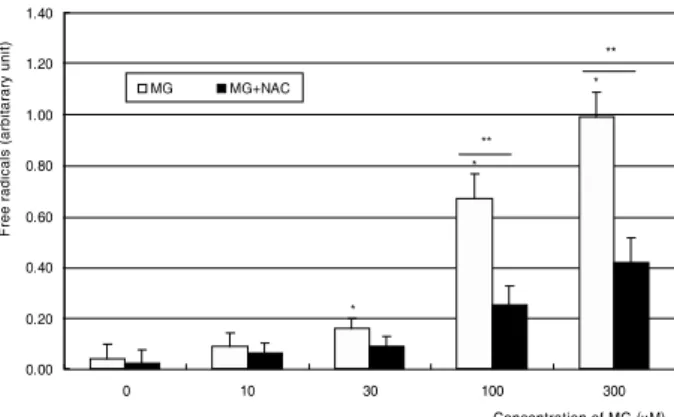
시켰으며 이러한 반응성산소종의 증가는 항산화제인 NAC 의 동시 투여에 의해 억제되었다(p<0.05, Fig. 5). 따라서 MG은 일산화질소의 생성을 억제하며 반응성산소종의 생성 을 증가시키는 경향이 있음을 알 수 있었다.
Figure 4. Effect of methylglyoxal on the generation of superoxide. Methylglyoxal increased superoxide pro- duction from 30 μM. (*p<0.05)
Figure 5. Effect of methylglyoxal (MG) on the genera- tion of reactive oxygen species. MG increased reactive oxygen species, which were abolished by co-exposed N-acetyl cysteine (NAC). (*,**p<0.05)
고 찰
본 연구의 결과는 MG이 섬유주세포에서 일산화질소의 생성저하와 반응성산소종의 증가에 의한 산화스트레스를 유발할 수 있음을 보여주고 있다.
AGE는 단백질의 비효소적 당화에 의해 형성되는 복합물 질로 다양한 종류가 있으며 단백질의 구조와 기능을 변화 시킨다. 이러한 단백질의 영구적인 당화는 산화스트레스와도 밀접하게 관련되어 있는데, AGE 자체뿐만 아니라 그 중간 대사물질인 MG, glyoxal 등도 지질 과산화 등의 과정에 의해 반응성유리기의 생성을 증가시키고 AGE의 막수용체를 활 성화시켜 세포 내에서 산화스트레스를 일으키며 염증반응 을 조장하여 노화와 당뇨, 혈관질환, 신경위축 등을 유발하 는 것으로 알려져 있으며35,36 당뇨망막증을 비롯한 다양한 안과질환에도 관련되어 있고37-41시신경유두에도 침착되는 것으로 알려져 있다.42
방수유출의 대부분을 담당하는 중요한 통로인 섬유주의 기능장애는 섬유주를 통한 방수유출로의 저항을 증가시켜 안압의 상승을 유발하여 결과적으로 녹내장을 초래하게 된 다. 섬유주 자체가 방수유출을 조절하는 기능을 가지고 있 으므로1,43섬유주의 생리적 기능을 정상적으로 유지하는 것 이 매우 중요할 것이다. 본 연구에서 AGE의 중간대사물질 인 MG이 섬유주세포에서 자유유리기의 생성에 미치는 영 향을 알아보기 위하여 18시간 동안 노출시켜 본 결과 MG 은 섬유주세포에서 일산화질소의 생성을 감소시켰으며 이 러한 일산화질소의 생성저하는 섬유주의 이완을 저해하여 방수유출을 감소시킬 수 있을 것이다. 또한 MG은 super- oxide와 반응성산소종의 생성을 증가시켰는데, 일산화질소 의 합성저하에 의해 superoxide의 생성이 더욱 증가되어 peroxynitrite 같은 유해한 자유유리기의 생성을 촉진하여 손상을 줄 수 있을 것이다.44 따라서 일산화질소의 생성을 촉진하거나 산화물질의 작용을 억제하면 섬유주의 손상을
방지하여 생리적 기능을 유지하는데 도움이 될 것인데 본 실험의 결과 항산화제인 NAC는 일산화질소의 생성을 증가 시켰을 뿐만 아니라 반응성산소종의 생성을 감소시켰다. 당 화산물은 일산화질소의 생성을 저하시킴으로써 작용을 나 타낼 수 있으므로45,46 일산화질소가 단순히 섬유주의 이완 에 의한 방수유출뿐만 아니라 방수의 순환에 의해 자유유 리기에 노출되어 있는 섬유주의 건전성을 유지하는 데에도 역할을 한다는 점을 시사하며 녹내장을 유발하는 기전을 연구하는 데 있어서 일산화질소의 역할과 산화스트레스의 중요성이 제시되고 있다.47,48
본 연구의 결과 당산화물인 MG은 섬유주세포에서 일산 화질소의 생성을 저하시키고 유발된 산화스트레스를 유발 하였으며, 반응성산소종은 섬유주세포의 손상뿐만 아니라 세포고사를 유발하기도 하며 세포의 노화에도 관여하므로
49-55
당산화물이 섬유주세포의 노화에 미치는 영향에 대해 서는 연구가 필요할 것으로 생각된다.
결론적으로 MG은 배양된 사람의 섬유주세포에서 일산화 질소의 생성을 저하시키고 반응성산소종의 생성을 증가시켜 산화스트레스를 유발할 수 있는 것으로 나타났다. 따라서 일산화질소의 생성을 생리적 수준으로 유지하거나 항산화 제를 사용함으로써 방수유출로로서의 섬유주의 기능을 유지 하며 산화스트레스에 의한 섬유주의 손상을 방지할 수도 있을 것이므로 향후 녹내장을 예방하거나 치료하는 방법을 개발하는데 있어서 이러한 점들을 고려해야 할 것으로 생 각된다.
참고문헌
1) Wiederholt M. Dirtect involvement of trabecular meshwork in the regulation of aqueous humor outflow. Curr Opin Ophthalmol 1998;9:46-9.
2) Alvarado J, Murphy C, Juster R. Trabecular meshwork cellu- larity in primary open-angle glaucoma and nonglaucomatous normals. Ophthalmology 1984;91:564-79.
3) Rohen JW, LÜtjen-drecoll E, FlÜgel C, et al. Ultrastructure of the trabecular meshwork in untreated cases of primary open-angle glaucoma. Exp Eye Res 1993;56:683-92.
4) Moncada S, Palmer RM, Higgs EA. Nitric oxide: physiology, pathophysiology, and pharmacology. Pharmacol Rev 1991;43:
109-42.
5) Bredt DS, Snyder SH. Nitric oxide: a physiologic messenger molecule. Annu Rev Biochem 1994;63:175-95.
6) Brüne B, Knethen A, Sandau KB. Nitric oxide and its role in apoptosis. Eur J Pharmacol 1998;351:261-72.
7) Nathanson JA, McKee M. Identification of an extensive system of nitric oxide-producing cells in the ciliary muscle and outflow pathway. Invest Ophthalmol Vis Sci 1995;36:1765-73.
8) Geyer O, Podos SM, Mittag T. Nitric oxide synthase activity in tissues of the bovine eyes. Graefes Arch Clin Exp Ophthalmol 1997;235:786-93.
9) Meyer P, Champion C, Schlotzer-Schrehardt U, et al. Localiza- tion of nitric oxide synthase isoforms in porcine ocular tissues.
Curr Eye Res 1999;18:375-80.
10) Schuman JS, Erickson K, Nathanson JA. Nitrovasodilator effects on intraocular pressure and ocular facility in monkeys. Exp Eye Res 1994;58:99-105.
11) Wana RF, Podos SM. Effect of the topical application of nitro- glycerin on intraocular pressure in normal and glaucomatous monkeys. Exp Eye Res 1995;60:337-9.
12) Nathanson JA, McKee M. Alterations of ocular nitric oxide syn- thase in human glaucoma. Invest Ophthalmol Vis Sci 1995;36:
1774-84.
13) Matsuo T. Basic nitric oxide production is enhanced by hydraulic pressure in cultured human trabecular cells. Br J Ophthalmol 2000;84:631-5.
14) von Zglinicki T. Oxidative stress shortens telomeres. Trends Biochem Sci 2002;27:339-44.
15) Kurz DJ, Decary S, Hong Y, et al. Chronic oxidative stress compromises telomere integrity and accelerates the onset of senescence in human endothelial cells. J Cell Sci 2004;117:2417-26.
16) Vasa M, Breitchopf K, Zeiher AM, Dimmeler S. Nitric oxide activates telomerases and delays endothelial cell senescence. Circ Res 2000;540-2.
17) Bucala R, Vlassara H, Cerami A. Advanced glycation end pro- ducts. Vol. 2. Boca Raton: CRS Press, 1992;53-9.
18) Bucala R, Tracey KJ, Cerami A. Advanced glycation end products quench nitric oxide and mediate defective endothelium-dependent vasodilatationin experimental diabetes. J Clin Invest 1991;87:
432-8.
19) Franke S, Dawczynski J, Strobel J, et al. Increase levels of advanced glycation end products in human cataractous lenses. J Cataract Refrac Surg 2003;29:998-1004.
20) Nakamura N, Hasegawa G, Obayashi H, et al. Increased concen- tration of pentosidine, an advanced glycation end product, and interleukin-6 in the vitreous of patients with proliferative diabetic retinopathy. Diabetes Res Clin Pract 2003;61:93-101.
21) Yim HS, Kang SO, Hah YC, et al. Free radicals generated during the glycation reaction of amino acids by methylglyoxal. J Biol Chem 1995;270:28228-33.
22) Wu L, Juurlink BH. Increased methylglyoxal and oxidative stress in hypertensive rat vascular smooth muscle cells. Hypertension 2002;39:809-14.
23) Kang JH. Oxidative damage of DNA induced by methylglyoxal in vitro. Toxicol Lett 2003;145:181-7.
24) Chang T, Wang R, Wu L. Methylglyoxal-induced nitric oxide and peroxynitrite production in vascular smooth muscle cells. Free Radi Biol Med 2005;38:286-93.
25) Valencia JV, Weldon SC, Quinn D, et al. Advanced glycation end product ligands for the receptor for advanced glycation end products: biochemical characterization and formation kinetics.
Anal Biochem 2004;324:68-78.
26) Shamsi FA, Lin K, Sady C, Nagaraj RH. Methylglyoxal-derived modifications in lens aging and cataract formation. Invet Oph- thalmol Vis Sci 1998;39:2355-64.
27) Mosmann T. Rapid colorimetric assay for cellular growth and survival: Application to proliferation and cytotoxicity assays. J Immunol Methods 1983;65:55-63.
28) Green LC, Wagner DA, Glogoski J, et al. Analysis of nitrate, nitrite and [15N]nitrate in biologic fluids. Anal Biochem 1982;
126:131-8.
29) Vermes I, Haanen C, Steffens-Nakken H, Reutelingsperger C.
Flow cytometric detection of phosphatidylserine expression on early apoptotic cells using fluorescein labeled Annexin V. J Immunol Methods 1995;184:39-51.
30) Beauchamp C, Fridovich I. Superoxide dismutase: improved assay and an assay applicable to acrylamide gels. Anal Biochem 1971;
44:276-87.
31) Teufelhofer O, Weiss R-M, Parzefall W, et al. Promyelocytic HL60 cells express NADPH oxidase and are excellent targets in a rapid spectrophotometric microplate assay for extracellular supe- roxide. Toxicolol Sci 2003;76:376-93.
32) Wang H, Joseph JA. Quantifying cellular oxidative stress by dichlorofluorescein assay using microplate reader. Free Radic Biol Med 1999;27:612-16.
33) Polansky JR, Weinreb RN, Baxter JD, Alvarado J. Human trabecularcells. I. Establishment in tissue culture and growth characteristics. Invest Ophthalmol Vis Sci 1979;18:1043-9.
34) Alvarado JA, Wood I, Polansky JR. Human trabecular cells. II.
Growth pattern and ultrastructural characteristics. Invest Oph- thalmol Vis Sci 1982;23:464-78.
35) Schimidt AM, Hori O, Brett J, et al. Cellular receptor for advanced glycation end products: implications for induction of oxidative stress and cellular dysfunction in the pathogenesis of vascular lesions. Atheroscler Thromb 1994;14:1521-28.
36) Ramasamy R, Vannucci S, Yan SSD, et al. Advanced glycation end products and RAGE: a common thread in aging, diabetes, neurodegeneration, and inflammation. Glycobiol 2005;15:16-28.
37) Stitt AW, Moore JE, Sharkey JA, et al. Advanced glycation end products in vitreous: structural and functional implications for diabetic vitreopathy. Invest Ophthalmol Vis Sci 1998;39:2517-23.
38) Stitt AW. Advanced glycation: An important pathological event in diabetic and age related ocular disease. Br J Ophthamol 2001;
85:746-53.
38) Stitt AW. Role of advanced glycation in the pathogenesis of diabetic retinopathy. Exp Mol Pathol 2003;57:95-108.
40) Kaji Y, Usui T, Oshika T, et al. Advanced glycation end products in diabetic corneas. Invest Ophthalmol Vis Sci 2000;41:362-8.
41) Pokupec R, Kalauz M, Turk N, Turk Z. Advanced glycation end products in human diabetic and non-diabetic cataractous lenses.
Graefes Arch Clin Exp Ophthalmol 2003;241:378-4.
42) Amano S, Kaji Y, Oshika T, et al. Advanced glycation end pro- ducts in human optic nerve head. Br J Ophthalmol 2001;85:52-5.
43) Alvarado JA, Alvarado RG, Yeh RF, et al. A new insight into the cellular regulation of aqueous outflow: how trabecular meshwork endothelial cells drive a mechanism that regulates the permeability of Schlemm’s canal endothelial cells. Br J Ophthalmol 2005;
89:1500-5.
44) Madamanchi NR, Vendov A, Runge MS. Oxidative stress and vascular disease. Arterioscler Thromb Vas Biol 2005;25:29-38.
45) Chakravarthy U, Hayes RG, Stitt RW, et al. Constitutive nitric oxide synthase expression in retinal vascular endothelial cells is suppressed by high glucose and advanced glycation end products.
Diabetes 1998;47:945-52.
46) Uhlmann S, Rezzoug K, Friedrichs U, et al. Advanced glycation end products quench nitric oxide in vitro. Graefes Arch Clin Exp Ophthalmol 2002;240:860-6.
47) Sagga S. Nitric oxide as a mediator of glaucoma pathogenesis. Med Sci Monit 2002;8:33-4.
48) Chen JZ, Kadlubar FF. A new clue to glaucoma pathogenesis. Am J Med 2003;114:697-8.
49) Schachtschabel DO, Binninger EA, Rohen JW. In vitro cultures of trabecular meshwork cells of the human eye as a model system for the study of cellular aging. Arch Gerontol Geriat 19899:
251-62.
50) Ferreira SM, Lerner SF, Brunzini R, et al. Oxidative stress markers in aqueous humor of glaucoma patients. Am J Oph- thalmol 2004;137:62-9.
51) Yildirim O, Ates NA, Ercan B, et al. A. Role of oxidativestress enzymes in open-angle glaucoma. Eye 2005;19: 580-3.
52) Izzotti A, Sacca SC, Cartiglia C, De Flora S. Oxidative deoxyribonucleic acid damage in the eyes of glaucoma patients.
Am J Med 2003;114:638-46.
53) Sacca SC, Pascotto A, Camicione P, et al. Oxidative DNA damage in human trabecular meshwork. Arch Ophthalmol 2005;123:
458-63.
54) Gabelt BT, Kaufman PL. Changes in aqueous humor dynamics with age and glaucoma. Prog Retin Eye Res 2005;24:612-37.
55) Liton PB, Challa P, Stinnett S, et al. Cellular senescence in the glaucomatous outflow pathway. Exp Gerontol 2005;40:745-8.
=ABSTRACT=
Effect of Methylglyoxal on the Oxidative Stress in Trabecular Meshwork Cells
Seung Hee Lee, MD, Sin Hoo Kim, MD, Jae Woo Kim, MD, PhD
Department of Ophthalmology, Catholic University of Daegu College of Medicine, Daegu, Korea
Purpose: To investigate the effect of methylglyoxal (MG), intermediate metabolite of advanced glycation end products(AGE), on the induction of oxidative stress in human trabecular meshwork cells (HTMC).
Methods: Primarily cultured HTMC were exposed to at concentrations of 0, 30, 100, and 300 μM of MG for 18 hours, with or without co-exposure to N-acetyl-cysteine. Cellular survival and apoptosis were assessed by MTT assay and flow cytometry using annexin-PI double staining. Production of nitric oxide (NO), superoxide, and reactive oxygen species (ROS) was assessed by Griess assay, cytochrome c assay, and dichlorofluorescein diacetate assay, respectively.
Results: MG did not affect cellular survival at concentrations under 100 μM, but induced apoptosis of HTMC at concentrations over 100 μM. MG decreased NO production, accompanied with increased superoxide production. In addition, MG increased ROS, which were abolished by N-acetylcysteine.
Conclusions: MG induced oxidative stress by decreasing NO production, accompanied by increasing superoxide and ROS productions in HTMC. AGE could induce trabecular meshwork dysfunction.
J Korean Ophthalmol Soc 2009;50(10):1569-1575
Key Words: Advanced glycation end products, Methylglyoxal, Oxidative stress, Trabecular meshwork cells
Address reprint requests to Jae Woo Kim, MD, PhD
Department of Ophthalmology, Catholic University of Daegu College of Medicine
#3056-6 Daemyeung 4-dong, Nam-gu, Daegu 705-718, Korea Tel: 82-53-650-4728, Fax: 82-53-627-0133, E-mail: [email protected]