J Korean Ophthalmol Soc 2009;50(10):1563-1568 DOI : 10.3341/jkos.2009.50.10.1563
= 증례보고 = 접수번호 : 50-10-04-28
고포도당이 섬유주세포의 산화스트레스에 미치는 영향
김재우⋅배창범⋅이정훈 대구가톨릭대학교 의과대학 안과학교실
목적: 고포도당이 섬유주세포의 산화스트레스에 미치는 영향을 알아보고자 하였다.
대상과 방법: 일차배양 섬유주세포를 고농도(25 mM)와 저농도(5 mM)의 포도당이 포함된 배지에 각각 7일간 노출시켰으며 L- arginine, DAHP, insulin, 아스코르빈산, sepiapterin에도 노출시켰다. 세포생존과 일산화질소 생성, superoxide 생성을 MTT, Griess, modified cytochrome c assay로 측정하였다.
결과: 고포도당은 섬유주세포의 생존에는 유의한 영향을 미치지 않았으나 일산화질소의 생성을 유의하게 억제하였고 동시에 노출한 DAHP는 일산화질소의 생성을 더욱 억제하였으나 insulin과 sepiapterin은 일산화질소의 생성을 증가시켰다. 고포도당은 superoxide의 생성을 증가시켰으며 insulin과 아스코르빈산, sepiapterin은 superoxide의 생성을 감소시켰다.
결론: 고포도당은 섬유주세포에서 일산화질소의 생성을 억제하는 동시에 superoxide의 생성을 증가시켰다. 고포도당은 섬유주세포에서 산화스트레스를 유발하여 섬유주의 손상과 기능저하를 초래할 수 있을 것이다.
<대한안과학회지 2009:50(10):1563-1568>
■ 접 수 일: 2009년 4월 21일 ■ 심사통과일: 2009년 7월 14일
■ 책 임 저 자: 김 재 우
대구시 남구 대명4동 3056-6 대구가톨릭대학교 의과대학 안과학교실 Tel: 053-650-4728, Fax: 053-627-0133 E-mail: [email protected]
섬유주의 기능저하로 인해 방수유출로의 저항이 증가되어 안압이 상승함으로써 원발개방각녹내장을 유발하는 것으로 알려져 있는데 섬유주를 구성하고 있는 섬유주세포는 방수 유출의 조절에 능동적으로 관여한다고 한다.1-3 섬유주를 통한 방수유출을 조절함에 있어서 일산화질소(nitric oxide, NO)가 중요한 역할을 하는데 녹내장이 있는 경우에는 섬유 주의 NO합성효소의 활성이 감소되어 있고, NO 공여약물을 투여하면 안압을 낮추는 효과가 있는 것으로 알려져 있다.4-7
자유유리기는 산화스트레스를 유발하여 인체의 노화를 유발하는 주요한 요인으로 알려져 있으며8 녹내장의 경우 에도 산화스트레스는 섬유주세포의 손상을 유발할 뿐만 아 니라 섬유주세포의 노화를 촉진할 수 있는 것으로 알려져 있고9안압상승과 시야손상의 정도가 섬유주에서의 산화스 트레스에 의한 핵산의 손상 정도와 비례하는 것으로도 알 려져 있다.10혈관내피세포의 경우 고농도의 포도당에 노출 되면 NO의 생성이 저하되며11산화스트레스가 유발되는 것 으로 알려져 있다.12 당뇨병이 있는 경우 녹내장이 발병할 위험이 높은 것으로 알려져 있고13 당뇨병이 있는 환자의 방수에서의 포도당 농도가 정상인에 비해 약 2배 정도 높다고 보고되어 있다.14따라서 고농도의 포도당(고포도당)에 섬유
주세포가 노출될 경우 섬유주세포의 정상적인 NO 생성에 영향을 미칠 수 있고 산화스트레스도 유발할 수 있을 것으로 생각해 볼 수 있으나, 인체의 섬유주세포에 대하여 고포도 당이 산화스트레스에 미치는 영향에 관해서는 아직 자세히 연구되지 않았다.
본 연구에서는 인체의 섬유주세포에서 고포도당이 NO의 생성에 미치는 영향과 산화스트레스를 유발하는지 알아보 고자 하였다.
대상과 방법
세포배양
안구은행에서 얻은 사후 6시간 이내에 적출한 안구의 앞 방각 주위 조직을 제거한 후 앞방각에서 섬유주를 벗겨내어 폴리라이신(Sigma, USA)로 처리한 배양접시에 옮긴 후 항 생제(Gibco, Carlsbad, USA)와 15% 우태아혈청(Gibco, USA)이 포함된 Dulbecco’s modified Eagle’s medium 배 지(DMEM, Gibco, Carlsbad, USA)를 사용하여 5% CO2
배양기에서 초대배양하였다. 섬유주세포가 이식된 조직편 주위로 자라나온 것을 확인한 후 섬유주조직의 이식편을 제거하고 배양을 계속하였으며 세포가 배양접시에 충만해 지면 10% 우태아혈청을 포함한 배지로 1:3의 비율로 트립신 처리하여 계대배양하였다.
약물처리
배양접시의 각 well에 일차배양한 섬유주세포를 분주한 후 24시간 동안 배양기에 넣어 세포를 부착시킨 후 배지를 제거하고 나서 저농도(5 mM, low glucose, LG)와 고농도 (25 mM, high glucose, HG)의 포도당을 각각 포함한 DMEM 배지에 10% 우태아혈청을 첨가하여 일주일간 배양하였다.
이때 삼투압의 영향을 알아보기 위하여 25 mM의 mannitol 을 포함한 배지에도 배양하였다. 또한 NO의 생성에 작용하 는 효소적 합성경로를 알아보기 위하여 각각 신합성경로에 작용하는 1 mM L-arginine, 5 mM DAHP (2,6-diamino- 6-hydroxypyrimidine), 10 g/ml insulin과 재합성경로에 작용하는 100 μM 아스코르빈산, 그리고 10, 100 μM의 sepiapterin에 동시에 노출시켰다.
MTT assay와 Griess assay
세포의 생존에 대한 효과는 세포생존과 세포독성의 sc- reening test로 흔히 이용되고 있는 발색검사법의 일종인 MTT (3-[4, 5–dimethylthiazol-2-yl]-2, 5-diphenyl- tetrazolium bromide) assay를15이용하였고 NO의 생성은 Griess assay를16 이용하였다. MTT assay는 약물처리한 세포의 배지에 MTT를 각 well 당 100 μl씩 투여한 후 4시간 동안 정치배양한 다음 염류용액으로 씻어낸 후 dimethyl- sulfoxide를 각 well 당 0.5 ml씩 넣어 10분 이상 흔든 다음 96-well plate에 200 μl씩 옮겨 spectrophotometer (Fluo- star Optima, BMG Labtech, Germany)로 570 nm에서 흡 광도를 측정하였다. 이때 세포의 생존 정도는 실험군의 값을 약물처리를 하지 않은 대조군의 비로 나누어 백분율로 나타 내었다. Griess assay는 3일 동안 약물처리한 세포의 배지 에 동량의 Griess 반응액을 섞은 후 96-well plate에 옮겨 NO생성의 반응물인 아질산염의 양을 spectrophotometer로 540 nm에서 흡광도를 측정하였다. 이때 표준치를 구하기 위해 sodium nitrite를 단계적으로 희석하여 사용하였다.
Superoxide의 생성 측정
산화스트레스에 미치는 영향을 알아보기 위하여 반응성 산소종의 일종인 superoxide의 생성을 modified cyto- chrome c assay로 측정하였다.17160 μM의 cytochrome c 와 100 unit/ml의 superoxide dismutase를 혼합하여 반응 액을 만든 후 96 well 배양접시에서 저농도와 고농도의 포 도당에서 각각 1주일간 배양한 섬유주세포에 노출시켜 한 시간 동안 환원된 cytochrome c를 550 nm의 파장에서 흡
광도의 변화를 측정하였다. 이때 2.1×10⁴ M/cm의 net extinction coefficient를 이용하였으며 흡광도의 변화치를 106개의 세포당 환원된 cytochrome c를 nmol로 변환하여 한 시간 동안 생성된 superoxide의 양을 nmol/106/cells/h로 나타내었다.
실험약품과 통계적 처리
세포배양 외에 실험에 사용된 약품은 Sigma사의 제품을 사용하였다. 모든 실험은 3계대에서 5계대 사이의 세포를 이용하였고 3회 이상 반복하여 시행하였다. 모든 실험에서 대조군은 약물처리를 하지 않은 군으로 하였으며, 실험군과 대조군의 비교는 unpaired t-test를 사용하였으며 유의수 준은 0.05%로 정하였다.
결 과
세포배양
초대배양 10일째부터 섬유주조직의 이식편 주위로 섬유 주세포가 자라 나오기 시작하였으며 섬유주세포의 확인은 특징적인 형태학적인 양상과 섬유주 조직의 이식편 주위에 서 위성양상으로 자라나는 섬유주세포의 특징적인 성장양 상으로 확인하였다.18,19
HG가 섬유주세포의 생존에 미치는 영향
HG를 7일간 섬유주세포에 노출시켰을 때 LG에 노출한 대조군에 비하여 9% 생존이 증가하여 세포의 생존에는 유 의한 영향을 미치지 않았다(p>0.05). 또한 삼투압의 영향 을 배제하기 위해 20 mM의 mannitol에 노출한 경우에서 유의한 차이를 보이지 않았다. 이러한 결과로써 본 실험에 사용된 약제에 의한 실험결과는 세포의 생존이나 삼투압의 변화에 의해 영향을 받지 않았다는 것을 알 수 있었다.
HG가 NO 생성에 미치는 영향
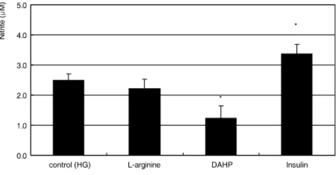
HG는 섬유주세포에 있어서 LG와 비교하여 유의하게 NO 의 생성을 감소시켰다(p<0.05)(Fig. 1). 또한 NO의 신합 성을 억제하는 DAHP에 의해 더욱 감소되었고(p=0.007) 신합성을 증가시키는 insulin에 의해 NO의 생성이 증가되 었다(p=0.015)(Fig. 2). NO의 재합성경로에 작용하는 약 물에 동시에 노출시킨 경우에는 100 μM의 고농도 sepiap- terin에 의해서만 NO의 생성이 증가되었다(p=0.005)(Fig. 3).
Figure 1. Effect of high glucose on the production of nitric oxide. One-week exposure to high glucose (25 mM) decreased nitric oxide production significantly compared to low glucose (5 mM). (*p<0.05)
Figure 2.Effect of 1 mM L-arginine, 5 mM DAHP, or 10 μg/ml insulin on the production of nitric oxide exposed to high glucose (HG) for 1 week. (*p<0.05)
Figure 3.Effect of 100 μM L-ascorbic acid and sepiap- terin on the production of nitric oxide exposed to high glucose (HG) for 1 week. (*p<0.05)
Figure 4. Effect of high glucose on the production of superoxide. One-week exposure to high glucose (25 mM) increased superoxide production significantly compared to low glucose (5 mM). (*p<0.05)
Figure 5. Effect of 1 mM L-arginine, 5 mM DAHP, or 10 μg/ml insulin on the production of superoxide exposed to high glucose (HG) for 1 week. (*p<0.05)
Figure 6.Effect of 100 μM L-ascorbic acid and sepiap- terin on the production of superoxide exposed to high glucose (HG) for 1 week. (*p<0.05)
HG가 superoxide의 생성에 미치는 영향
HG는 섬유주세포에 1주일간 노출시켰을 때 LG와 비교 하여 유의하게 superoxide의 생성을 증가시켰다(p=0.018) (Fig. 4). 이러한 superoxide의 생성은 DAHP에 의해 증가 되었고(p=0.491) insulin에 의해 유의하게 감소되었으며 (p=0.001)(Fig. 5) L-ascorbic acid와 고농도의 sepiapterin 에 의해서 증가되었다(p=0.001, 0.002)(Fig. 6).
고 찰
섬유주는 섬유주를 통한 방수유출을 능동적으로 조절하며 endothelin과 NO가 이에 관여한다고 알려져 있다.3,20 산화 스트레스는 섬유주에서 endothelin과 NO 분비의 불균형을 초래하여 안압을 상승시키기도 하는데 HG는 망막의 혈관 내피세포를 비롯한 다양한 종류의 세포에서 NO의 생성을 저하시키고 산화스트레스를 유발한다고 한다.12,21
자유유리기인 NO는 고농도에서는 반응성 산화물질로 전환 되어 세포에 병적인 손상을 유발하기도 하지만 저농도에서는 인체에서 중요한 생리적인 조절인자로 작용하여 다양한 역
할을 하는데22-24NO 생성에는 tetrahydrobiopterin이 필수 요소일 뿐만 아니라 중요한 조절인자로도 작용하는 데 이의 합성 경로는 신합성경로와 재합성경로가 있다.25섬유주세포 에서도 NO합성효소가 발현될 뿐만 아니라26-28 글루코코티 코이드 수용체가 나타나므로29HG가 NO의 생성을 저하시킬 가능성이 있으며 반응성산소종의 생성을 증가시켜 산화스트 레스를 유발할 수 있을 것이다.
본 연구의 결과는 HG가 배양된 사람의 섬유주세포에서 NO의 생성을 감소시키며 동시에 superoxide의 생성을 증가 시켜 산화스트레스를 유발할 수 있다는 것을 보여주고 있다.
HG는 NO의 생성을 유의하게 억제하였으며 tetrahydro- biopterin의 생성억제재로 작용하는 DAHP에 의해 생성이 더욱 저하되었고, 또한 tetrahydrobiopterin의 신합성경로에 관여하는 효소인 GTP cyclohydrolase I의 작용을 증가시키는 insulin에 의해 NO의 생성 감소가 억제되는 것으로 보아25,30 HG가 NO의 생성을 억제하는 기전은 주로 tetrahydro- biopterin의 신합성경로에 작용하여 NO의 생성을 억제하는 것으로 생각된다.
또한 HG는 반응성산소종인 superoxide의 생성을 유의하 게 증가시켰는데 NO의 생성감소와 동반되어 superoxide의 생성증가가 나타나는 것으로 보아 HG가 산화스트레스를 유발하는 기전은 생리적인 활성을 나타내는 NO의 농도가 감소함에 따라 증가된 superoxide가 NO와 주로 결합하면 서 유해한 반응성산소종인 peroxynitrite를 생성함으로써 산화스트레스를 유발하는 eNOS uncoupling의 기전 관여할 것으로 생각된다.12 그리고 NO의 생성을 촉진하는 약물에 함께 노출시켰을 때 superoxide의 생성이 감소하는 것으로 보아 NO의 생성을 생리적인 활성농도까지 유지하면 산화 스트레스를 억제할 수 있다는 점을 시사하고 있다.
한편 NO 합성의 기질로 작용하는 L-arginine은 NO와 superoxide의 생성 변화에 유의한 영향을 끼치지 않아 HG 에 의한 영향은 tetrahydrobiopterin에 의한 것임을 짐작할 수 있다.
본 연구의 결과에 의하면 인체의 섬유주세포에서 HG는 NO의 생성을 억제함으로써 섬유주를 구조적으로 위축시켜 결과적으로 섬유주를 통한 방수유출을 저하시킬 뿐만 아니라 NO의 생리적 활성을 저하시켜 다양한 병적 결과를 초래할 수 있다. 즉 NO의 생리적 활성저하에 따라 해로운 supero- xide나 peroxynitrite 같은 해로운 반응성산소종의 생성이 오히려 증가하게 되는데12,30이러한 반응성산소종은 산화스 트레스를 유발할 뿐만 아니라 세포의 노화도 초래하는 것 으로 알려져 있다.31-35따라서 섬유주의 NO 생성감소에 의 해 산화스트레스가 지속될 경우 섬유주세포의 변성을 유발 하며 섬유주세포의 노화를 초래할 수도 있을 것이다.10,36
연령이 증가할수록 섬유주세포의 숫자가 감소하고37 또 원발성개방각녹내장의 조직소견이 노화한 섬유주세포와 유 사한 점을38-41고려해보면, NO는 섬유주를 통한 방수유출 을 증가시킬 뿐만 아니라 섬유주세포의 노화를 방지하는 작용도 나타낼 가능성이 있기 때문에 섬유주에서 생리적 농도의 NO 생성을 유지하는 것이 매우 중요하다고 할 수 있다. 그러므로 본 연구의 결과와 같이 HG에 의해 NO의 생 성이 감소되며 섬유주의 위축을 유발하는 endothelin의 발 현을 촉진시키므로42 단기간에는 섬유주를 통한 방수유출이 저하되어 안압을 상승시킬 수 있을 뿐만 아니라, 장기간에 걸쳐 산화스트레스가 유발되면 섬유주세포가 노화되어 그 기능이 떨어짐으로써 섬유주를 통한 방수유출이 감소되어 결과적으로 녹내장을 유발하거나 악화시킬 수 있는 요인이 될 수 있을 것이다.43
한편 본 연구의 결과와 같이 HG에 단기간 노출시킬 경우 섬유주세포의 생존에는 별 다른 영향을 미치지 않으나 섬 유주세포에 다양한 구조적 변화를 유발할 수 있고44장기적 으로는 최총당화산물이 축척되어 산화스트레스뿐만 아니라 세포의 노화와 세포고사를 유발할 가능성이 있으며,10,21,36,45
HG가 섬유주세포에 미치는 장기간의 영향에 대해서는 향 후 좀 더 자세한 연구가 필요할 것이다.
결론적으로 HG는 배양된 사람의 섬유주세포에 대해 세 포의 생존에는 영향을 주지 않았으나 NO의 생성을 저하시 켰으며 superoxide의 생성을 증가시켜 산화스트레스를 유발 하였다. 따라서 HG에 의해 유발되는 산화스트레스는 섬유 주의 기능저하를 초래하여 안압이 상승하는 기전의 하나가 될 수 있을 것이며, NO의 생성을 유지하거나 항산화제를 사용함으로써 산화스트레스에 대해 섬유주세포를 보호할 수 있을 것으로 생각된다.
참고문헌
1) Alvarado J, Murphy C, Juster R. Trabecular meshwork cellularity in primary open-angle glaucoma and nonglaucomatous normals.
Ophthalmology 1984;91:564-79.
2) Rohen JW, LÜtjen-drecoll E, FlÜgel C, et al. Ultrastructure of the trabecular meshwork in untreated cases of primary open-angle glaucoma. Exp Eye Res 1993;56:683-92.
3) Alvarado JA, Alvarado RG, Yeh RF, et al. A new insight into the cellular regulation of aqueous outflow: how trabecular meshwork endothelial cells drive a mechanism that regulates the permeability of Schlemm’s canal endothelial cells. Br J Ophthalmol 2005;
89:1500-5.
4) Schuman JS, Erickson K, Nathanson JA. Nitrovasodilator effects on intraocular pressure and ocular facility in monkeys. Exp Eye Res 1994;58:99-105.
5) Wana RF, Podos SM. Effect of the topical application of nitro- glycerin on intraocular pressure in normal and glaucomatous
monkeys. Exp Eye Res 1995;60:337-9.
6) Nathanson JA, McKee M. Alteration of ocular nitric oxide syn- thase in human glaucoma. Invest Ophthalmol Vis Sci 1995;36:
1774-84.
7) Matsuo T. Basic nitric oxide production is enhanced by hydraulic pressure in cultured human trabecular cells. Br J Ophthalmol 2000;84:631-5.
8) Wei YH. Oxidativestress and mitochondrial DNA mutations in human aging. Proc Soc Exp Biol Med 1998;217:53-63.
9) Sacca SC, Izzotti A, Rossi P, Traverso C. Glaucomatous outflow pathway and oxidative stress. Exp Eye Res 2007;84:389-99.
10) Sacca SC, Pascotto A, Camicione P, et al. Oxidative DNA damage in the human trabecular meshwork: clinical correlation in patients with primary open-angle glaucoma. Arch Ophthalmol 2005;123:
458-63.
11) Brodsky SV, Morrishow AM, Dharia N, et al. Glucose scavenging of nitric oxide. Am J Physiol 2001;280:480-6.
12) El-Remessy AB, Abou-Mohamed G, Caldwell RW, Caldwell RB.
High glucose-induced tyrosine nitration in endothelial cells: role of eNOS uncoupling and aldose reductase activation. Invest Ophthalmol Vis Sci 2003;44:3135-43.
13) Becker B. Diabetes and primary open-angle glaucoma. Am J Ophthalmol 1971;1:1-16.
14) Davies PD, Duncan G, Pynsent PB, et al. Aqueous humor glucose concentration in cataract patients and its effect on the lens. Exp Eye Res 1984;39:605-9.
15) Mosmann T. Rapid colorimetric assay for cellular growth and survival: Application to proliferation and cytotoxicity assays. J Immunol Methods 1983;65:55-63.
16) Green LC, Wagner DA, Glogoski J, et al. Analysis of nitrate, nitrite and [15N]nitrate in biologic fluids. Anal Biochem 1982;
126:131-8.
17) Teufelhofer O, Weiss R-M, Parzefall W, et al. Promyelocytic HL60 cells express NADPH oxidase and are excellent targets in a rapid spectrophotometric microplate assay for extracellular superoxide. Toxicol Sci 2003;76:376-83.
18) Polansky JR, Weinreb RN, Baxter JD, Alvarado J. Human trabecular cells. I. Establishment in tissue culture and growth characteristics. Invest Ophthalmol Vis Sci 1979;18:1043-9.
19) Alvarado JA, Wood I, Polansky JR. Human trabecular cells. II.
Growth pattern and ultrastructural characteristics. Invest Ophthalmol Vis Sci 1982;23:464-78.
20) Haefliger IO, Dettman E, Liu R, et al. Potential role of nitric oxide and endothelin in the pathogenesis of glaucoma. Surv Ophthalmol 1999;43:S51-8.
21) Chakravarthy U, Hayes RG, Stitt AW, et al. Constitutive nitric oxide synthase expression in retinal vascular endothelial cells is suppressed by high glucose and advanced glycation end products.
Diabetes 1998;47:945-52.
22) Moncada S, Palmer RM, Higgs EA. Nitric oxide: physiology, pathophysiology, and pharmacology. Pharmacol Rev 1991;43:
109-42.
23) Bredt DS, Snyder SH. Nitric oxide: a physiologic messenger molecule. Annu Rev Biochem 1994;63:175-95.
24) Brüne B, Knethen A, Sandau KB. Nitric oxide and its role in apoptosis. Eur J Pharmacol 1998;351:261-72.
25) Gross SS, Levi R. Tetrahydrobiopterin synthesis. J Biol Chem 1992;267:25722-9.
26) Nathanson JA, McKee M. Identification of an extensive system of nitric oxide-producing cells in the ciliary muscle and outflow pathway. Invest Ophthalmol Vis Sci 1995;36:1765-73.
27) Geyer O, Podos SM, Mittag T. Nitric oxide synthase activity in tissues of the bovine eyes. Graefes Arch Clin Exp Ophthalmol 1997;235:786-93.
28) Meyer P, Champion C, Schlotzer-Schrehardt U, et al. Locali- zation of nitric oxide synthase isoforms in porcine ocular tissues.
Curr Eye Res 1999;18:375-80.
29) Weinreb RN, Bloom E, Baxter JD, et al. Detection of glucocortocoid receptors in cultured human trabecular cells.
Invest Ophthalmol Vis Sci 1981;21:403-7.
30) Alp NJ, Channon KM. Regulation of endothelial nitric oxide synthase by tetrahydrobiopterin in vascular disease. Arterioscler Thromb Vasc Biol 2004;24:413-20.
31) VasaM, Breitschopf K, Zeiher AM, Dimmeler S. Nitric oxide activates telomerase and delays endothelial cell senescence. Circ Res 2000;87:540-2.
32) Zhou L, Li Y,Yue BY. Oxidative stress affects cytoskeletal structure and cell-matrix interactions in cells from ocular tissue:
the trabecular meshwork. J Cell Physiol 1999;180:182-9.
33) Kurz DJ, Decary S, Hong Y, et al. Chronic oxidative stress compromises telomere integrity and accelerates the onset of senescence in human endothelial cells. J Cell Sci 2004;117:2417-26.
34) von Zglinicki T. Role of oxidative stress in telomere length regulation and replicative senescence. Ann N Y Acad Sci 2000;
908:99-110.
35) Furumoto K, Inoue E, Nagao N, et al. Age- dependent telomere shortening is slowed down by enrichment of intracellular vitamin C viasuppression of oxidative stress. Life Sci 1998;63:935-48.
36) Sato T, Roy S. Effect of high glucose on fibronectin expression and cell proliferation in trabecular meshwork cells. Invest Ophthalmol Vis Sci 2002;43:170-5.
37) Grierson I, Howes RC. Age-related depletion of the cell popula- tion in the human trabecular meshwork. Eye 1987;1:204-10.
38) Alvarado J, Murphy C, Juster R.Trabecular meshwork cellularity in primary open-angle glaucoma and nonglaucomatous normals.
Ophthalmology 1984;91:564-79.
39) Millard CB, Tripathi BJ, Tripathi RC. Age-related changes in protein profiles of the normal human trabecular meshwork. Exp Eye Res 1987;45:623-31.
40) Schchschabel DO, Binninger E. Aging of trabecular meshwork cells of the human eye in vitro. Z Gerontol 1990;23:133-5.
41) Horstmann HJ, Rohen JW, Sames K. Age-related changes in the composition of proteins in the trabecular meshwork of the human eye. Mech Ageing Dev 1983;21:121-36.
42) Park J-Y, Takahara N, Gabriele A, et al. Induction of endothelin-1 expression by glucose. An effect of protein kinase c activation.
Diabetes 2000;49:1239-48.
43) Chen JZ, Kadlubar FF. A new clue to glaucoma pathogenesis. Am J Med 2003;114:697-8.
44) Wordinger RJ, Clark AF. Effects of glucocortocoids on the trabecular meshwork: Towards a better understanding of glaucoma. Prog Ret Eye Res 1999;5:629-67.
45) Barber AJ, Lieth E, Khin SA, et al. Neuronal apoptosis in the retina during experimental and human diabetes. J Clin Invest 1998;102:783-91.
=ABSTRACT=
Effect of High Glucose on the Oxidative Stress in Trabecular Meshwork Cells
Jae Woo Kim, MD, PhD, Chang Beum Bae, MD, Jeong Hun Lee, MD
Department of Ophthalmology, Catholic University of Daegu College of Medicine, Daegu, Korea
Purpose: To investigate the effect of high glucose (HG) on the oxidative stress in cultured human trabecular meshwork cells (HTMC).
Methods: Primarily cultured HTMC were exposed to low glucose (5 mM) and HG (25 mM) for 7 days. Additionally, 1 mM L- arginine, 5 mM DAHP, 10 μg/ml insulin, 100 μM L-ascorbic acid, 10, and 100 μM sepiapterin were co-exposed. The cellular survival and nitric oxide (NO) production were assessed by MTT assay and Griess assay, respectively. Superoxide production was measured by modified cytochrome c assay.
Results: HG did not affect the survival of cultured HTMC significantly. HG decreased NO production. Co-exposed DAHP decreased but DAHP and insulin increased NO production. In addition, HG increased superoxide production, which was decreased by insulin, L-ascorbic acid, and sepiapterin.
Conclusions: HG decreased NO production accompanied with increased superoxide production in HTMC. Thus HG induces oxidative stress in HTMC and may cause cellular dysfunction and damage of the trabecular meshwork.
J Korean Ophthalmol Soc 2009;50(10):1563-1568
Key Words: Glucose, Nitric oxide, Oxidative stress, Superoxide, Trabecular meshwork cells
Address reprint requests to Jae Woo Kim, MD, PhD
Department of Ophthalmology, Catholic University of Daegu College of Medicine
#3056-6 Daemyeung 4-dong, Nam-gu, Daegu 705-718, Korea Tel: 82-53-650-4728, Fax: 82-53-627-0133, E-mail: [email protected]