ISSN 0378-6471 (Print)⋅ISSN 2092-9374 (Online)
http://dx.doi.org/10.3341/jkos.2015.56.12.1939
Original Article
배양된 사람 코점막 섬유아세포에 대한 양막추출물의 효과
Effect of Amniotic Membrane Extract on Cultured Human Nasal Mucosa Fibroblasts
황석진⋅김근해
Seok Jin Hwang, MD, Keun Hae Kim, MD, PhD
대구가톨릭대학교 의과대학 안과학교실
Department of Ophthalmology, Catholic University of Daegu School of Medicine, Daegu, Korea
Purpose: To investigate the effects of amniotic membrane extract (AME) on the survival of cultured human nasal mucosa fibroblasts.
Methods: Primary cultured human nasal mucosa fibroblasts were exposed to 0, 10, 20, or 30 μg/mL AME for 3 days. The survival of the human nasal mucosa fibroblasts was measured using the MTT assay and apoptosis was evaluated with flow cytometry us- ing annexin-V/propidium iodide double staining.
Results: AME decreased significantly in fibroblast proliferation after exposure to 10 μg/mL (p = 0.000), and caused significant apoptosis of the fibroblasts after exposure to 10 μg/mL (p = 0.024).
Conclusions: AME decreased fibroblast proliferation in vitro at least through induction of apoptosis. Therefore, adjuvant use of AME during endonasal dacryocystorhinostomy may improve clinical outcomes.
J Korean Ophthalmol Soc 2015;56(12):1939-1944
Key Words: Amniotic membrane extract, Apoptosis, Nasal mucosa fibroblast
■Received: 2015. 7. 3. ■ Revised: 2015. 8. 7.
■Accepted: 2015. 9. 25.
■Address reprint requests to Keun Hae Kim, MD, PhD Department of Ophthalmology, Daegu Catholic University Medical Center, #33 Duryugongwon-ro 17-gil, Nam-gu, Daegu 42472, Korea
Tel: 82-53-650-4148, Fax: 82-53-627-0133 E-mail: [email protected]
ⓒ2015 The Korean Ophthalmological Society
This is an Open Access article distributed under the terms of the Creative Commons Attribution Non-Commercial License (http://creativecommons.org/licenses/by-nc/3.0/) which permits unrestricted non-commercial use, distribution, and reproduction in any medium, provided the original work is properly cited.
코눈물관폐쇄 치료의 수술방법인 코경유 눈물주머니코 안연결술 후 수술 실패의 가장 흔한 원인은 코안 골공 부위 의 폐쇄로 골공 부위에 점차적인 반흔이나 육아종 형성으 로 인해 발생하는 것으로 알려져 있다.1-3 골공 부위를 가능 한 한 크게 만드는 것이 재발의 위험성을 감소시킬 수 있지 만, 섬유아세포의 반응과 이로 인한 반흔 형성을 줄이는 것 이 주요 관건이다.4,5 그래서 수술 중 0.4 mg/mL 마이토마 이신 C나 5 mg/mL 5-플루오로우라실을 코안 골공 부위에
5분간 처치하거나, 수술이 끝날 때 Sodium hyaluronate와 sodium carboxymethylcellulose를 혼합하여 제조한 용액형 유착방지제를 골공 부위에 사용하기도 한다.6-8 또한 일부에 서는 수술 직후 양막충전술을 시행하기도 하는데, 사용된 양막이 창상의 염증을 줄이고 초기 육아종 발생을 줄인다 고 한다.9
양막은 1940년 De Rötth10가 결막붙음증과 결손의 치료 를 위해 처음 안과 영역에 사용한 이후 최근까지 다양한 안 과 영역에서 널리 사용되고 있다. 양막은 태반의 가장 안쪽 에 있는 막으로 두꺼운 기저막과 풍부한 무혈관성 기질로 구성되며, 조직적합성 항원이 표시되지 않아 이식을 하여 도 거부반응이 없고 충분한 양을 손쉽게 얻을 수 있다.11,12 임상적으로 양막은 상피화를 촉진시키고 염증을 감소시키 며 신생혈관과 반흔형성을 억제하는 효과가 있다고 알려져 있다.11,13-16
본 연구는 코경유 눈물주머니코안연결술 중 양막추출물 의 보조적인 사용이 코점막 섬유아세포에 미치는 영향과 창상의 수축에 미치는 영향을 알아보기 위해 일차배양한 사람의 코점막 섬유아세포를 0, 10, 20, 30 μg/mL 농도의 양막추출물에 3일간 노출시킨 후 섬유아세포의 증식에 대 해 억제 효과를 가지는지 측정하고 그 기전에 세포 자가소 멸(apoptosis)이 관련되어 있는지 알아보았다.
대상과 방법
사람 코점막 섬유아세포의 배양
알레르기 비염, 용종 등의 이비인후과적 질환이나 코 수 술의 과거력이 없이 원발 후천 코눈물관폐쇄로 수술할 예 정인 10명의 환자에서 코내시경을 이용한 코경유 눈물주머 니코안연결술 도중 중간코선반의 부착지점 바로 앞쪽의 코 점막 조직 일부를 절제하였다. 절제한 코점막 조직을 phos- phate-buffered saline (PBS, Gibco, Carlsbad, CA, USA) 용 액으로 세척한 후 10% 우태아 혈청(fetal bovine serum, Gibco, Carlsbad, CA, USA)과 함께 1% 항생제와 항진균제 (antibiotic-antimycotic, Gibco, Carlsbad, CA, USA)가 포함 된 Dulbecco's modified Eagle's medium (DMEM, Gibco, Carlsbad, CA, USA) 배지로 하여 5% 이산화탄소가 공급되 는 37℃ 배양기에서 일차배양하였다. 세포가 조직 주위로 바닥에 자라나오는 것을 확인한 후 조직을 제거하고 배양 을 계속해 세포가 배양접시에 밀집하게 되면 0.05% tryp- sin-EDTA (Gibco, Carlsbad, CA, USA)를 처리하여 계대배 양하였고, 모든 실험에는 4차 계대 세포를 이용하였다.
양막추출물의 제조와 처치
양막 채취를 위해 제왕절개술이 예정되어 있으며 B형 간 염, C형 간염, 후천성면역결핍증, 매독 등에 대한 혈청검사 상 음성인 산모에게 제왕절개술을 시행하기 전날 미리 양막 사용에 대한 피험자 동의서를 받았다. 제왕절개술 직후 나온 태반을 즉시 항생제가 포함된 PBS로 여러 번 세척한 후 융모 막에서 양막을 분리하였다. 이 양막을 -80℃ 냉장고에서 얼 린 후 분쇄하여 균질화시켰다. 이후 4℃에서 하룻밤 동안 교 반시킨 후 원심분리하여 주사기 필터로 여과시켰다. 양막추 출물에 포함된 단백질의 양은 595 nm 흡광도에서 Bradford assay17로 측정하였으며, 표준단백질은 우혈청 알부민(bovine serum albumin, Sigma, St Louis, MO, USA)을 사용하였다.
배양된 섬유아세포를 형태학적으로 확인한 다음 24 well plate의 각 well에 1×10⁵ cells/mL의 농도로 분주한 후 10%
우태아 혈청과 함께 1% 항생제와 항진균제가 포함된 DMEM 배지를 공급하여 24시간 동안 37℃, 5% 이산화탄
소 배양기에 넣어 배양시켜서 세포를 바닥에 부착시켰다. 그 리고 부착된 섬유아세포에 양막추출물을 10, 20, 30 μg/mL의 농도로 3일간 노출시켰다.
MTT assay
양막추출물이 섬유아세포의 증식에 영향을 주는지 알아 보기 위하여 MTT assay를 시행하였다.17 여러 농도의 양막 추출물로 처치한 섬유아세포를 3일 동안 배양한 후 각 well 당 5 mg/mL 농도의 MTT (Sigma, St Louis, MO, USA) 100 μL와 DMEM 배지 400 μL를 넣고 4시간 동안 배양하 였다. 상층액을 버린 후 각 well을 다시 PBS로 1회 씻어낸 후 Dimethyl sulfoxide (DMSO, Sigma, St Louis, MO, USA)를 well당 500 μL씩 넣고 실온에서 24 well plate를 15분간 흔들어 반응이 잘 일어나게 하였다. 각 well에서 200 μL씩을 96 well plate로 옮긴 후 분광광도계(FLUOstar OPTIMD, BMG Labtech, Offenburg, Germany)를 이용하여 540 nm에서 흡광도를 측정하였다. 이때 섬유아세포의 증 식 정도는 실험군의 값을 양막추출물로 처치하지 않은 대 조군의 비로 나누어 백분율로 나타내었다.
Annexin-propidium iodide (PI) 이중염색을 이용한 유세포분석
양막추출물에 유발되는 세포 자가소멸의 정도를 정량적 으로 분석하기 위하여 상용의 kit (TACS Annexin V-FITC apoptosis detection kit, R&D systems, Minneapolis, MN, USA)를 사용하여 annexin-V와 propidium iodide (PI)로 이 중염색한 후 유세포분석을 실시하였다. Trypsin 처리한 세 포를 원심분리하고 차가운 PBS로 세척한 다음 5 μL의 Annexin V와 1 μL의 PI를 100 μL의 세포부유액에 넣었다.
이를 상온에서 15분간 배양한 다음 400 μL의 완충액에서 부드럽게 섞은 후 유세포분석기(Gallios, Beckman Coulter, Brea, CA, USA)를 이용하여 형광 emission 530 nm와 575 nm 이상의 파장에서 유세포분석을 시행하였다.
통계학적 처리
모든 실험은 총 3회 이상 반복하여 시행하였다. 모든 실 험에서 대조군은 양막추출물에 노출되지 않은 군으로 하였 고, 실험군과 대조군의 비교는 unpaired t 검정을 사용하였 으며, 통계학적 유의수준은 p<0.05로 정하였다.
결 과
MTT assay
섬유아세포를 양막추출물에 3일간 노출시켰을 때 10, 20,
Figure 1. Effect of amniotic membrane extract (AME) on the
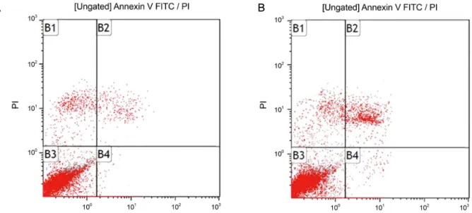
survival of the cultured human nasal mucosa fibroblasts. AME decreased fibroblast proliferation significantly in a dose-de- pendent manner compared to non-exposed control. *p < 0.05.Figure 2. Flow cytometric analysis of apoptosis using annexin-PI double staining. Cells in quadrant B1, B2, B3, B4 represents ne-
crotic cells, late apoptotic cells, living cells and early apoptotic cells, respectively. (A) Unstained control. (B) Exposed to 20 μg/mL amniotic membrane extract. FITC = fluorescein isothiocyanate; PI = propidium iodide.Figure 3. Flow cytometric analysis of apoptosis using annex-
in-PI double staining. 10, 20, 30 μg/mL amniotic membrane extract increased the degree of apoptosis significantly com- pared to non-exposed control. PI = propidium iodide.*p < 0.05.30 μg/mL의 모든 농도에서 양막추출물에 노출시키지 않은 대조군에 비하여 섬유아세포의 증식을 유의하게 감소시켰 다(p=0.000) (Fig. 1). 10 μg/mL의 농도에서는 91.71%, 20 μg/mL 의 농도에서는 88.13%, 30 μg/mL의 농도에서는 84.24%로 섬유아세포의 생존을 유의하게 감소시켰다(p=0.000, p=0.000, p=0.000).
Annexin-propidium iodide (PI) 이중염색을 이용한 유세포분석
Annexin-V/PI 이중염색 후 유세포분석에서 살아있는 정 상 세포는 annexin-V에 음성이면서 PI에도 음성으로, 세포 자가소멸의 경우는 annexin-V에 양성이면서 PI에 음성으 로, 세포 괴사(necrosis)의 경우는 annexin-V에 양성이면서 PI에도 양성으로 나타난다(Fig. 2). 양막추출물에 3일간 노
출시켰을 때, 10, 20, 30 μg/mL 농도의 양막추출물 모두에 서 세포 자가소멸의 정도를 유의하게 증가시켰다(p=0.024, p=0.002, p=0.002) (Fig. 3). 그리고 20, 30 μg/mL 농도의 양 막추출물은 대조군에 비하여 세포 괴사를 유발하는 정도에 있어서 유의한 차이를 나타냈다(p=0.005, p=0.000) (Fig. 4).
고 찰
양막은 Davis18가 1910년 최초로 피부이식을 위해 사용 하였고, 안과 영역에서는 1940년 De Rötth10가 결막붙음증 과 결손의 치료를 위해 처음 사용하였다. 1995년 이후부터 안과 영역에서 다양한 안구표면질환의 적응증에 사용되고 있는데, 널리 사용되기 시작한 것은 1997년 냉동보존방법 이 알려지면서부터이다.
A B
Figure 4. Flow cytometric analysis of necrosis using annex-
in-PI double staining. 20, 30 μg/mL amniotic membrane ex- tract affected the degree of necrosis significantly compared to non-exposed control. PI = propidium iodide. *p < 0.05.양막은 표피성장인자(epidermal growth factor), 각질세포성 장인자(keratinocyte growth factor), 간세포성장인자(hepatocyte growth factor) 등의 각종 성장인자들을 함유하고 있어 상피 세포의 이동, 분화, 유착을 강화시키고 창상의 상피화를 증 진시키며,19,20 IL-1 receptor antagonist, IL-10 등의 항염증단 백을 함유하여 항염증작용을 한다.21 또한 양막은 조직적합 항원이 표현되지 않아서 이식 거부반응이 없고,22,23 TGF-β 신호전달체계를 억제하여 반흔 형성을 줄여준다.24,25
이러한 상피화 촉진 효과를 이용하여 지속각막상피결손, 노출성 각막염, 헤르페스성 각막염, 수포각막병증, 각막화 상, 각막궤양 및 각막천공 등의 다양한 각막표면질환에서 양막이식이 이용되고 있다.26,27 또한 군날개의 재발률을 낮 추기 위해 사용하는 항대사물질을 양막이식으로 대체하기 도 하며,28 엑시머레이저 각막절제술 후 양막연고를 도포하 여 염증과 반흔을 줄이고 근시 진행을 억제하려는 보고도 있었다.29
Choi et al9은 코경유 눈물주머니코안연결술 시 양막으로 감싼 Merocel® 충전술을 시행하여 창상의 염증을 감소시키 고 치유속도를 증가시키며, 초기 2개월까지 육아종 발생을 줄일 수 있었다고 보고한 바 있다. 하지만 이 방법은 양막 으로 감싼 Merocel®을 10-14일간 충전하고 있어야 하기 때 문에 환자의 불편함과 감염관리를 철저히 해야 한다는 단 점이 있고, Merocel® 제거 후에는 양막의 영향이 감소하여 실리콘관의 지속적인 기계적 자극으로 인한 만성적인 염증 반응을 줄이지 못하므로 결국 6개월째에는 육아종 발생을 줄이지 못했다는 제한점이 있었다.
본 연구는 양막의 섬유아세포에 대한 영향에 의거하여 코경유 눈물주머니코안연결술 중 양막추출물의 보조적인 사용이 코점막 섬유아세포에 대해 항증식 효과를 가지는지 확인하고, 세포 자가소멸과 창상의 수축에 미치는 영향을 알아보았다. 그 결과 양막추출물은 낮은 농도에서도 섬유
아세포의 증식을 억제시켰으며, 농도가 높아지면서 점차 섬유아세포의 증식을 많이 억제시키는 농도 의존성 변화를 보였다. 양막추출물은 10 μg/mL의 낮은 농도에서부터 세 포 자가소멸을 유발시키는 것으로 나타났는데, 이는 섬유 아세포의 증식을 억제시키는 데 세포 자가소멸이 연관되어 있을 가능성을 시사한다. 또한 20, 30 μg/mL의 높은 농도 에서는 오히려 세포괴사를 유발하는 것으로 나타났는데, 세포배양상태에서는 세포 자가소멸을 일으킨 세포 성분을 탐식할 세포들이 주위에 없으므로 세포 자가소멸을 일으킨 세포들이 세포괴사의 양상을 나타낼 수도 있다는 점을 고 려해야 한다.30-32 그러나 고농도의 양막추출물을 사용하게 되면 코점막에 손상을 유발할 가능성이 낮게나마 있을 수 도 있으므로 이에 대한 추가적인 생체 내 실험이 필요할 것 으로 보인다.
본 연구의 결과를 바탕으로 눈물주머니코안연결술의 성 공률을 높이기 위하여 수술 시 Choi et al9이 시행한 대로 양막을 감싼 Merocel®을 만들어 3일간만 유지한 후 제거하 여 코를 막는 기간을 줄여서 시행해 보거나 양막추출물을 상품화하여 창상 부위에 뿌리는 새로운 방법을 시도해 볼 수도 있을 것이다. 이는 Choi et al9의 보고에서처럼 장시간 의 Merocel® 충전이 불필요하며, 코에 직접 뿌리는 방법은 효과가 저하될 때 다시 시술하기가 간편하다는 장점이 있 을 것으로 보인다. 그러나 이는 신선한 양막을 사용할 때에 해당되는 방법들로 기존에 상품화되어 나와있는 양막을 위 와 같은 방법으로 코점막에 처치하게 된다면 상이한 결과 가 나올 수 있을 것으로 보인다.
본 연구는 생체 외 실험이므로 생체 내에서 유효성을 가 지는지에 대한 태생적인 한계가 있으며, 양막추출물이 섬 유아세포의 증식을 억제하는 효과를 노출 시간에 따라 장 기적으로 관찰하지 못하였다는 한계점이 있다. 따라서 임 상적인 적용에 앞서 6개월 이상의 장기간 동안 양막추출물 의 영향 및 재노출 시에도 초기 노출과 같은 효과를 나타내 는지에 대한 연구가 필요할 것이다.
결론적으로 양막추출물은 생체 외 환경에서 배양된 사 람 코점막 섬유아세포의 증식을 억제하였으며, 세포 자가 소멸이 어느 정도 그 기전에 연관성이 있는 것으로 보인 다. 그러므로 코경유 눈물주머니코안연결술 시 양막추출 물의 보조적인 사용은 수술 결과를 향상시킬 수 있을 것으 로 보인다.
REFERENCES
1) Allen K, Berlin AJ. Dacryocystorhinostomy failure: association with nasolacrimal silicone intubation. Ophthalmic Surg 1989;20:486-9.
2) Zolli CL, Shannon GM. Dacryocystorhinostomy: a review of 119 cases. Ophthalmic Surg 1982;13:905-10.
3) McLachlan DL, Shannon GM, Flanagan JC. Results of dacryocys- torhinostomy: analysis of the reoperations. Ophthalmic Surg 1980;
11:427-30.
4) Tarbet KJ, Custer PL. External dacryocystorhinostomy, Surgical success, patient satisfaction, and economic cost. Ophthalmology 1995;102:1065-70.
5) Walland MJ, Rose GE. Factors affecting the success rate of open lacrimal surgery. Br J Ophthalmol 1994;78:888-91.
6) Kao SC, Liao CL, Tseng JH, et al. Dacryocystorhinostomy with in- traoperative mitomycin C. Ophthalmology 1997;104:86-91.
7) Yalaz M, Firinciogullari E, Zeren H. Use of mitomycin C and 5-flu- orouracil in external dacryocystorhinostomy. Orbit 1999;18:239-45.
8) Kim JH, Kim KS, Yoon HC, et al. Anti-adhesive effect of GUARDIX-SL(R) after endoscopic sinus surgery. Korean J Otolaryngol-Head Neck Surg 2005;48:1478-83.
9) Choi YJ, Hwang SJ, Lee TS. Short-term clinical results of amniotic membrane application to endonasal dacryocystorhinostomy. J Korean Ophthalmol Soc 2008;49:384-9.
10) De Rötth A. Plastic repair of conjunctival defects with fetal membranes.
Arch Ophthalmol 1940;23:522-5.
11) Tseng SC, Prabhasawat P, Lee SH. Amniotic membrane trans- plantation for conjunctival surface reconstruction. Am J Ophthalmol 1997;124:765-74.
12) Houlihan JM, Biro PA, Harper HM, et al. The human amnion is a site of MHC class Ib expression: evidence for the expression of HLA-E and HLA-G. J Immunol 1995;154:5665-74.
13) Kim JC, Tseng SC. The effects on inhibition of corneal neo- vascularization after human amniotic membrane transplantation in severely damaged rabbit corneas. Korean J Ophthalmol 1995;9:
32-46.
14) Prabhasawat P, Barton K, Burkett G, Tseng SC. Comparison of conjunctival autografts, amniotic membrane grafts, and primary closure for pterygium excision. Ophthalmology 1997;104:974-85.
15) Tseng SC, Prabhasawat P, Barton K, et al. Amniotic membrane transplantation with or without limbal allografts for corneal sur- face reconstruction in patients with limbal stem cell deficiency.
Arch Ophthalmol 1998;116:431-41.
16) Lee SH, Tseng SC. Amniotic membrane transplantation for persis- tent epithelial defects with ulceration. Am J Ophthalmol 1997;
123:303-12.
17) Mosmann T. Rapid colorimetric assay for cellular growth and sur- vival: application to proliferation and cytotoxicity assays. J Immunol Methods 1983;65;55-63.
18) Davis JW. Skin transplantation with a review of 550 cases at the Johns Hopkins Hospital. Johns Hopkins Med J 1910;15:307.
19) Meller D, Pires RT, Tseng SC. Ex vivo preservation and expansion of human limbal epithelial stem cells on amniotic membrane cultures. Br J Ophthalmol 2002;86:463-71.
20) Meller D, Tseng SC. Conjunctival epithelial cell differentiation on amniotic membrane. Invest Ophthalmol Vis Sci 1999;40:878-86.
21) Hao Y, Ma DH, Hwang DG, et al. Identification of antiangiogenic and antiinflammatory proteins in human amniotic membrane.
Cornea 2000;19:348-52.
22) van Herendael BJ, Oberti C, Brosens I. Microanatomy of the hu- man amniotic membranes. A light microscopic, transmission, and scanning electron microscopic study. Am J Obstet Gynecol 1978;
131:872-80.
23) Dua HS, Azuara-Blanco A. Amniotic membrane transplantation.
Br J Ophthalmol 1999;83:748-52.
24) Li DQ, Lee SB, Tseng SC. Differential expression and regulation of TGF-beta1, TGF-beta2, TGF-beta3, TGF-betaRI, TGF-betaRII and TGF-betaRIII in cultured human corneal, limbal, and con- junctival fibroblasts. Curr Eye Res 1999;19:154-61.
25) Tseng SC, Li DQ, Ma X. Suppression of transforming growth fac- tor-beta isoforms, TGF-beta receptor type II, and myofibroblast differentiation in cultured human corneal and limbal fibroblasts by amniotic membrane matrix. J Cell Physiol 1999;179:325-35.
26) Lee KH, Lee DW, Kim IC. The clinical effect of amniotic mem- brane transplantation for various ocular surface diseases. J Korean Ophthalmol Soc 2007;48:7-12.
27) Oh DH, Kwon MS, Kim JC. Five layered reinforcing amniotic membrane transplantation for treatment of deep corneal ulcer or perforation. J Korean Ophthalmol Soc 2011;52:1232-7.
28) Jang JH, Choi TH. The effect of amniotic membrane trans- plantation for pterygium excision. J Korean Ophthalmol Soc 2005;
46:597-604.
29) Lee DY, Park WC, Rho SH. The effect of amniotic membrane oint- ment application on photore fractive keratectomized cornea in rabbits. J Korean Ophthalmol Soc 2000;41:2069-77.
30) Portera-Cailliau C, Hedreen JC, Price DL, Koliatsos VE. Evidence for apoptotic cell death in Huntington disease and excitotoxic ani- mal models. J Neurosci 1995;15(5 Pt 2):3775-87.
31) Charriaut-Marlangue C, Aggoun-Zouaoui D, Represa A, Ben-Ari Y. Apoptotic features of selective neuronal death in ischemia, epi- lepsy and gp 120 toxicity. Trends Neurosci 1996;19:109-14.
32) Leist M, Gantner F, Bohlinger I, et al. Tumor necrosis factor-in- duced hepatocyte apoptosis precedes liver failure in experimental murine shock models. Am J Pathol 1995;146:1220-34.
= 국문초록 =
배양된 사람 코점막 섬유아세포에 대한 양막추출물의 효과
목적: 양막추출물이 배양된 사람의 코점막 섬유아세포의 생존에 미치는 효과에 대해 알아보고자 하였다.
대상과 방법: 일차배양한 사람의 비점막 섬유아세포를 0, 10, 20, 30 μg/mL의 양막추출물에 3일간 노출시킨 후 섬유아세포의 생존은 MTT assay로, 세포의 자가소멸은 annexin-V/propidium iodide 이중염색 후 유세포분석으로 평가하였다.
결과: 양막추출물은 10 μg/mL의 농도에서부터 섬유아세포의 증식을 유의하게 억제시켰으며(p=0.000), 유세포분석에서 10 μg/mL의 농도에서부터 섬유아세포의 자가소멸을 유의하게 증가시켰다(p=0.024).
결론: 양막추출물은 생체 외 환경에서 섬유아세포의 증식을 억제하였으며, 세포 자가소멸이 어느 정도 그 기전에 연관성이 있는 것으 로 보인다. 그러므로 코경유 눈물주머니코안연결술 시 양막추출물의 보조적인 사용은 수술 결과를 향상시킬 수 있을 것으로 보인다.
<대한안과학회지 2015;56(12):1939-1944>