ISSN 0378-6471 (Print)⋅ISSN 2092-9374 (Online)
https://doi.org/10.3341/jkos.2018.59.6.543
Original Article
섬유주세포에서 발프로익산이 일산화질소의 생성과 일산화질소합성효소의 발현에 미치는 영향
Effect of Valproic Acid on Nitric Oxide and Nitric Oxide Synthase in Trabecular Meshwork Cell
손명서1⋅박철호2⋅김재우1
Myung Seo Son, MD1, Cheul Ho Park, MD2, Jae Woo Kim, MD, PhD1
대구가톨릭대학교 의과대학 안과학교실1, 현대안과의원2
Department of Ophthalmology, Catholic University of Daegu School of Medicine1, Daegu, Korea Hyundai Eye Clinic2, Kumi, Korea
Purpose: To investigate the effects of valproic acid on the production of nitric oxide (NO) and expression of endothelial nitric ox- ide synthase (eNOS) in cultured human trabecular meshwork cells (HTMC).
Methods: Primarily cultured HTMC were exposed to 0.25, 0.5, and 1.0 mM valproic acid for 6, 12, and 24 hours. Expression of eNOS mRNA was assessed with Reverse transcription-polymerase chain reaction, and production of NO was assessed with Griess assay. Cellular survival was assessed with the 3-[4, 5–dimethylthiazol-2-yl]-2, 5-diphenyltetrazolium bromide (MTT) assay.
Results: Valproic acid at concentrations of 0.25, 0.5, 1.0 mM did not affect the cellular survival of HTMC significantly after ex- posure for 24 hours. Valproic acid increased NO production in a dose- and time-dependent manner. Also, valproic acid in- creased the degree of eNOS mRNA expression in a dose-dependent manner in HTMC.
Conclusions: Valproic acid increases production of NO and expression of eNOS mRNA in HTMC. Thus, valproic acid might in- crease aqueous outflow through the trabecular meshwork.
J Korean Ophthalmol Soc 2018;59(6):543-548
Keywords: Endothelial nitric oxide synthase (eNOS), Nitric oxide, Trabecular meshwork, Valproic acid
■Received: 2017. 7. 6. ■ Revised: 2017. 8. 16.
■Accepted: 2018. 5. 31.
■Address reprint requests to Jae Woo Kim, MD, PhD Department of Ophthalmology, Daegu Catholic University School of Medicine, #33 Duryugongwon-ro 17-gil, Nam-gu, Daegu 42472, Korea
Tel: 82-53-650-4728, Fax: 82-53-627-0133 E-mail: [email protected]
* This work was supported by the grant of purpose of donation, department of Ophthalmology, Daegu Catholic University School of Medicine.
* Conflicts of Interest: The authors have no conflicts to disclose.
ⓒ2018 The Korean Ophthalmological Society
This is an Open Access article distributed under the terms of the Creative Commons Attribution Non-Commercial License (http://creativecommons.org/licenses/by-nc/3.0/) which permits unrestricted non-commercial use, distribution, and reproduction in any medium, provided the original work is properly cited.
발프로익산(valproic acid, 2-propylpentanoic acid, VPA) 은 짧은 사슬의 지방산으로 간질의 치료제로 사용되어 왔 으며,1 신경세포의 분화 및 보호, 항암작용 등 다양한 약리 작용을 나타내는 것으로 알려져 있다.2 히스톤 디아세틸레 이즈(histone deacetylases, HDAC) 저해제인 VPA의 또 다 른 작용은 일산화질소(nitric oxide, NO)의 생성을 감소시키 는 것이므로3 이에 따라 세포의 생존을 저하시킬 수 있지만 이에 반해 VPA가 NO의 생성을 증가시킨다는 보고도 있 다.4,5
섬유주세포는 녹내장에서 방수유출로의 조절에 중요한 역할을 하는데, 섬유주의 변성으로 인해 방수유출로의 저
항이 증가되면 개방각녹내장을 유발하는 기전이 된다.6,7 자 유유리기인 NO는 세포의 종류에 따라 다양한 역할을 나타 낼 수 있으며, 평활근 이완효과도 나타내는 것으로 알려져 있다. 섬유주세포는 형태학적 연구와 전기생리학적 연구에 서 평활근과 유사한 성질을 가진 것으로 알려져 있으며8,9 endothelial nitric oxide synthase (eNOS)에서 생산하는 NO 는 섬유주를 이완시켜 방수유출을 촉진하는 것으로 알려져 있다.10-12
안구 내에서 VPA는 eNOS의 발현과 NO의 생성을 증가 시켜 섬유주를 이완시킴으로써 섬유주를 통한 방수유출을 증가시켜 안압하강을 유발할 가능성이 있으나 VPA가 NO 의 생성에 미치는 영향에 대해서는 다양한 보고가 있으며3-5 특히 인체의 섬유주세포에서 NO의 생성에 관한 연구는 아 직 자세히 되어 있지 않다. 따라서 본 연구에서는 사람의 섬유주세포를 일차배양하여 VPA가 섬유주에서 NO의 생 성에 미치는 영향과 eNOS의 발현에 미치는 영향을 알아보 고자 하였다.
대상과 방법
세포배양
본 연구는 대구가톨릭대학교병원 의학윤리심의위원회 (institutional review board, IRB)의 승인을 받았고(승인번호:
CR-16-114) 헬싱키 선언에 따라 시행되었다. 안구은행에서 얻은 사후 6시간 이내에 적출한 안구의 전방각에서 섬유주를 벗겨내어 폴리라이신(Sigma, St. Louis, MO, USA)으로 처리 한 배양접시에 옮긴 후 항생제(Gibco, Invitrogen, Carlsbad, CA, USA)와 15% 우태아혈청(Hyclone, Thermoscientific, Carlsbad, CA, USA)이 포함된 Dulbecco’s modified Eagle’s medium 배지(DMEM, Gibco, Invitrogen, Carlsbad, CA, USA)를 사용하여 5% CO2 배양기에서 초대배양하였다. 섬 유주세포가 이식된 조직편 주위로 자라나온 것을 확인한 후 섬유주조직의 이식편을 제거하고 배양을 계속하였으며 세포가 배양접시에 충만해지면 10% 우태아혈청(Gibco, Invitrogen, Carlsbad, CA, USA)을 포함한 배지로 1:3의 비 율로 트립신 처리하여 계대배양하였다.
약물처리
24 well 배양접시에 2 × 10⁴ cells/mL의 농도로 각 well에 세포를 분주한 후 24시간 동안 배양기에 넣어 세포를 부착시 킨 후 배지를 제거하고 나서 10% 혈청이 포함된 DMEM 배 지를 이용하여 VPA (Sigma, St. Louis, MO, USA)를 0.25, 0.5, 1.0 mM의 농도로 단계적으로 희석한 후 6시간, 12시간, 24시간 각각 노출시킨 다음 아래의 실험을 시행하였다.
MTT assay
세포의 생존에 대한 효과는 세포생존과 세포독성의 선별 검사로 흔히 이용되고 있는 발색검사의 일종인 MTT (3-[4, 5 –dimethylthiazol-2-yl]-2, 5-diphenyltetrazolium bromide, Sigma, St. Louis, MO, USA) assay13를 이용하였다. 약물처 리한 세포의 배지에 MTT를 각 well당 100 μL씩 투여한 후 4시간 동안 정치배양한 다음 염류용액으로 씻어낸 후 di- methylsulfoxide를 각 well당 0.5 mL씩 넣어 10분 이상 흔 든 다음 96-well 배양접시에 200 μL씩 옮겨 분광광도계 (FLUOstar OPTIMA, BMG labtech, Germany)로 570 nm에 서 흡광도를 측정하였다. 이때 세포의 생존 정도는 실험군 의 값을 약물처리를 하지 않은 대조군의 비로 나누어 백분 율로 나타내었다.
Griess assay
섬유주세포에서 NO의 생성은 Griess assay를 이용하여 측정하였다.14 일차배양한 섬유주세포를 VPA에 농도와 시 간별로 노출시킨 다음 배지에 동량의 Griess reagent (Sigma, St. Louis, MO, USA)를 섞은 후 96-well plate에 옮겨 spec- trophotometer (FLUOstar Optima, BMG Labtech, Offenburg, Germany)로 540 nm에서 흡광도를 측정하였다. 이때 표준 치를 구하기 위해 sodium nitrite (Sigma, St. Louis, MO, USA)를 단계적으로 희석하여 사용하였다.
eNOS mRNA 발현을 측정하기 위한 Reverse transcription-polymerase chain reaction
VPA에 노출시킨 섬유주세포에서 Trizol (Invitrogen, Carlsbad, CA, USA)을 이용하여 RNA를 분리한 후 분리된 RNA에서 eNOS mRNA의 발현을 RT-PCR을 이용해 확인 하였다. 섬유주세포에서 분리한 RNA와 Oligo dT primer, nuclease-free water를 혼합하여 만든 RNA denaturation mix를 70℃에 5분간 denaturation 시키고 얼음에 5분간 둔 다음 Prime RT premix (Genet bio, Seoul, Korea)와 혼합하 여 42oC에서 1시간, 70oC 10분간 반응시켜 cDNA로 합성 하였다. 합성한 cDNA에 2XGoTaq Green Master Mix (Promega, Fitchburg, WI, USA)와 10 pmol의 forward pri- mer (ctg gct ttc cct tcc agt tc, 225 bp), reverse primer (cct tcc aga tta agg cgg ac, 225 bp)를 각각 혼합하여 DNAEngine cycler (Bio-Rad, Hercules, CA, USA)를 사용 하여 94℃에서 5분, 94℃ 30초, 54oC 30초, 72oC 30초로 총 30 cycles를 시행한 후 57oC에서 5분간 반응시켰다. 증폭된 polymerase chain reaction product를 1% agarose gel에 전 기 영동하여 DNA band를 multi Gauge v2.02 (Fujifilm, Tokyo, Japan)를 이용하여 분석하였다. 이때 β–action을 in-
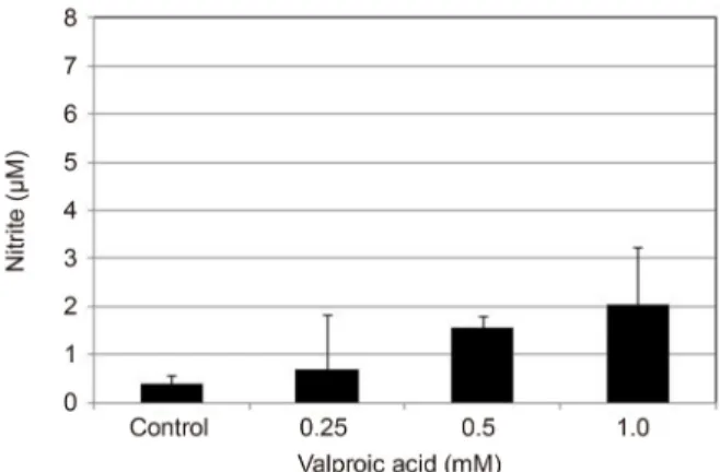
Figure 1. Effects of valproic acid (VPA) on the survival of tra-
becular meshwork cells. VPA did not affect the survival sig- nificantly compared to non-exposed control (p> 0.05).Figure 2. Effects of valproic acid on the production of nitric ox-
ide in trabecular meshwork cells after exposure for 6 hours.Valproic acid did not affect the production of nitric oxide sig- nificantly (p> 0.005).
ternal standard로 사용하였다.
통계적 처리
모든 실험은 3계대에서 5계대 사이의 세포를 이용하였고 대조군은 약물처리를 하지 않은 군으로 3회 이상 시행하였 으며, NO의 생성은 평균 ± 표준오차로 나타내어 unpaired t-test를 사용하여 유의성을 비교하였으며 유의수준은 0.05%
로 정하였다.
결 과
세포배양
초대배양 7일째부터 섬유주조직의 이식편 주위로 섬유주 세포가 자라 나오기 시작하였으며 섬유주세포의 확인은 세 포들이 밀집해서 단층을 형성하며 세포들 사이에 분지를 내어 서로 연접하며 약간 길다란 모양의 세포체를 가지는 편평한 모양의 특징적 형태학적인 양상과 섬유주 조직의 이식편 주위에서 위성양상으로 자라나는 섬유주세포의 특 징적인 성장양상으로 확인하였다.15-17
VPA가 섬유주세포의 생존에 미치는 영향
0.25, 0.5, 1.0 mM 농도의 VPA에 24시간 노출시킨 후 시 행한 MTT assay에서 모든 농도에서 약물에 노출되지 않은 대조군에 비해 세포의 생존율에 유의한 차이를 나타내지 않았다(p<0.05) (Fig. 1).
VPA가 섬유주세포에서 NO의 생성에 미치는 영향 섬유주세포에서 VPA는 약물처리 후 6시간 후에는 각 농 도에서 약물처리를 하지 않은 대조군에 비하여 배지에서의
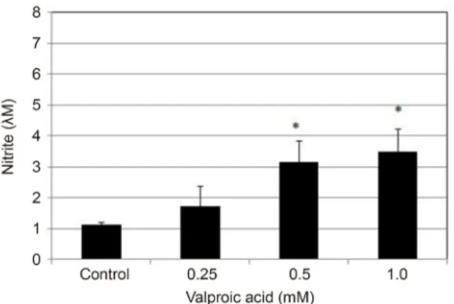
nitrite 생성량에서 유의한 차이를 나타내지 않았다(p=0.914, 0.788, 0.200) (Fig. 2). 그러나 약물처리 후 12시간 후에는 0.5 mM과 1.0 mM에 노출시킨 경우 약물처리를 하지 않은 대조군에 비하여 유의하게 배지에서의 nitrite 생성량을 증 가시켰으며(p=0.012, 0.006) (Fig. 3), 24시간 후에도 0.5 mM과 1.0 mM에 노출시킨 경우 배지에서의 nitrite 생성량 을 유의하게 증가시켰다(p=0.020, 0.029) (Fig. 4).
VPA가 eNOS mRNA의 발현에 미치는 영향
약물처리를 하지 않은 대조군에 비하여 VPA는 약물처리 후 6시간부터 약물에 노출되지 않은 대조군에 비해 eNOS mRNA의 발현을 증가시켰다(Fig. 5). 이러한 eNOS mRNA 의 발현은 노출한 VPA의 농도가 증가할수록 발현의 정도 가 증가하였다.
고 찰
본 연구의 결과는 사람의 섬유주세포에서 VPA는 eNOS 의 발현을 증가시키면서 NO의 생성을 촉진하는 작용을 나 타낼 수 있음을 보여주고 있다. VPA는 신경계의 흥분을 억 제하는 다양한 약리작용을 나타내는데 VPA는 세포분화를 조절하는 HDAC에 대한 저해작용을 나타내어18,19 종양세포 의 증식을 억제할 뿐만 아니라 세포고사를 유발하며,20-23 기내실험과 생체 내 실험에서 혈관내피세포의 증식과 이동, 그리고 신생혈관생성을 억제하는 것이 보고되었다.3 안과 영역에서 VPA는 글루탐산에 의한 신경흥분을 조절하여 안 구내 망막신경절세포를 보호하는 작용을 나타내며24 만성 고안압증 모델을 이용한 실험에서 망막신경절세포의 구조 와 기능을 유지하는 작용을 나타낸다.25
Figure 3. Effects of valproic acid on the production of nitric
oxide in trabecular meshwork cells after exposure for 12 hours.Exposure to 0.5 and 1.0 mM valproic acid increased the pro- duction of nitric oxide significantly (*p< 0.05).
Figure 4. Effects of valproic acid on the production of nitric ox-
ide in trabecular meshwork cells after exposure for 24 hours.Exposure to 0.5 and 1.0 mM valproic acid increased the pro- duction of nitric oxide significantly (*p< 0.05).
A B C
Figure 5. Expression of endothelial nitric oxide synthase (eNOS) mRNA after exposed to valproic acid (VPA) in trabecular mesh-
work cells. Exposure to 0.25, 0.5, 1.0 mM for 6, 12, 24 hours increased the degree of eNOS mRNA expression in a dose- and time-dependent manners. (A) After 6 hours. (B) After 12 hours. (C) After 24 hours of exposure. β-actin was used as internal standard.0.5 mM의 VPA는 인체에서 사용할 수 있는 VPA의 적정 범위 내에 해당하는데19 VPA가 섬유주세포의 생존에 영향 을 미칠 수 있으므로 0.25 mM에서 1.0 mM 농도에 노출시 킨 다음 24시간 후에 섬유주세포의 생존에 미치는 영향을 알아본 결과 본 실험에 사용한 농도에서 섬유주세포의 생 존에는 유의한 영향을 미치지 않았다. 따라서 본 실험에서 측정한 NO의 생성량 차이는 세포의 생존변화에 의한 것이 아님을 알 수 있다.
HDAC 저해제로서 VPA의 또 다른 작용으로 혈관내피세 포를 이용한 연구에서 세포의 이동과 증식을 억제하면서 일산화질소의 생성을 감소시키는 것으로 보고하였으나3 다 른 연구에서는 NO의 생성을 저하시킨다는 보고도 있었다.4
혈관내피세포에서 NO의 생성에 대한 이러한 상반된 결과 를 구명하기 위해 시행한 연구에서 VPA가 NO 생성을 증 가시키며 그 기전은 SH2 domain-containing protein ty- rosine phosphatase 1 (SH-PTP1)의 활성을 조절함에 의한 것이라고 밝혀졌다.5 본 연구에서 VPA가 섬유주세포에서 NO의 생성에 미치는 영향을 알아본 결과 VPA는 노출 초 기인 6시간 후에는 NO의 생성에 유의한 영향을 미치지 않 았으나 12시간과 24시간 노출시킨 경우에는 0.5 mM과 1.0 mM 농도에서 VPA에 노출시키지 않은 대조군에 비하여 농도에 비례하여 NO의 생성을 유의하게 증가시켰다. 또한 VPA에 노출시킨 후 시행한 eNOS mRNA의 발현을 조사한 결과에서 VPA의 농도에 따라 eNOS mRNA의 발현이 증가
하였는데, 만일 이러한 eNOS mRNA 발현의 증가가 나타 나지 않으면서 NO의 생성이 증가되었다면 VPA가 전사과 정에 관여하지 않고 eNOS의 안정성을 증가시켜 NO의 생 성을 증가시켰을 가능성이 있을 것이다. 비록 본 실험에서 는 eNOS 단백을 정량하지는 못했으나 본 실험의 결과는 VPA가 인체의 섬유주세포에서 전사과정에 관여하여 eNOS 의 발현을 증가시켜 NO의 생성을 증가시킬 가능성이 있음 을 보여주고 있다.
VPA는 망막에서 허혈성 손상이나 산화스트레스에 대해 신경세포보호작용을 나타내는 것으로알려져 있으며,26-29 정 상안압녹내장 모델의 동물실험에서도 신경세포보호작용이 있는 것으로 보고되었다.30 따라서 녹내장에서 VPA는 망막 신경절세포에 대한 보호작용을 나타낼 뿐만 아니라 본 연 구의 결과에 의하면 NO의 생성 증가에 의해 섬유주를 통 한 방수 유출을 촉진시켜 안압하강작용도 나타낼 가능성이 있으나 실제 인체에 대한 임상적 효과는 아직 자세히 보고 되지 않았다.
결론적으로 VPA는 인체의 섬유주세포에서 eNOS mRNA 의 발현과 NO 생성을 증가시킴으로써 섬유주를 이완시켜 섬유주를 통한 방수유출을 증가시켜 안압하강작용을 나타 낼 가능성이 있다. 그러나 VPA는 다양한 작용을 나타내며, 실제 생체 내에서 안압에 대한 효과 및 신경세포보호작용 에 대해서는 동물 및 임상 실험을 비롯한 보다 자세한 연구 가 필요할 것으로 생각된다.
REFERENCES
1) Löescher W. Basic pharmacology of valproate: a review after 35 years of clinical use for the treatment of epilepsy. CNS Drugs 2002;16:669-94.
2) Monti B, Polazzi E, Contestabile A. Biochemical, molecular and epigenetic mechanisms of valproic acid neuroprotection. Cur Mol Pharmacol 2009;2:95-109.
3) Michaelis M, Michaelis UR, Fleming I, et al. Valproic acid inhibits angiogenesis in vitro and in vivo. Mol Pharmacol 2004;65:520-7.
4) Hyndman KA, Ho DH, Sega MF, Pollock JS. Histone deacetylase 1 reduces NO production in endothelial cells via lysine deacetylation of NO synthase 3. Am J Physiol Heart Circ Physiol 2014;307:
H803-9.
5) Cho DH, Park JH, Joo Lee E, et al. Valproic acid increases NO pro- duction via the SH-PTP1–CDK5–eNOS-Ser(116) signaling cas- cade in endothelial cells and mice. Free Radic Biol Med 2014;76:96-106.
6) Alvarado J, Murphy C, Juster R. Trabecular meshwork cellularity in primary open-angle glaucoma and nonglaucomatous normals.
Ophthalmology 1984;91:564-79.
7) Rohen JW, Lütjen-drecoll E, Flügel C, et al. Ultrastructure of the trabecular meshwork in untreated cases of primary open-angle glaucoma (POAG). Exp Eye Res 1993;56:683-92.
8) Wiederholt M, Dörschner N, Groth J. Effect of diuretics, channel modulators and signal interceptors on contractility of the tra- becular meshwork. Ophthalmologica 1997;211:153-60.
9) Wiederholt M, Stumpff F. The trabecular meshwork and aqueous humor reabsorption. In: Civan MM, ed. Current Topics in Membranes. The Eye's Aqueous Humor: From Secretion To Glaucoma, 1st ed. San Diego: Academic Press, 1998; v. 45. chap.
7.
10) Wiederholt M, Sturm A, Lepple-Wienhues A. Relaxation of tra- becular meshwork and ciliary muscle by release of nitric oxide.
Invest Ophthalmol Vis Sci 1994;35:2515-20.
11) Behar-Cohen FF, Goureau O, D’Hermies F, Courtois Y. Decreased intraocular pressure induced by nitric oxide donors is correlated to nitrite production in the rabbit eye. Invest Ophthalmol Vis Sci 1996;37:1711-5.
12) Dismuke WM, Mbadugha CC, Ellis DZ. NO-induced regulation of human trabecular meshwork cell volume and aqueous humor out- flow facility involve the BKCa ion channel. Am J Physiol Cell Physiol 2008;294:C1378-86.
13) Mosmann T. Rapid colorimetric assay for cellular growth and sur- vival: application to proliferation and cytotoxicity assays. J Immunol Methods 1983;65:55-63.
14) Green LC, Wagner DA, Glogowski J, et al. Analysis of nitrate, ni- trite and [15N]nitrate in biologic fluids. Anal Biochem 1982;126:131-8.
15) Stamer WD, Roberts BC, Epstein DL, Allingham RR. Isolation of primary open-angle glaucomatous trabecular meshwork cells from whole eye tissue. Curr Eye Res 2000;20:347-50.
16) Polansky JR, Weinreb RN, Baxter JD, Alvarado J. Human tra- becular cells. I. Establishment in tissue culture and growth characteristics. Invest Ophthalmol Vis Sci 1979;18:1043-9.
17) Alvarado JA, Wood I, Polansky JR. Human trabecular cells. II.
Growth pattern and ultrastructural characteristics. Invest Ophthalmol Vis Sci 1982;23:464-78.
18) Göttlicher M, Minucci S, Zhu P, et al. Valproic acid defines a novel class of HDAC inhibitors inducing differentiation of transformed cells. EMBO J 2001;20:6969-78.
19) Phiel CJ, Zhang F, Huang EY, et al. Histone deacetylase is a direct target of valproic acid, a potent anticonvulsant, mood stabilizer, and teratogen. J Biol Chem 2001;276:36734-41.
20) Kawagoe R, Kawagoe H, Sano K. Valproic acid induces apoptosis in human leukemia cells by stimulating both caspase-dependent and -independent apoptotic signaling pathway. Leuk Res 2002;26:495-502.
21) Phillips A, Bullock T, Plant N. Sodium valproate induces apoptosis in the rat hepatoma cell line, FaO. Toxicology 2003;192:219-27.
22) Tang R, Faussat AM, Majdak P, et al. Valproic acid inhibits pro- liferation and induces apoptosis in acute myeloid leukemia cells expressing P-gp and MRP1. Leukemia 2004;18:1246-51.
23) Witt D, Burfeind P, von Hardenberg S, et al. Valproic acid inhibits the proliferation of cancer cells by re-expressing cyclin D2.
Carcinogenesis 2013;34:1115-24.
24) Biermann J, Grieshaber P, Goebel U, et al. Valproic acid–mediated neuroprotection and regeneration in injured retinal ganglion cells.
Invest Ophthalmol Vis Sci 2010;51:526-34.
25) Alsarraf O, Fan J, Dahrouj M, et al. Acetylation preserves retinal ganglion cell structure and function in a chronic model of ocular hypertension. Invest Ophthalmol Vis Sci 2014;55:7486-93.
26) Laufs U, Liao JK. Post-transcriptional regulation of endothelial ni-
= 국문초록 =
섬유주세포에서 발프로익산이 일산화질소의 생성과 일산화질소합성효소의 발현에 미치는 영향
목적: 섬유주세포에서 발프로익산이 일산화질소의 생성과 일산화질소합성효소(endothelial nitric oxide synthase, eNOS)의 발현에 미치는 영향을 알아보고자 하였다.
대상과 방법: 사람의 섬유주세포를 일차배양한 후 발프로익산에 0.25, 0.5 1.0 mM의 농도로 노출시킨 후 6시간, 12시간, 24시간에 Reverse transcription-polymerase chain reaction을 이용하여 eNOS mRNA의 발현을 조사하였다. 또한 Griess assay를 이용하여 일산화질소의 생성량을 측정하였으며 세포의 생존은 3-[4, 5–dimethylthiazol-2-yl]-2, 5-diphenyltetrazolium bromide (MTT) assay 로 측정하였다.
결과: 섬유주세포에서 0.25, 0.5 1.0 mM 농도의 발프로익산은 24시간 노출 후에 섬유주세포의 생존에 영향을 미치지 않았다. 발프로 익산은 섬유주세포에서 일산화질소의 생성을 증가시켰으며, 노출 시간과 농도에 따라 일산화질소의 생성량이 증가하였다. 또한 발프 로익산은 eNOS mRNA의 발현을 함께 증가시켰으며 농도에 비례하여 eNOS mRNA의 발현의 정도가 증가하였다.
결론: 섬유주세포에서 발프로익산은 일산화질소의 생성과 eNOS mRNA의 발현을 증가시켰다. 따라서 발프로익산은 섬유주를 통한 방수유출을 증가시키는 작용을 나타낼 가능성이 있다.
<대한안과학회지 2018;59(6):543-548>
tric oxide synthase mRNA stability by Rho GTPase. J Biol Chem 1998;273:24266-71.
27) Bose P, Dai Y, Grant S. Histone deacetylase inhibitor (HDACI) mechanisms of action: emerging insights. Pharmacol Ther 2014;143:323-36.
28) Zhang Z, Qin X, Zhao X, et al. Valproic acid regulates antioxidant enzymes and prevents ischemia/reperfusion injury in the rat retina.
Curr Eye Res 2012:37:429-37.
29) Kimura A, Namekata K, Guo X, et al. Valproic acid prevents NMDA-induced retinal ganglion cell death via stimulation of neu- ronal TrkB receptor signaling. Am J Pathol 2015;185:756-64.
30) Kimura A, Guo X, Noro T, et al. Valproic acid prevents retinal de- generation in a murine model of normal tension glaucoma.
Neurosci Lett 2015:588:108-13.