© 2017 The Korean Ophthalmological Society
This is an Open Access article distributed under the terms of the Creative Commons Attribution Non-Commercial License (http://creativecommons.org/licenses /by-nc/3.0/) which permits unrestricted non-commercial use, distribution, and reproduction in any medium, provided the original work is properly cited.
Original Article
Comparative Study of the Effects of Trabecular Meshwork Outflow Drugs on the Permeability and Nitric Oxide Production
in Trabecular Meshwork Cells
Jae Woo Kim
Department of Ophthalmology, Catholic University of Daegu School of Medicine, Daegu, Korea
Purpose: To compare the effects of the barrier function in human trabecular meshwork (TM) cells monolayer and the production of nitric oxide (NO) between trabecular outflow drugs, Rho-associated kinase (ROCK) in- hibitors, adenosine, and statin.
Methods: Primary cultured TM cells were exposed to 10 or 25 mM Y-27632, 0.1 or 1 mM N6-cyclohexyladenos- ine (CHA), or 15 or 30 mM simvastatin for 24 hours. NO production and expression of endothelial nitric oxide synthase mRNA were measured by Griess assay and reverse transcription polymerase chain reaction, re- spectively. Barrier functions of the TM cell monolayer were measured by carboxyfluorescein and trans-endo- thelial electrical resistance. The expression of matrix metalloproteinase-2 mRNA was assessed with reverse transcription polymerase chain reaction.
Results: In TM cells, treatment with each drug increased endothelial nitric oxide synthase mRNA expression.
Treatment with 25 mM Y-27632 and 1.0 mM CHA increased NO production significantly (p = 0.035 and p = 0.043, respectively). Treatment with each drug increased the permeability (all p = 0.001) and decreased the trans-en- dothelial electron resistance of the TM cell monolayer. Treatment with 0.1 mM and 1.0 mM CHA significantly increased matrix metalloproteinase-2 mRNA expression, but simvastatin inhibited its expression.
Conclusions: Since treatment with ROCK inhibitor more greatly increased NO production and permeability than did adenosine or statin, ROCK inhibitor seems to be more effective for lowering intraocular pressure.
Key Words: Adenosine, Permeability, Rho-associated kinase inhibitor, Statin, Trabecular meshwork
Glaucoma is a progressive optic neuropathy caused by death of the retinal ganglion cells and is the leading cause of irreversible blindness worldwide. The mechanism by which this progressive retinal ganglion cell death occurs is
not fully understood. It is clear that multiple causes can give rise to the common effect of ganglion cell death. The ciliary secretion of aqueous humor usually remains normal in glaucoma; therefore, it is thought that impaired drainage through the trabecular pathway caused by increased resis- tance is the primary cause of increased intraocular pres- sure (IOP) in primary open-angle glaucoma [1,2]. While most glaucoma medications approved for clinical use act either on the uveoscleral pathway and/or aqueous humor
Received: February 8, 2017 Accepted: March 13, 2017
Corresponding Author: Jae Woo Kim, MD. Department of Ophthalmol- ogy, Catholic University of Daegu School of Medicine, #33 Duryugong- won-ro 17-gil, Nam-gu, Daegu 42472, Korea. Tel: 82-53-650-4728, Fax:
82-53-627-0133, E-mail: [email protected]
formation, none target the major site of resistance to aque- ous humor outflow in glaucoma, namely, the trabecular meshwork/Schlemm’s canal (TM/SC; a conventional out- flow pathway). Therefore, novel pharmacological targets that can affect the conventional outflow pathway are high- ly desirable. Recently, several new drugs targeting the tra- becular outflow pathway have entered clinical development [3], such as Rho-associated kinase (ROCK) inhibitors, ade- nosine agonist, and statin [4].
Rho GTPase activity is regulated by physiological li- gands and growth factors that engage membrane bound receptors; upon conversion to its GTP-bound (activated) form, Rho GTPase then stimulates Rho kinase activity. In- hibition of Rho GTPase activity in TM and SC cells induc- es changes in cell shape and leads to decreases in actin stress fibers and cell adhesion and in myosin light-chain (MLC) phosphorylation [5]. ROCK inhibitors Y-27632, H-1152, and fasudil have been demonstrated to induce very rapid and long-lasting decreases in IOP [6-9]. Adenosine agonists increase trabecular out flow by changing TM cell volume and ion transport and by remodeling the extracel- lular matrix (ECM) [10]. Cholesterol-lowering statin sig- nificantly increased aqueous outflow facility in an or- gan-cultured porcine anterior eye segment perfusion model [11], and it has been reported that human patients who were administered statin drugs had a reduced risk of glaucoma [12].
Free radical nitric oxide (NO) has multiple functions in different cells. TM cells have shown smooth muscle cell- like activity in morphological and electrophysiological studies [13,14]. NO increases aqueous outflow by relaxing TM, and levels of NO and nitric oxide synthase (NOS) ex- pression are reduced in glaucomatous human eyes [15-17].
Although it is known that ROCK inhibitor and statin in- crease the expression of endothelial nitric oxide synthase (eNOS) [18,19], their effects on the expression of eNOS in human TM cells are still unknown.
The significance of matrix metalloproteinases (MMPs) in outflow resistance was shown when treatment of anteri- or segments in perfusion culture with MMPs was found to increase outflow, while specific inhibition of MMP activity decreased outflow facility [20]. Although ROCK inhibitors do not affect the expression of MMP-2, which are involved in the remodeling of ECM [21], treatment with adenosine significantly increases its expression [22], and the effect of statin on its expression has not been studied in TM cells.
This study was performed to compare the effects of ROCK inhibitors, adenosine, or statin on the in vitro tra- becular outflow by measuring the barrier function of a TM cell monolayer. The effects of these trabecular outflow drugs on the production of NO and expression of eNOS and MMP-2 mRNA were also studied.
Materials and Methods
Cell culture and experimental treatment
This study followed the tenets of the Declaration of Hel- sinki and was approved by the institutional review board/
ethics committee of Daegu Catholic University Hospital (No. CR-16-117-L).
TM cell cultures were established from an enucleated human eye obtained from the eye bank and transported on ice within 2 hours after exsanguination, as previously de- scribed [23]. Briefly, TM tissues were excised by dissecting a continuous strand of tissue between the line of Schwalbe and the scleral spur by teasing away the TM tissue using a curette. The excised TM tissues were placed in a sterile culture dish with Dulbecco's modified Eagle's medium (DMEM) containing 15% fetal bovine serum (FBS), 2 mM glutamine, 50 μg/mL gentamicin, and 2.5 mg/mL fungizone and left undisturbed for 3 to 5 days in a 37°C incubator with a 5% CO2 atmosphere. After observation of initial cell growth, the explants were removed, and the cul- tures were maintained in media containing 10% FBS.
Cultures approaching confluency were trypsinized and inoculated into culture plates. After attachment, the cells were washed three times with serum-free medium and then cultured for 24 hours in media supplemented with 10% FBS containing the following drugs. ROCK inhibitor (10 mM or 25 mM Y-27632; Sigma, St. Louis, MO, USA), adenosine A1 agonist (0.1 mM or 1 mM N6-cyclohexylade- nosine [CHA], Sigma), or statin (15 or 30 mM simvastatin, Sigma) was added to the cultures, as in previous studies [7,19,22].
MTT (3-[4, 5–dimethylthiazol-2-yl]-2, 5-diphenyltetra- zolium bromide) assay
Cell survival was determined by a rapid MTT colorimet- ric assay. The MTT assay is based on the tetrazolium salt
MTT that reacts with living but not dead cells. As signals are generated, the optical density is directly proportional to the number of cells [23]. For the assay, 100 μL of a MTT stock solution (5 mg MTT/mL phosphate buffered saline) was added to each well and incubated for 4 hours at 37°C, after which all media was removed from the well. After 0.5 mL of dimethyl sulfoxide was added to each well, 100 μL of solution from each well was transferred to a 96-well plate and analyzed on a multi-well scanning spectropho- tometer (λ = 570 nm, Fluostar Optima; BMG Labtech, Of- fenburg, Germany).
Measurement of monolayer cell permeability with car- boxyfluorescein
A permeability study of the TM cell monolayer was per- formed as previously described with minor modification [24]. Briefly, primary cultured human TM cells were incu- bated in the inner chamber (insert diameter, 12 mm; pore size, 0.4 mm) of a 12-well plate (No. 3460, Transwell;
Corning, Tewksbury, MA, USA) as 2×10⁴ cells/mL supple- mented with 10% FBS. After the cells reached confluency, the media was changed to 1% serum-containing DMEM to avoid the effects of growth factors and proteins in serum;
then, the TM cells were exposed to each drug for 24 hours.
After washing three times with PBS, 50 mM of the tracer carboxyfluorescein (Sigma-Aldrich, St. Louis, MO, USA) was added to each well. The media was collected from the outer well to analyze fluorescence after 2 hours, and the concentration of carboxyfluorescein in the collected media was measured using a spectrofluorometer (Fluostar Opti- ma, BMG Labtech) with an excitation wavelength of 490 nm and an emission wavelength of 530 nm.
Measurement of monolayer trans-endothelial electron resistance
TM cells were seeded at 2×10⁴ cells/well and were grown until confluent in the inner well (pore size, 0.4 mm; diam- eter, 12 mm) in 12-well culture plates (Transwell, Corning) [7,25]. Twenty-four hours after exposure to each drug, trans-endothelial electron resistance (TEER) was measured with an electrical resistance system (epithelial voltohmme- ter, EVOM2; World Precision Instruments, Sarasota, FL, USA) according to the manufacturer’s instructions, and the results were recorded as net value (Ω cm2).
Measurement of NO production
Nitrite concentrations in the media were measured using the Griess reaction [26]. Briefly, media samples were col- lected from each well following appropriate treatment and reacted with modified Griess reagent by mixing equal vol- umes at room temperature for 15 minutes. Optical density was then measured on a multi-well scanning spectropho- tometer at 540 nm. The nitrite concentration was deter- mined from a comparison of absorbance with that of a standard solution of sodium nitrite in media. The back- ground absorbance, measured using the media alone, was subtracted from all values.
RT-PCR for measurement of the expression of eNOS and MMP-2 mRNA
After exposure to each drug, total RNA was extracted with Trizol (Invitrogen, Carlsbad, CA, USA). An RNA de- naturation mix consisting of isolated RNA, oligo dT prim- ers, and nuclease-free water was denatured. RT-PCR was performed using oligonucleotide primers specific to eNOS and MMP-2. Two tubes were run in parallel with the sec- ond tube containing only Platinum Taq polymerase to as- sure that the source of the RT-PCR product was mRNA.
cDNA was synthesized by adding prime RT premix. Taq Green Master Mix and 10 pM each of forward and reverse primers (eNOS: forward primer, ctg gct ttc cct tcc agt tc, 225 bp; reverse primer, cct tcc aga tta agg cgg ac, 225 bp) (MMP-2: forward primer, ctc gtg cct tcct aag tct gg, 251 bp; reverse primer, ggc gtt ccc ata ctt cac ac, 251 bp) were added to the synthesized cDNA. The amplification reaction was carried out for 30 cycles on a DNA Engine Cycler (Bio-Rad, Hercules, CA, USA). The amplified PCR prod- ucts were analyzed using Multi-gauge software (Fujifilm, Tokyo, Japan) after electrophoresis. b-actin was used as an internal standard.
Statistical analysis
Every experiment was performed in triplicate, and the data are expressed as mean ± standard error of the mean of the number of wells analyzed. Experimental differences between the results of control cultures and a single treat- ment group were evaluated using Student’s t-test. A p-val- ue less than 0.05 was considered statistically significant.
Results
Cell culture
The TM cells started to grow into the surrounding tissue explants as a monolayer after 2 or 3 weeks of primary cul- ture. The presence of TM cells was confirmed by their characteristic morphology and characteristic growth pat- tern as satellite colonies distant from the tissue explants [27].
Effects of trabecular outflow drugs on TM cell survival and NO production
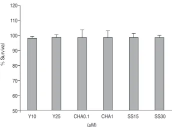
Treatment with each drug tested did not significantly in- hibit the survival of TM cells (p > 0.05) (Fig. 1). Thus, the drugs did not affect cell survival and proliferation.
Effects of trabecular outflow drugs on the NO produc- tion and eNOS mRNA expression
After 24 hours of exposure, treatment with 25 mM Y-26732 or 1.0 mM CHA significantly increased the nitrite concentration in the media in a dose-dependent manner (p
= 0.035, p = 0.043) (Fig. 2). Treatment with each drug also significantly increased the expression of eNOS mRNA (p
< 0.05)(Fig. 3). Treatment with Y-26732 increased the eNOS mRNA expression to a level higher than that pro- duced by CHA or simvastatin.
Effects of trabecular outflow drugs on barrier function The permeability assay using a flux of carboxyfluoresce- in showed that treatment with each drug tested except 0.1 mM CHA increased the concentration of carboxyfluoresce- in in the outer well in a dose-dependent manner compared to non-exposed controls (p < 0.05) (Fig. 4). Treatment with 10 mM or 25 mM Y-26732 produced 12.74% and 21.25%
higher concentrations of carboxyfluorescein, respectively, in a dose-dependent fashion compared to treatment with the other drugs.
The TEER of the TM cell monolayers was significantly lower after adding 10 μM or 25 mM Y-27632, 1.0 mM CHA, or 30 mM simvastatin compared with that of controls (p <
0.05) (Fig. 5). Treatment with 25 mM Y-26732 produced a significantly greater 28.91 Ω cm2 decrease in TEER com- pared to treatment with the other drugs.
Effects of trabecular outflow drugs on the expression of MMP-2 mRNA
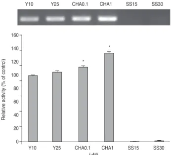
Treatment of the TM cells with 0.1 mM or 1.0 mM CHA resulted in an increase in the level of MMP-2 mRNA up to 11.98% or 32.61%, respectively (p = 0.012, p = 0.001) (Fig.
6). On the contrary, treatment of the TM cells with Y-27632 did not affect MMP-2 mRNA expression, while the addition of simvastatin inhibited its expression.
% Survival
(µM)
Y10 Y25 CHA0.1 CHA1 SS15 SS30
120 110 100 90 80 70 60 50
Fig. 1. Effects of trabecular outflow drugs on the survival of tra- becular meshwork cells. Treatment with each drug did not signifi- cantly affect survival compared to nonexposed controls (p > 0.05).
Y = Y-27632; CHA = N6-cyclohexyladenosine; SS = simvastatin.
Nittite (µM)
(µM)
Control Y10 Y25 CHA0.1 CHA1 SS15 SS30 10.0
8.0 6.0 4.0 2.0 0.0
⁎
†
Fig. 2. Effects of trabecular outflow drugs on the production of nitric oxide in trabecular meshwork cells after exposure for 24 hours. Treatment with 25 mM Y-27632 or 1.0 mM CHA signifi- cantly increased the production of nitric oxide in a dose-depen- dent manner. Y = Y-27632; CHA = N6-cyclohexyladenosine; SS
= simvastatin. *p = 0.035; †p = 0.043.
Discussion
The TM/SC complex is considered the conventional aqueous humor outflow pathway and is a primary site of increased resistance to aqueous humor outflow in glauco- ma. There is a significant need for glaucoma drugs that
specifically target the physiologic cause of elevated IOP and thereby enhance trabecular outflow. The most widely prescribed glaucoma drugs, prostaglandin analogues, low- er IOP by increasing the aqueous drainage through the un- conventional uveoscleral outflow pathway. The older, non-prostaglandin drug classes, b-blockers, a-agonists,
Relative activity (% of control)
(µM) Y10
Y10
Y25 Y25
CHA0.1 CHA0.1
CHA1 CHA1
SS15 SS15
SS30 SS30
350 300 250 200 150 100 50 0
⁎ ⁎
⁎
⁎ ⁎
⁎
Fig. 3. Effects of trabecular outflow drugs on the expression of endothelial nitric oxide synthase mRNA in trabecular meshwork cells. Treatment with each drug significantly increased the ex- pression of endothelial nitric oxide synthase mRNA (*p < 0.05).
b-actin was used as an internal standard. Y = Y-27632; CHA = N6-cyclohexyladenosine; SS = simvastatin.
Ω cm2
Control Y10 Y25 CHA0.1 CHA1 (µM) SS15 SS30 200
180 160 140 120 100 80 60
⁎
⁎
⁎ ⁎
Fig. 5. Effects of trabecular outflow drugs on the trans-endotheli- al electrical resistance of the trabecular meshwork cell monolayer.
Treatment with Y-27632, 1.0 mM CHA, or simvastatin significant- ly increased the resistance compared with nonexposed controls (*p < 0.05). Y = Y-27632; CHA = N6-cyclohexyladenosine; SS = simvastatin.
Relative activity (% of control)
Y10 (µM) Y10
Y25 Y25
CHA0.1 CHA0.1
CHA1 CHA1
SS15 SS15
SS30 SS30
160 140 120 100 80 60 40 20 0
⁎
⁎
Fig. 6. Effects of trabecular outflow drugs on the expressin of matrix metalloproteinases-2 (MMP-2) mRNA in trabecular meshwork cells. Treatment with 0.1 mM or 1.0 mM CHA signifi- cantly increased the expression of MMP-2 mRNA (*p < 0.05).
Simvastatain did not affect the expression of MMP-2. b-actin was used as an internal standard. Y = Y-27632; CHA = N6-cyclohex- yladenosine; SS = simvastatin.
% Permeability
(µM)
Y10 Y25 CHA0.1 CHA1 SS15 SS30
160 140 120 100 80 60
⁎ ⁎
⁎ ⁎
⁎
⁎
Fig. 4. Effects of trabecular outflow drugs on the permeability of carboxyfluorescein through the trabecular meshwork cell mono- layer. Treatment with 10 μM or 25 µM Y-27632, 1.0 µM CHA, or 15 mM or 30 mM simvastatin significantly increased the permea- bility of carboxyfluorescein compared with non-exposed controls (*p < 0.05). Carboxyfluorescein intensity of the outer chamber normalized to the mean value of non-exposed controls (permea- bility 100%). Y = Y-27632; CHA = N6-cyclohexyladenosine; SS = simvastatin.
and carbonic anhydrase inhibitors, lower IOP by decreas- ing the production of aqueous humor. Recently, several new classes of drugs targeting the trabecular outflow path- way have been introduced, and their diverse effects are under investigation [4].
Rho kinase has been suggested as a potential therapeutic target for the development of drugs to modulate IOP in glaucoma patients. Considering ROCK inhibitor-induced changes in MLC phosphorylation and actin-myosin orga- nization, it seems that cellular relaxation and loss of cell-substratum adhesions in TM and SC cells could result in either increased paracellular fluid flow across the SC or an altered flow pathway through the juxtacanalicular tis- sue, thereby lowering resistance to outflow [7,24,28].
The statin drugs induce changes in cell shape and actin cytoskeletal organization and decrease MLC phosphoryla- tion in TM cells, all of which are events that are likely to lead to cellular and tissue relaxation. In addition, these ef- fects of statins appear to be mediated by inhibition of iso- prenylation of the small GTP-binding proteins such as Rho-GTPase [11]. Compared to ROCK inhibitors or statin, adenosine increases trabecular outflow by inducing ECM change [10]. Adenosine A1 receptor exist in TM cells, and CHA modulates MMP-2 secretion in TM cells [22,29,30].
It is known that ROCK inhibitor, adenosine, and statin all increase trabecular outflow, although no comparative study of their effects on the increase in trabecular outflow had been reported. Thus, this study was performed to compare their effects on in vitro trabecular outflow by measuring the permeability and resistance of TM cell monolayers. The results showed that treatment with each drug tested in the present study increased permeability, except 0.1 mM CHA. Treatment with the ROCK inhibitor Y-26732 more greatly increased the permeability compared to adenosine or statin. Furthermore, treatment with 25 mM Y-26732 most significantly lowered the TEER of TM cell monolayer.
NO is involved in the regulation of trabecular outflow, and external administration of NO results in decreased IOP [31]. There has been evidence that treatment with ROCK inhibitor or statin increased eNOS expression [18,19], but their effects on the expression of eNOS in hu- man TM cells had not yet been investigated. Moreover, ad- enosine’s effects on the expression of eNOS and NO pro- duction were still not explored. In the present study, treatment with the ROCK inhibitor Y-26732 increased the
expression of eNOS and NO production more significantly than adenosine or statin. Thus, increased NO production by ROCK inhibitor might have an additive effect on in- creasing outflow.
Treatment with CHA significantly increased MMP-2 ex- pression. On the contrary, treatment with Y-27632 did not significantly increase MMP-2 expression, and treatment with simvastatin inhibited MMP-2 expression. Thus, it is likely that adenosine has another mechanism through which it increases trabecular outflow by remodeling of the ECM in addition to cellular morphological changes, as produced by ROCK inhibitor or statin. Treatment with sta- tin had no effect on MMP-2 expression.
ROCK inhibitors show multiple effects other than de- creasing IOP, such as antioxidant and anti-inflammatory activities [32,33]. Furthermore, ROCK inhibitors have an antifibrotic effect and decrease scar formation after trabe- culectomy [33].
Clinical trabecular outflow modulators based on preclin- ical efficacy models are currently being explored. Results of clinical trials of the ROCK inhibitor ripasudil (K-115) have been reported [34,35]. The neuroprotective effect of another ROCK inhibitor, fasudil, has been evaluated [36,37]. Among adenosine agonists, clinical trials on the favorable IOP-lowering effect of trabodenoson (INO-8875) has been reported recently [38]. These clinical studies re- vealed that ROCK inhibitors showed a stronger IOP-lower- ing effect than adenosine or statin, in agreement with our present in vitro study. However, there are limitations of these newly proposed mechanisms. For example, the inci- dence of ocular hyperemia with ROCK inhibitors is higher than that with latanoprost, while the magnitude of IOP lowering is not superior. Therefore, the risk to benefit ratio for patients seems unfavorable, and we must continue to identify new mechanisms through which to lower IOP [39].
In conclusion, ROCK inhibitor more highly increased the permeability and decreased the resistance of a TM cell monolayer than adenosine or statin. In addition, increased expression of eNOS mRNA and NO production by ROCK inhibitor might result in further IOP reduction. Thus, tra- becular outflow might be more enhanced by ROCK inhibi- tor than by adenosine or statin. In the future, more clinical studies will be needed to explore and compare IOP-lowing effects in humans.
Conflict of Interest
No potential conflict of interest relevant to this article was reported.
Acknowledgements
This work was supported by the 2015 Cheil-Nammyung Foundation Research Fund.
References
1. Alvarado J, Murphy C, Juster R. Trabecular meshwork cel- lularity in primary open-angle glaucoma and nonglauco- matous normals. Ophthalmology 1984;91:564-79.
2. Rohen JW, Lutjen-Drecoll E, Flugel C, et al. Ultrastructure of the trabecular meshwork in untreated cases of primary open-angle glaucoma (POAG). Exp Eye Res 1993;56:683- 92.
3. Schmidl D, Schmetterer L, Garhofer G, Popa-Cherecheanu A. Pharmacotherapy of glaucoma. J Ocul Pharmacol Ther 2015;31:63-77.
4. Kopczynski CC, Epstein DL. Emerging trabecular outflow drugs. J Ocul Pharmacol Ther 2014;30:85-7.
5. Inoue T, Tanihara H. Rho-associated kinase inhibitors: a novel glaucoma therapy. Prog Retin Eye Res 2013;37:1-12.
6. Thieme H, Nuskovski M, Nass JU, et al. Mediation of cal- cium-independent contraction in trabecular meshwork through protein kinase C and rho-A. Invest Ophthalmol Vis Sci 2000;41:4240-6.
7. Rao PV, Deng PF, Kumar J, Epstein DL. Modulation of aqueous humor outflow facility by the Rho kinase-specific inhibitor Y-27632. Invest Ophthalmol Vis Sci 2001;42:1029- 37.
8. Honjo M, Tanihara H, Inatani M, et al. Effects of rho-asso- ciated protein kinase inhibitor Y-27632 on intraocular pres- sure and outf low facility. Invest Ophthalmol Vis Sci 2001;42:137-44.
9. Novack GD. Rho kinase inhibitors for the treatment of glaucoma. Drugs Future 2013;38:107-13.
10. Zhong Y, Yang Z, Huang WC, Luo X. Adenosine, adenos- ine receptors and glaucoma: an updated overview. Biochim Biophys Acta 2013;1830:2882-90.
11. Song J, Deng PF, Stinnett SS, et al. Effects of cholester-
ol-lowering statins on the aqueous humor outflow pathway.
Invest Ophthalmol Vis Sci 2005;46:2424-32.
12. Stein JD, Newman-Casey PA, Talwar N, et al. The relation- ship between statin use and open-angle glaucoma. Oph- thalmology 2012;119:2074-81.
13. Wiederholt M, Dorschner N, Groth J. Effect of diuretics, channel modulators and signal interceptors on contractility of t he t rabecula r meshwork. O phthalmologica 1997;211:153-60.
14. Wiederholt M, Stumpff F. The trabecular meshwork and aqueous humor reabsorption. In: Civan MM, editor. Cur- rent topics in membranes. Vol. 45. The eye's aqueous hu- mor: from secretion to glaucoma. San Diego: Academic Press; 1998. p. 163-202.
15. Wiederholt M, Sturm A, Lepple-Wienhues A. Relaxation of trabecular meshwork and ciliary muscle by release of ni- tric oxide. Invest Ophthalmol Vis Sci 1994;35:2515-20.
16. Behar-Cohen FF, Goureau O, D’Hermies F, Courtois Y.
Decreased intraocular pressure induced by nitric oxide do- nors is correlated to nitrite production in the rabbit eye. In- vest Ophthalmol Vis Sci 1996;37:1711-5.
17. Dismuke WM, Mbadugha CC, Ellis DZ. NO-induced reg- ulation of human trabecular meshwork cell volume and aqueous humor outflow facility involve the BKCa ion channel. Am J Physiol Cell Physiol 2008;294:C1378-86.
18. Rikitake Y, Liao JK. Rho GTPases, statins, and nitric ox- ide. Circ Res 2005;97:1232-5.
19. Noma K, Oyama N, Liao JK. Physiological role of ROCKs in the cardiovascular system. Am J Physiol Cell Physiol 2006;290:C661-8.
20. Bradley JM, Vranka J, Colvis CM, et al. Effect of matrix metalloproteinases activity on outflow in perfused human organ culture. Invest Ophthalmol Vis Sci 1998;39:2649-58.
21. Sanka K, Maddala R, Epstein DL, Rao PV. Influence of ac- tin cytoskeletal integrity on matrix metalloproteinase-2 ac- tivation in cultured human trabecular meshwork cells. In- vest Ophthalmol Vis Sci 2007;48:2105-14.
22. Shearer TW, Crosson CE. Adenosine A1 receptor modula- tion of MMP-2 secretion by trabecular meshwork cells. In- vest Ophthalmol Vis Sci 2002;43:3016-20.
23. Mosmann T. Rapid colorimetric assay for cellular growth and survival: application to proliferation and cytotoxicity assays. J Immunol Methods 1983;65:55-63.
24. Kameda T, Inoue T, Inatani M, et al. The effect of Rho-asso- ciated protein kinase inhibitor on monkey Schlemm’s canal endothelial cells. Invest Ophthalmol Vis Sci 2012;53:3092-
103.
25. Alvarado JA, Betanzos A, Franse-Carman L, et al. Endo- thelia of Schlemm’s canal and trabecular meshwork: dis- tinct molecular, functional, and anatomic features. Am J Physiol Cell Physiol 2004;286:C621-34.
26. Green LC, Wagner DA, Glogowski J, et al. Analysis of ni- trate, nitrite, and [15N]nitrate in biological fluids. Anal Bio- chem 1982;126:131-8.
27. Alvarado JA, Wood I, Polansky JR. Human trabecular cells. II. Growth pattern and ultrastructural characteristics.
Invest Ophthalmol Vis Sci 1982;23:464-78.
28. Zhang M, Maddala R, Rao PV. Novel molecular insights into RhoA GTPase-induced resistance to aqueous humor outflow through the trabecular meshwork. Am J Physiol Cell Physiol 2008;295:C1057-70.
29. Husain S, Shearer TW, Crosson CE. Mechanisms linking adenosine A1 receptors and extracellular signal-regulated kinase 1/2 activation in human trabecular meshwork cells.
J Pharmacol Exp Ther 2007;320:258-65.
30. Fleischhauer JC, Mitchell CH, Stamer WD, et al. Common actions of adenosine receptor agonists in modulating hu- man trabecular meshwork cell transport. J Membr Biol 2003;193:121-36.
31. Kim HY, Kim JW. Effect of nitric oxide on the permeabili- ty of trabecular meshwork cell monolayer. J Korean Oph- thalmol Soc 2015;56:771-5.
32. Satoh K, Fukumoto Y, Shimokawa H. Rho-kinase: import- ant new therapeutic target in cardiovascular diseases. Am J Physiol Heart Circ Physiol 2011;301:H287-96.
33. Honjo M, Tanihara H, Kameda T, et al. Potential role of Rho-associated protein kinase inhibitor Y-27632 in glaucoma filtration surgery. Invest Ophthalmol Vis Sci 2007;48:5549-57.
34. Tanihara H, Inoue T, Yamamoto T, et al. Intra-ocular pres- sure-lowering effects of a Rho kinase inhibitor, ripasudil (K-115), over 24 hours in primary open-angle glaucoma and ocular hypertension: a randomized, open-label, cross- over study. Acta Ophthalmol 2015;93:e254-60.
35. Kaneko Y, Ohta M, Inoue T, et al. Effects of K-115 (Ripa- sudil), a novel ROCK inhibitor, on trabecular meshwork and Schlemm’s canal endothelial cells. Sci Rep 2016;6:19640.
36. Ichikawa M, Yoshida J, Saito K, et al. Differential effects of two ROCK inhibitors, Fasudil and Y-27632, on optic nerve regeneration in adult cats. Brain Res 2008;1201:23-33.
37. Hata Y, Miura M, Nakao S, et al. Antiangiogenic proper- ties of fasudil, a potent Rho-kinase inhibitor. Jpn J Oph- thalmol 2008;52:16-23.
38. Novack GD. Eyes on new product development. J Ocul Pharmacol Ther 2016;32:401-2.
39. Prasanna G, Li B, Mogi M, Rice DS. Pharmacology of novel intraocular pressure-lowering targets that enhance conventional outflow facility: pitfalls, promises and what lies ahead? Eur J Pharmacol 2016;787:47-56.