pISSN: 0378-6471 eISSN: 2092-9374 http://dx.doi.org/10.3341/jkos.2011.52.9.1089
= 증례보고 =
동공 산동제와 축동제가 섬유주세포에 미치는 영향
이정훈⋅김재우
대구가톨릭대학교 의과대학 안과학교실
목적: 임상에서 사용되는 동공 산동제와 축동제가 배양된 섬유주세포의 생존과 일산화질소의 생성에 미치는 영향을 알아보고자 하였다.
대상과 방법: 섬유주세포를 일차배양한 후 트로피카미드, 싸이클로펜톨레이트, 아트로핀, 필로카핀에 0, 0.01, 0.1, 1.0 mg/ml의 농도로 2시간 동안 각각 노출시킨 후 MTT assay와 Griess assay를 이용하여 세포의 생존율과 일산화질소의 생성량을 각각 측정하였다.
결과: 트로피카미드, 싸이클로펜톨레이트, 아트로핀, 필로카핀 모두 0.1 mg/ml의 농도에서부터 섬유주세포의 생존을 저하시켰다. 0.01 mg/ml의 농도에서 트로피카미드, 싸이클로펜톨레이트, 아트로핀은 일산화질소의 생성을 증가시키고 필로카핀은 일산화질소의 생성을 저하시켰으나 통계적으로 유의하지는 않았다.
결론: 임상적으로 사용되는 동공 산동제와 축동제는 고농도 또는 단기간에 자주 점안할 경우 사용할 경우 섬유주세포의 생존을 저하시 킬 수 있으나 일산화질소의 생성에는 영향을 주지 않는 것으로 보아 단시간에 섬유주를 통한 방수유출에는 영향을 끼치지 않을 것으로 생각한다.
<대한안과학회지 2011;52(9):1089-1093>
■ 접 수 일: 2011년 2월 17일 ■ 심사통과일: 2011년 4월 19일
■ 게재허가일: 2011년 7월 12일
■ 책 임 저 자: 김 재 우
대구시 남구 대명 4동 3056-6 대구가톨릭대학교병원 안과
Tel: 053-650-4728, Fax: 053-627-0133 E-mail: [email protected]
안과영역에서 임상적인 검사를 위해 트로피카미드, 싸이 클로펜톨레이트 같은 산동제를 사용하여 동공을 확대 또는 마비시키거나 치료목적으로 아트로핀 점안제를 사용하고 있 으며, 검사나 치료목적으로 필로카핀 같은 축동제를 흔히 사 용하고 있는데 대부분의 경우 이러한 약제는 안전한 것으로 알려져 있으나 안압 상승을 비롯한 여러 부작용을 야기할 수 도 있으며1,2산동제를 사용했을 때 나타나는 안압상승의 정도 가 녹내장의 진행을 예측할 수 있는 인자라는 보고도 있다.3 섬유주를 통한 방수유출을 조절함에 있어서 자유유리기인 일산화질소(nitric oxide, NO)가4-7중요한 역할을 하며 섬유 주세포에서도 NO합성효소가 발현될 뿐만 아니라8-10녹내장 이 있는 경우에는 NO합성효소의 활성이 감소되어 있는 것으 로 알려져 있다.11-14NO는 섬유주를 이완시켜 섬유주를 통한 방수유출을 촉진시키는 작용을 나타내므로15-17 이러한 약제 가 NO의 생성을 감소시키거나 섬유주세포의 기능을 약화시 킨다면 일부에서 부작용으로 나타나는 안압 상승의 원인이 될 수 있을 것이며, 이런 약제를 단기간 자주 점안하거나 고 농도로 사용한다면 부작용이 더 흔하게 나타날 것이다.
그러나 이런 약제가 섬유주의 생존에 미치는 영향은 아
직 알려져 있지 않으며, NO의 생성에 미치는 영향도 알려 져 있지 않다.
본 연구에서는 인체의 섬유주세포를 일차배양하여 동공 산동제와 축동제에 노출시켜 섬유주세포의 생존과 NO의 생성에 미치는 영향에 대해 알아보고자 하였다.
대상과 방법
세포배양
안구은행에서 얻은 사후 6시간 이내에 적출한 안구의 앞 방각 주위 조직을 제거한 후 앞방각에서 섬유주를 벗겨내 어 폴리라이신(Sigma, St. Louis, MO, USA)로 처리한 배 양접시에 옮긴 후 항생제(Gibco, Carlsbad, CA, USA)와 10% 우태아혈청(Gibco)이 포함된 Dulbecco’s modified Eagle’s medium 배지(DMEM, Gibco)를 사용하여 5% CO2
배양기에서 초대배양하였다. 섬유주세포가 이식된 조직편 주위로 자라나온 것을 확인한 후 섬유주조직의 이식편을 제거하고 배양을 계속하였으며 세포가 배양접시에 충만해 지면 10% 우태아혈청을 포함한 배지로 1:3의 비율로 트립 신 처리하여 계대배양하였다.
약물처리
일차배양한 사람의 섬유주세포를 24 well 배양접시에 분
0 20 40 60 80 100 120
0.01 0.1 1
concentration (mg/ml)
*
*
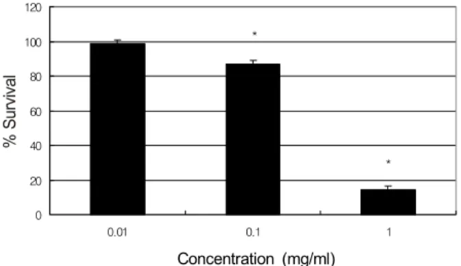
Concentration (mg/ml)
Figure 1. Effects of tropicamide on the survival of trabecular
meshwork cells. Tropicamide decreased cellular survival. *p < 0.05.0 20 40 60 80 100 120
0.01 0.1 1
*
*
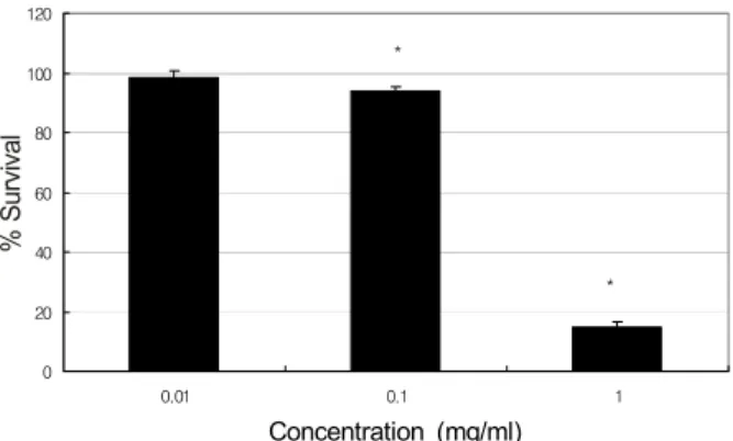
Concentration (mg/ml)
Figure 2. Effects of cyclopentolate on the survival of trabecular mesh-
work cells. Cyclopentolate decreased cellular survival. *p < 0.05.0 20 40 60 80 100 120
0.01 0.1 1
*
*
Concentration (mg/ml)
Figure 3. Effects of atropine on the survival of trabecular mesh-
work cells. Atropine decreased cellular survival. *p < 0.05.주한 후 24시간 동안 배양기에 넣어 세포를 부착시킨 후 배 지를 제거하고 나서 혈청 단백질에 의한 항산화효과를 배 제하기 위하여 1%의 저농도 혈청과 5 mM의 저농도 포도 당이 포함된 DMEM배지로 교환하였다. 각 1%의 트로피카 미드, 싸이클로펜톨레이트, 필로카핀과 아트로핀 점안제 (Alcon, Fort Worth, Texas, USA)를 1%의 저농도 혈청이 포함된 DMEM 배지로 0, 0.01, 0.1, 1.0 mg/ml의 농도로 단계적으로 희석하여 2시간 동안 노출시켰다.
MTT assay와 Griess assay
세포의 생존에 대한 효과는 세포생존과 세포독성의 선별 검사로 흔히 이용되고 있는 발색검사의 일종인 MTT (3-[4, 5–dimethylthiazol-2-yl]-2, 5-diphenyltetrazolium bromide, Sigma) assay를18 이용하였고 NO의 생성은 Griess assay 를19이용하였다. MTT assay는 약물처리한 세포의 배지에 MTT를 각 well당 100 μl씩 투여한 후 4시간 동안 정치배 양한 다음 염류용액으로 씻어낸 후 dimethylsulfoxide (Sigma)를 각 well당 0.5 ml씩 넣어 10분 이상 흔든 다음 96-well 배양접시에 200 μl씩 옮겨 분광광도계(FLUOstar OPTIMA, BMG labtech, Offenburg, Germany)로 570 nm 에서 흡광도를 측정하였다. 이 때 세포의 생존정도는 실험 군의 값을 약물처리를 하지 않은 대조군의 비로 나누어 백 분율로 나타내었다. Griess assay는 0.01 mg/ml의 농도로 2시간 동안 각각의 약물을 처리한 세포의 배지에 동량의 Griess 반응액(Sigma)을 섞은 후 96-well 배양접시에 옮 겨 NO생성의 반응물인 아질산염의 양을 분광광도계로 540 nm에서 흡광도를 측정하였다. 이 때 표준치를 구하기 위해 sodium nitrite (Sigma)를 단계적으로 희석하여 사용하였다.
통계적 처리
모든 실험은 3계대에서 5계대 사이의 세포를 이용하였 다. 모든 실험에서 대조군은 약물처리를 하지 않은 군으로 하였으며, 실험군과 대조군의 비교는 unpaired t-test를 사 용하였으며 유의수준은 0.05%로 정하였다.
결 과
세포배양
초대배양 7일째부터 섬유주조직의 이식편 주위로 섬유주 세포가 자라 나오기 시작하였으며 섬유주세포의 확인은 세 포들이 밀집해서 단층을 형성하며 세포들 사이에 분지를
내어 서로 연접하며 약간 길다란 모양의 세포체를 가지는 편평한 모양의 특징적 형태학적인 양상과 섬유주 조직의 이식편 주위에서 위성양상으로 자라나는 섬유주세포의 특 징적인 성장양상으로 확인하였다.20,21
산동제와 축동제가 섬유주세포의 생존에 미치는 영향 산동제인 트로피카미드와 싸이클로펜톨레이트, 아트로핀 은 0.1 mg/ml의 농도에서부터 모두 유의하게 섬유주세포의
0 20 40 60 80 100 120
0.01 0.1 1
*
*
Concentration (mg/ml)
Figure 4. Effects of pilocarpine on the survival of trabecular
meshwork cells. Pilocarpine decreased cellular survival. *p < 0.05.80 85 90 95 100 105 110
Tropicamide Cyclopentolate Atropine Pilocarpine
Figure 5. Effects of 0.01 mg/ml tropicamide, cyclopentolate,
atropine, and pilocarpine on the production of nitric oxide in trabecular meshwork cells. All agents did not affect the pro- duction of nitric oxide significantly compared to the non-ex- posed control. p > 0.05.생존을 저하시키기 시작하여 1.0 mg/ml의 농도에서는 생존 을 급격하게 감소시켰다(p<0.05) (Fig. 1, 2, 3). 이와 마 찬가지로 축동제인 필로카핀도 0.1 mg/ml의 농도에서는 세 포의 생존을 감소시키기 시작하여 1.0 mg/ml의 농도에서는 섬 유주 세포의 생존을 급격하게 감소시켰다(p<0.05) (Fig. 4).
산동제와 축동제가 섬유주세포의 NO의 생성에 미치는 영향
위의 생존실험을 근거로 하여 섬유주세포의 생존에 영향 을 미치지 않는 0.01 mg/ml의 농도를이용하여 각 약물을 의한 NO의 생성 정도를 조사하였다. 그 결과 트로피카미드 와 싸이클로펜톨레이트, 그리고 아트로핀은 NO의 생성을 증가시켰고, 필로카핀은 NO의 생성을 감소시켰다(Fig. 5).
그러나 산동제는 NO의 생성을 증가시키고 축동제는 NO의 생성을 저하시키는 경향을 나타내기는 하였으나 약물을 처 리하지 않은 대조군에 비해 통계적으로 유의한 차이를 나 타내지는 않았다(p>0.05).
고 찰
본 연구의 결과는 동공 산동제와 축동제가 섬유주세포의 생존에 유의한 영향을 미칠 수 있으나 NO의 생성에는 유의 한 영향을 미치지 않는다는 것을 보여주고 있다.
항콜린계제재인 트로피카미드와 싸이클로펜톨레이트, 그 리고 아트로핀은 동공 확장 및 마비를 통한 검사와 치료용 점안약으로 흔히 사용되고 있는데 일부에서는 이러한 동공 확장제가 안압 상승 등의 부작용을 유발할 수 있다고 보고 되어 있다.1-3본 연구의 결과에서 항콜린계 약물은 섬유주 세포의 생존을 저하시켰는데 이러한 생존저하는 고농도로 사용할수록 더욱 뚜렷이 나타났다.
녹내장 약제가 섬유주세포에 독성을 나타낼 수 있으나22 필로카핀을 점안할 경우 방수 내 농도는 점안 농도의 0.1%
정도가 되므로23 실제 점안할 경우 본 실험에 사용된 필로 카핀의 최대 농도인 1 mg/ml가 섬유주세포에 작용할 것으 로 생각한다. 콜린계 동공축동제인 필로카핀의 경우에도 역 시 섬유주세포의 생존을 저하시키는 것으로 나타났다. 임상 적으로 동공확장제나 동공수축제를 사용할 경우 단기간에 너무 자주 점안하거나 고농도로 사용할 경우 섬유주세포의 생존에 영향을 미치며 그 결과 섬유주의 기능을 약화시킬 수 있으므로 주의해야 할 것이다.
본 연구에서는 보존제가 포함된 점안제를 단계적으로 희 석하여 실험을 시행하였으므로 보존제인 benzalkonium chloride가 세포의 생존에 영향을 미칠 가능성도 생각해 볼 수 있다. 보존제는 대개 각막에 고농도로 침착되는 데 반해 섬유주세포에서는 농도가 저하될 것으로 생각하며 0.005%
의 농도에서는 섬유주세포의 생존에 영향을 미치지 않는다 는 보고도 있다.24비록 본 실험에서 보존제 단독으로 사용 했을 때 섬유주세포의 생존에 미치는 영향은 조사하지 않 았으나 본 실험에 사용된 보존제의 최대 농도는 0.001%이 므로 약제에 포함된 보조제가 세포의 생존에 미치는 영향 은 크지 않을 것으로 생각한다. 또한 보존제는 점안제의 각 막투과성을 높이는 효과를 나타내므로 실제 점안제의 방수 내 농도는 실험에 사용한 농도보다 높게 나타날 수 있다.23
NO의 생성저하에 의해 섬유주가 수축함으로써 섬유주가 수축되어 안압이 상승할 수 있는데11-13산동제의 부작용으 로 나타나는 안압상승의 원인의 하나로 NO의 생성이 저하 되어 유발될 가능성을 생각해 볼 수 있다. 섬유주세포의 생 존을 저하시키는 원인으로 약제의 직접적인 독성을22비롯 해서 다양한 원인이 있겠으나 섬유주세포의 생존 저하가 NO의 생성에 영향을 받을 수 있으므로 이를 배제하기 위해 본 연구에서는 섬유주세포의 생존에 영향을 미치지 않는 저농도에서 NO의 생성에 미치는 영향을 조사하였다. 그 결
과 비록 산동제는 NO의 생성증가를 나타내었고, 축동제는 NO의 생성 저하를 나타내기는 하였으나 NO의 생성에 통계 적으로 유의한 영향을 미치지는 않았다. 이러한 결과는 산 동제의 단기간 사용에 의한 안압상승이 섬유주세포의 생존 이나 NO의 생성과는 관련이 없으며 다른 해부학적 또는 기 능적 원인에 의해 안압 상승이 유발될 것이라는 점을 시사 한다. 그러나 비록 저농도인 경우에도 장기간 사용할 경우 섬유주의 기능 저하나 섬유주세포의 노화를 야기할 가능성 도 있으므로25,26장기간의 사용효과에 대해서는 좀 더 자세 한 연구가 필요할 것이다.
결론적으로 실험실 내 세포배양의 조건에서 산동제와 축 동제는 고농도에서는 모두 섬유주세포의 생존을 저하시켰 으므로 임상적으로 이러한 약제를 너무 자주 점안하거나 고농도로 사용하지 않도록 주의해야 한다. 그리고 산동제에 의해 안압상승이 유발되는 경우는 NO의 생성저하와는 무 관할 것으로 생각하며 그 기전에 대해서는 향후 더 자세한 연구가 필요할 것으로 생각한다.
참고문헌
1) Abraham SV. Mydriatic glaucoma: a statistical study. Arch Ophthalmol 1933;10:757-62.
2) Nelson ME, Orton HP. Counteracting the effects of mydriatics:
Does it benefit the patient? Arch Ophthalmol 1987;105:486-9.
3) Siam GA, de Barros DS, Gheith ME, et al. The amount of intra- ocular pressure rise during pharmacological pupillary dilatation is an indicator of the likelihood of future progression of glaucoma. Br J Ophthalmol 2007;91:1170-2.
4) Wiederholt M. Direct involvement of trabecular meshwork in the regulation of aqueous humor outflow. Curr Opin Ophthalmol 1998;9:46-9.
5) Moncada S, Palmer RM, Higgs EA. Nitric oxide: physiology, path- ophysiology, and pharmacology. Pharmacol Rev 1991;43:109-42.
6) Bredt DS, Snyder SH. Nitric oxide: a physiologic messenger molecule. Annu Rev Biochem 1994;63:175-95.
7) Brüne B, Knethen A, Sandau KB. Nitric oxide and its role in apoptosis. Eur J Pharmacol 1998;351:261-72.
8) Nathanson JA, McKee M. Identification of an extensive system of nitric oxide-producing cells in the ciliary muscle and outflow path- way of the human eye. Invest Ophthalmol Vis Sci 1995;36:1765-73.
9) Geyer O, Podos SM, Mittag T. Nitric oxide synthase activity in tis- sues of the bovine eye. Graefes Arch Clin Exp Ophthalmol 1997;
235:786-93.
10) Meyer P, Champion C, Schlotzer-Schrehardt U, et al. Localization of nitric oxide synthase isoforms in porcine ocular tissues. Curr
Eye Res 1999;18:375-80.
11) Schuman JS, Erickson K, Nathanson JA. Nitrovasodilator effects on intraocular pressure and outflow facility in monkeys. Exp Eye Res 1994;58:99-105.
12) Wang RF, Podos SM. Effect of the topical application of nitro- glycerin on intraocular pressure in normal and glaucomatous monkeys. Exp Eye Res 1995;60:337-9.
13) Nathanson JA, McKee M. Alterations of ocular nitric oxide syn- thase in human glaucoma. Invest Ophthalmol Vis Sci 1995;36:
1774-84.
14) Matsuo T. Basic nitric oxide production is enhanced by hydraulic pressure in cultured human trabecular cells. Br J Ophthalmol 2000;84:631-5.
15) Schneemann A, Dijkstra BG, van den Berg TJ, et al. Nitric ox- ide/guanylate cyclase pathways and flow in anterior segment perfusion. Graefes Arch Clin Exp Ophthalmol 2002;240:936-41.
16) Galassi F, Renieri G, Sodi A, et al. Nitric oxide proxies and ocular perfusion pressure in primary open angle glaucoma. Br J Ophthalmol 2004;88:757-60.
17) Ellis DZ, Dismuke WM, Chokshi BM. Characterization of soluble guanylate cyclase in NO-induced increases in aqueous humor out- flow facility and in the trabecular meshwork. Invest Ophthalmol Vis Sci 2009;50:1808-13.
18) Mosmann T. Rapid colorimetric assay for cellular growth and sur- vival: Application to proliferation and cytotoxicity assays. J Immunol Methods 1983;65:55-63.
19) Green LC, Wagner DA, Glogowski J, et al. Analysis of nitrate, ni- trite and [15N]nitrate in biological fluids. Anal Biochem 1982;
126:131-8.
20) Polansky JR, Weinreb RN, Baxter JD, Alvarado J. Human tra- becular cells. I. Establishment in tissue culture and growth characteristics. Invest Ophthalmol Vis Sci 1979;18:1043-9.
21) Alvarado JA, Wood I, Polansky JR. Human trabecular cells. II.
Growth pattern and ultrastructural characteristics. Invest Ophthalmol Vis Sci 1982;23:464-78.
22) Kawa JE, Higginbotham EJ, Chang IL, Yue BY. Effects of anti- glaucoma medications on bovine trabecular meshwork cells in vitro. Exp Eye Res 1993;57:557-65.
23) Green K, Downs SJ. Ocular penetration of pilocarpine in rabbits.
Arch Ophthalmol 1975;93:1165-8.
24) Yu AL, Fuchshofer R, Kampik A, Welge-Lűssen U. Effects of oxi- dative stress in trabecular meshwork cells are reduced by prosta- glandin analogues. Invest Ophthalmol Vis Sci 2008;49:4872-80.
25) Vasa M, Breitschopf K, Zeiher AM, Dimmeler S. Nitric oxide acti- vates telomerase and delays endothelial cell senescence. Circ Res 2000;87:540-2.
26) Scalera F, Borlak J, Beckmann B, et al. Endogenous nitric oxide synthesis inhibitor asymmetric dimethyl L-arginine accelerates en- dothelial cell senescence. Arterioscler Thromb Vasc Biol 2004;24:
1816-22.
=ABSTRACT=
Effects of Pupil Dilation and Constriction Agents on Trabecular Meshwork Cells
Jeong Hun Lee, MD, Jae Woo Kim, MD, PhD
Department of Ophthalmology, Catholic University of Daegu College of Medicine, Daegu, Korea
Purpose: To investigate the effects of pupil dilation and constriction agents on the survival and production of nitric oxide (NO) in cultured human trabecular meshwork cells (HTMC).
Methods: Primarily cultured HTMC were exposed to 0, 0.01, and 0.1 mg/ml of tropicamide, cyclopentolate, atropine, or pilocarpine for 2 hours. Cellular survival and production of NO were assessed using the MTT assay and Griess assay, respectively.
Results: Tropicamide, cyclopentolate, atropine, and pilocarpine decreased cellular survival at the concentration of 0.1 mg.
At the concentration of 0.01 mg/ml, all agents decreased production of NO to some extent, although the reduction was not statistically significant.
Conclusions: Pupil dilation and constriction agents may be toxic to HTMC if used at high concentrations or if used fre- quently in the short-term but may not affect trabecular outflow.
J Korean Ophthalmol Soc 2011;52(9):1089-1093
Key Words: Atropine, Cyclopentolate, Pilocarpine, Trabecular meshwork cells, Tropicamide
Address reprint requests to Jae Woo Kim, MD, PhD
Department of Ophthalmology, Daegu Catholic University Medical Center
#3056-6 Daemyeong 4-dong, Nam-gu, Daegu 705-718, Korea Tel: 82-53-650-4728, Fax: 82-53-627-0133, E-mail: [email protected]