© 2019 The Korean Ophthalmological Society
This is an Open Access article distributed under the terms of the Creative Commons Attribution Non-Commercial License (http://creativecommons.org/licenses /by-nc/3.0/) which permits unrestricted non-commercial use, distribution, and reproduction in any medium, provided the original work is properly cited.
Original Article
Refractive Outcomes of 4-Year-old Children after Intravitreal Anti- vascular Endothelial Growth Factor versus Laser Photocoagulation for
Retinopathy of Prematurity
Hyun Goo Kang1, Tae Young Kim1, Jinu Han1, Sueng-Han Han2
1Department of Ophthalmology, Institute of Vision Research, Gangnam Severance Hospital, Yonsei University College of Medicine, Seoul, Korea
2Department of Ophthalmology, Institute of Vision Research, Severance Hospital, Yonsei University College of Medicine, Seoul, Korea
Purpose: To compare long-term refractive outcomes associated with intravitreal anti-vascular endothelial growth factor (VEGF) versus laser photocoagulation treatment for retinopathy of prematurity (ROP).
Methods: A total of 52 eyes from 27 ROP patients treated at two tertiary referral-based hospitals from August 2006 to December 2013 were reviewed. The primary outcome was refractive error measured at the age of 4 years, accounting for within-patient inter-eye correlation. Secondary outcomes included the recurrence rate and treatment complications.
Results: The mean age at refraction was 4.7 ± 0.3 years in the laser group (n = 30) and 4.4 ± 0.3 years in the an- ti-VEGF group (n = 22). No significant differences were noted in gestational age, birthweight, post-menstrual age at treatment, or ROP stage/zone distribution between groups. Mean spherical equivalent was also not significant- ly different (-1.0 diopters in the laser group and -0.3 diopters in the injection group, p = 0.603). Clustered regres- sion analysis revealed that only gestational age was significantly correlated with mean spherical equivalent (p <
0.001; 95% confidence interval, -0.007 to -0.002). Recurrence was noted in four eyes (13.3%) in the laser group, but this difference was not significant (p = 0.128). There were no major systemic complications reported in either group.
Conclusions: Treatment type, whether laser or anti-VEGF injection, does not appear to influence long-term re- fractive outcomes in ROP. Concern regarding refractive outcomes should not be the most important factor when selecting ROP treatment modality.
Key Words: Intravitreal injections, Laser therapy, Refractive errors, Retinopathy of prematurity, Vascular endotheli- al growth factor A
Retinopathy of prematurity (ROP) is a proliferative reti- nal vascular disease that develops in premature infants [1,2]. The incidence of childhood blindness due to ROP ranges from 3% to 10% worldwide [3-6]. Normal retinal vascular growth and development are disrupted by fetal environmental changes. Physiological hyperoxia causes
Received: February 7, 2019 Final revision: February 18, 2019 Accepted: February 27, 2019
Corresponding Author: Sueng-Han Han, MD. Department of Oph- thalmology, Institute of Vision Research, Severance Hospital, Yon- sei University College of Medicine, 50-1 Yonsei-ro, Seodaemun-gu, Seoul 03722, Korea. Tel: 82-2-2228-3570, Fax: 82-2-312-0541, E-mail:
vascular endothelial growth factor (VEGF)-mediated ces- sation of retinal vascular development [2,7]. The release of VEGF also causes vitreoretinal neovascularization, which leads to retinal traction, detachment, and hemorrhage, with profound loss of visual function [8-10].
Early detection and appropriately-timed intervention are critical to treatment and long-term outcomes of this dis- ease [11-13]. Current treatment strategies include early treatment with laser photocoagulation based on the 2004 Early Treatment for Retinopathy of Prematurity study [14], or with intravitreal anti-VEGF injections, based on groundbreaking studies such as the 2011 Bevacizumab Eliminates the Angiogenic Threat ROP study [12], either alone or in combination [15,16]. Although there are many benefits to anti-VEGF therapy, several studies have found temporary systemic suppression of VEGF and other growth factors after intravitreal injection in neonatal in- fants [17,18], triggering concern regarding potential neuro- logical or developmental delays [19].
One particularly concerning long-term consequence of ROP treatment is the elevated rate of myopia and high my- opia in these infants, especially with the already high prev- alence in Korean and Japanese populations [20]. Although several studies have noted greater prevalence and severity of myopia in laser-treated eyes over anti-VEGF, there are striking limitations due to the wide range of ages at refrac- tion and variation in treatment zone, which were not al- ways taken into account [21,22].
Therefore, in this study we examined the long-term re- fractive outcomes of infants treated for ROP by collecting a large number of cases and adjusting for age at the time of refraction in analysis. We also noted the rate of recurrence and the incidence of systemic complications.
Materials and Methods
This retrospective case study was performed at two ter- tiary referral-based hospitals, Severance Hospital and Gangnam Severance Hospital, which are affiliated with Yonsei University College of Medicine. Consecutive in- fants diagnosed with ROP and treated from August 2006 to December 2013 were included, and their medical charts were reviewed. This study was conducted with institution- al review board approval from Gangnam Severance Hospi- tal (3-2018-0050) and adhered to the tenets of the Declara-
tion of Helsinki. Informed consent was obtained from the parents or guardians of all subjects.
Infants were screened for ROP if they were born at ges- tational age <32 weeks and their birthweight was <1,500 g;
more mature and larger infants were examined if the clini- cal course was complex and/or unstable, as determined by the primary neonatologist. All ophthalmic examinations were performed by qualified ophthalmologists using the revised guidelines of the 2005 International Committee for the Classification of ROP to determine the stage and zone [23]. The indications for treatment were infants who met the criteria for type 1 ROP used in the Early Treatment for Retinopathy of Prematurity study [14], although earlier treatment was performed at the primary ophthalmologists’
discretion in cases involving signs of clinical pre-plus dis- ease, which was defined as abnormal vascular changes in- sufficient for the diagnosis of plus disease.
For laser photocoagulation, infants were treated under general anesthesia using an indirect diode laser system with a handheld aspheric lens using scleral depression. For anti-VEGF injection, either bevacizumab (0.625 mg/0.025 mL; off-label Avastin, Genentech, San Francisco, CA, USA) or ranibizumab (0.2 mg/0.02 mL; Lucentis, Novartis, Basel, Switzerland) [18,24] was injected into the diseased eye aseptically at the pars plana, 0.5 to 1 mm posterior to the limbus, using a sterile 30-gauge needle. The patients were re-examined the next day and then every week to monitor disease progression. Dilated fundus examinations were performed with indentation indirect ophthalmoscopy at every visit to confirm disease regression and vascular- ization up to the ora serrata.
All infants underwent regular follow-up visits and eye examination including initial cycloplegic retinoscopy or subsequent manifest refraction by certified ophthalmolo- gists. If hyperopia was noted at the initial cycloplegic re- fraction, subsequent examinations were done with cyclo- plegic refraction. The spherical and cylinder power, as well as the spherical equivalent (SE) are all noted in diopters (D). Patients who underwent vitrectomy or scleral buckling due to retinal detachment (five eyes from three patients), who underwent both laser and anti-VEGF injection (five eyes from three patients), or who lacked refraction data (54 eyes from 28 patients) were excluded.
The patients were grouped according to the type of treatment received: intravitreal anti-VEGF or laser photo- coagulation. The main outcome was long-term refractive
error measured at the age of 4 years. Secondary outcomes included recurrence rate and treatment complications.
Statistical analyses were performed using Stata ver. 13.1 (Stata Corp., College Station, TX, USA). Values are ex- pressed as mean ± standard deviation, with ranges given when appropriate. Chi-square test (Fisher exact test) and analysis of variance were used to compare the two treat- ment groups. A generalized estimating equation was used to compare refractive error between groups. Generalized estimating equation models that accounted for age at the time of refractive error measurement and within-patient inter-eye correlations were used to compare SE between the two treatment groups. Clustered regression analysis was used with each individual representing one cluster to consider similar characteristics between the right and left eyes in a single child and was performed to assess the im- pact of various factors on refractive outcomes. A p-value less than 0.05 was considered statistically significant.
Results
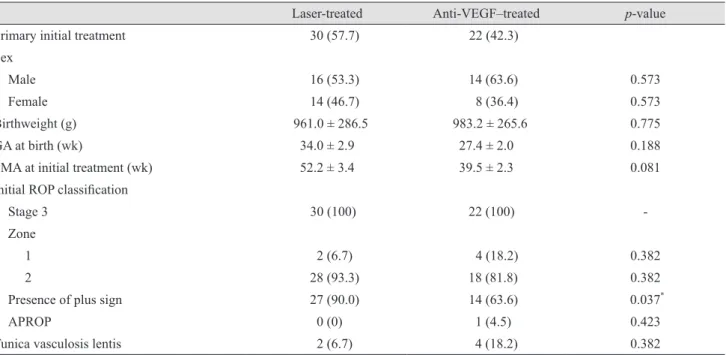
Refractions were available for 27 of 55 (49.1%) eligible patients. The baseline characteristics of each treatment group are summarized in Table 1. A total of 30 eyes (15 subjects) were treated with laser and 22 eyes (12 subjects) with anti-VEGF. In the anti-VEGF group, 20 (90.9%) eyes were treated with bevacizumab and 2 (9.1%) with ranibi- zumab. There were no significant differences between the two groups in sex ratio, gestational age, birthweight, post-menstrual age at treatment, and ROP stage/zone distri- bution. The mean age at refraction was 4.7 ± 0.3 years for the laser group and 4.4 ± 0.3 years for the anti-VEGF group.
Refractive outcomes
Refractive error grouped according to the treatment zone is summarized in Table 2. No significance was noted in mean SE between treatment groups (-1.0 D in the laser group vs. -0.3 D in the anti-VEGF group, p = 0.603). Simi- larly, no differences were found in mean spherical or cyl- inder power.
The distribution of refractive outcomes, categorized as
Table 1. Baseline patient characteristics
Laser-treated Anti-VEGF–treated p-value
Primary initial treatment 30 (57.7) 22 (42.3)
Sex
Male 16 (53.3) 14 (63.6) 0.573
Female 14 (46.7) 8 (36.4) 0.573
Birthweight (g) 961.0 ± 286.5 983.2 ± 265.6 0.775
GA at birth (wk) 34.0 ± 2.9 27.4 ± 2.0 0.188
PMA at initial treatment (wk) 52.2 ± 3.4 39.5 ± 2.3 0.081
Initial ROP classification
Stage 3 30 (100) 22 (100) -
Zone
1 2 (6.7) 4 (18.2) 0.382
2 28 (93.3) 18 (81.8) 0.382
Presence of plus sign 27 (90.0) 14 (63.6) 0.037*
APROP 0 (0) 1 (4.5) 0.423
Tunica vasculosis lentis 2 (6.7) 4 (18.2) 0.382
Values are presented as number (%) or mean ± standard deviation.
VEGF = vascular endothelial growth factor; GA = gestational age; PMA = post-menstrual age; ROP = retinopathy of prematurity; AP- ROP = aggressive posterior retinopathy of prematurity.
*p < 0.05.
high hyperopia to very high myopia, can be seen in Fig. 1.
These refractive categories were chosen based on previous studies [25,26]. There were no significant differences in the distribution between the two groups. When combining all myopia (≥-1 D), there were 11 eyes (36.7%) in the laser group and 8 (36.4%) in the injection group, which was not a significant difference (p = 0.607). Additionally, no dif-
ferences in the prevalence of myopia between the treat- ment groups were found when grouped according to the treatment zones.
Clustered regression analysis revealed that only gesta- tional age showed significant correlation with the mean SE (p < 0.001; 95% confidence interval, -0.007 to -0.002). Oth- er factors such as the treatment type, ROP treatment zone, and birthweight were not significant factors.
Complications and recurrences
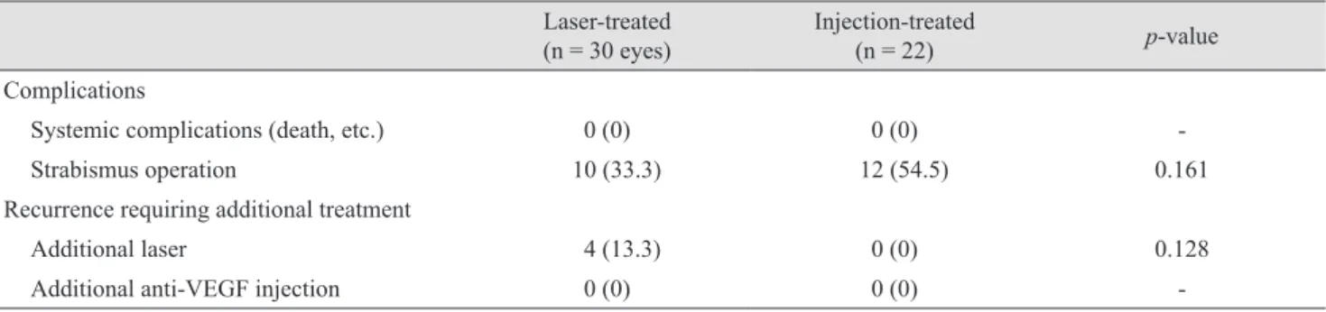
Systemic complications and recurrences requiring addi- tional treatment can be found in Table 3. No systemic complications were reported in either treatment group. The rate of recurrences requiring additional treatment was 13.3% (n = 4) for the laser group and 0% (n = 0) for the an- ti-VEGF group (p = 0.128). Fully vascularized retinas to the ora serrata were noted in 22 (100%) eyes in the an- ti-VEGF group.
Discussion
Despite a recent trend in treating ROP primarily with intravitreal anti-VEGF injections, due in part to potential systemic complications, a large proportion of infants are Table 2. Refractive error after laser indirect ophthalmoscopy therapy or intravitreal anti-VEGF injection for treating retinopathy of prematurity
Laser-treated
(n = 30 eyes) Anti-VEGF–treated
(n = 22) p-value
Mean spherical power
Zone 1 (2 vs. 4) -0.75 ± 2.83 -0.88 ± 2.49 -
Zone 2 (28 vs. 18) -0.29 ± 3.50 0.57 ± 5.33 0.496
Mean cylinder power
Zone 1 (2 vs. 4) -3.88 ± 0.88 -0.69 ± 0.38 -
Zone 2 (28 vs. 18) -1.00 ± 3.52 -0.32 ± 5.51 0.294
Mean spherical equivalence
Zone 1 (2 vs. 4) -2.69 ± 3.27 -1.22 ± 2.63 -
Zone 2 (28 vs. 18) -1.00 ± 3.52 -0.32 ± 5.51 0.603
Mean age at last refraction (yr) 4.7 ± 0.3 4.4 ± 0.3 0.007*
Zone 1 (2 vs. 4) 4.9 ± 0.0 4.1 ± 0.0 <0.001*
Zone 2 (28 vs. 18) 4.7 ± 0.3 4.5 ± 0.3 0.113
VEGF = vascular endothelial growth factor.
*p < 0.05.
Fig. 1. Distribution of the refractive outcome in spherical equiv- alences after treatment for retinopathy of prematurity. Refractive error was divided into six categories: very high myopia (≥-8 di- opters [D]), high myopia (<-8 to -5 D), low myopia (<-5 to -1 D), emmetropia (<-1 to +1 D), low hyperopia (>+1 to +4 D), and high hyperopia (>+4 D). No significant difference was noted between treatment groups in children at the age of 4 years. VEGF = vas- cular endothelial growth factor.
20 15 10 5 0
High hyperopia Low hyperopia Emmetropia Low myopia High myopia
Very high myopia
No. of eyes
Anti-VEGF–treated Laser-treated
still treated via laser therapy [27,28]. Owing to several studies reporting an association between myopia and laser treatment, concern regarding long-term refractive out- comes may influence primary ophthalmologists’ recom- mendations regarding treatment method [25,29]. In our study, we found that in children aged 4 years, the type of treatment (laser or injection) did not appear to influence long-term refractive outcomes of ROP.
Conflicting results have been reported in a few studies regarding the long-term refractive outcomes of ROP treat- ment [30], with major limitations acknowledged due to the wide range of ages and low number of cases involved. A literature review by Aghdam et al. [20] in 2016 found that mean final refractive error in 9 studies ranged from -4.4 to -10.1 D in laser-treated eyes and +0.4 to -3.7 D in injec- tion-treated eyes, only one study reporting that outcomes were similar. However, age at the time of refraction ranged from 7 to 76 months with potential observer bias and small sample sizes, which made drawing definitive conclusions difficult. In our study of children aged 4 years, we noted no statistically significant differences in spherical/cylinder power and calculated SE between laser- or anti-VEGF-treat- ed eyes.
When we compared the effects of treatment zones in the development of myopia, we noted that there was an overall trend toward more severe myopia in eyes treated closer to the posterior pole (zone 1 vs. zone 2), which varies from a previous report where no difference in myopia severity was noted between zone 1 and posterior zone 2 ROP [25].
Unfortunately, the number of cases treated in Zone 1 in our study was too small to draw meaningful statistical conclusions. Additionally, we noted no statistically signifi- cant differences when myopia severity was compared be-
tween laser and anti-VEGF in each treatment zone. Thus, our results suggest that the greater proportion of myopia noted in children treated for ROP is an outcome intrinsic to the disease [31], and not necessarily influenced by type of treatment.
In terms of safety, we found no reports of systemic com- plications such as neurodevelopmental delay or death in ei- ther treatment group. The recurrence rate was 0% for la- ser- and 13.3% for anti-VEGF-treated eyes, which is low compared to previously published studies with rates rang- ing from 13% to 35.4% [32-34]. In cases with zone 2 or 3 ROP, laser therapy appears to be a viable treatment option due to its lower rate of recurrence, especially in cases in which timely follow-up is difficult or there may be low compliance to additional therapies. However, the an- ti-VEGF group was observed to have fully vascularized retinas up to the ora serrata in 100% of eyes on the most recent follow-up.
This study has several limitations. First, it was a retro- spective study with a variable follow-up period and with- out appropriate controls, such as those for treatment indi- cations that might call for earlier treatment for type 1 ROP.
Second, our study population was limited to Asian patients in a tertiary university hospital setting. Third, not all sys- temic complications may have been properly observed or reported to us from the neonatologists. Finally, we did not routinely perform fluorescein angiography to document fully vascularized retinas in these children; however, a ret- ina specialist performed thorough fundus examination at each follow-up outpatient visit. The strengths of this study include statistical comparison of long-term refractive out- comes of similarly-aged children treated at two affiliated institutions using the same treatment protocols.
Table 3. Systemic complications and recurrence after laser therapy or intravitreal anti-VEGF injection for retinopathy of prema- turity
Laser-treated
(n = 30 eyes) Injection-treated
(n = 22) p-value
Complications
Systemic complications (death, etc.) 0 (0) 0 (0) -
Strabismus operation 10 (33.3) 12 (54.5) 0.161
Recurrence requiring additional treatment
Additional laser 4 (13.3) 0 (0) 0.128
Additional anti-VEGF injection 0 (0) 0 (0) -
Values are presented as number (%).
VEGF = vascular endothelial growth factor.
In conclusion, our study demonstrated that long-term re- fractive outcomes in infants treated for ROP did not vary between laser and anti-VEGF injection treatment groups.
Concern regarding refractive outcome should not be the most important factor when selecting treatment modality.
Considering the low rate of recurrence and established safety, conventional laser therapy remains a viable prima- ry therapeutic option in select cases.
Conflict of Interest
No potential conflict of interest relevant to this article was reported.
Acknowledgements
This study was supported by a new faculty research seed money grant of Yonsei University College of Medicine for 2017 (2017-32-0038).
References
1. Blencowe H, Lawn JE, Vazquez T, et al. Preterm-asso- ciated visual impairment and estimates of retinopathy of prematurity at regional and global levels for 2010. Pediatr Res 2013;74 Suppl 1:35-49.
2. Kushner BJ, Essner D, Cohen IJ, Flynn JT. Retrolental fibroplasia. II. Pathologic correlation. Arch Ophthalmol 1977;95:29-38.
3. Darlow BA. Retinopathy of prematurity: new develop- ments bring concern and hope. J Paediatr Child Health 2015;51:765-70.
4. Goggin M, O'Keefe M. Childhood blindness in the Repu- blic of Ireland: a national survey. Br J Ophthalmol 1991;75:425-9.
5. Hoogerwerf A, Schalij-Delfos NE, van Schooneveld MJ, Termote JU. Incidence of retinopathy of prematurity over the last decade in the Central Netherlands. Neonatology 2010;98:137-42.
6. Isaza G, Arora S, Bal M, Chaudhary V. Incidence of reti- nopathy of prematurity and risk factors among premature infants at a neonatal intensive care unit in Canada. J Pe- diatr Ophthalmol Strabismus 2013;50:27-32.
7. Tsui I, Chu A. Hot topics in retinopathy of prematurity. Pe- diatr Ann 2017;46:e415-22.
8. Gilbert C, Rahi J, Eckstein M, et al. Retinopathy of prema- turity in middle-income countries. Lancet 1997;350:12-4.
9. Palmer EA, Flynn JT, Hardy RJ, et al. Incidence and early course of retinopathy of prematurity. The Cryotherapy for Retinopathy of Prematurity Cooperative Group. Ophthal- mology 1991;98:1628-40.
10. Schaffer DB, Palmer EA, Plotsky DF, et al. Prognostic fac- tors in the natural course of retinopathy of prematurity.
The Cryotherapy for Retinopathy of Prematurity Coopera- tive Group. Ophthalmology 1993;100:230-7.
11. Early Treatment for Retinopathy of Prematurity Coopera- tive Group. Revised indications for the treatment of retino- pathy of prematurity: results of the early treatment for reti- nopathy of prematurity randomized trial. Arch Ophthalmol 2003;121:1684-94.
12. Mintz-Hittner HA, Kennedy KA, Chuang AZ; BEAT-ROP Cooperative Group. Efficacy of intravitreal bevacizumab for stage 3+ retinopathy of prematurity. N Engl J Med 2011;364:603-15.
13. Repka MX, Tung B, Good WV, et al. Outcome of eyes de- veloping retinal detachment during the Early Treatment for Retinopathy of Prematurity Study (ETROP). Arch Oph- thalmol 2006;124:24-30.
14. Good WV; Early Treatment for Retinopathy of Prematu- rity Cooperative Group. Final results of the Early Treat- ment for Retinopathy of Prematurity (ETROP) randomized trial. Trans Am Ophthalmol Soc 2004;102:233-48.
15. Ahmed AE, Channa R, Durrani J, et al. Early experience with intravitreal bevacizumab combined with laser treat- ment for retinopathy of prematurity. Middle East Afr J Ophthalmol 2010;17:264-7.
16. Chung EJ, Kim JH, Ahn HS, Koh HJ. Combination of laser photocoagulation and intravitreal bevacizumab (Avastin) for aggressive zone I retinopathy of prematurity. Graefes Arch Clin Exp Ophthalmol 2007;245:1727-30.
17. Kong L, Bhatt AR, Demny AB, et al. Pharmacokinetics of bevacizumab and its effects on serum VEGF and IGF-1 in infants with retinopathy of prematurity. Invest Ophthalmol Vis Sci 2015;56:956-61.
18. Chen SN, Lian I, Hwang YC, et al. Intravitreal anti-vascu- lar endothelial growth factor treatment for retinopathy of prematurity: comparison between ranibizumab and beva- cizumab. Retina 2015;35:667-74.
19. Morin J, Luu TM, Superstein R, et al. Neurodevelopmental
outcomes following bevacizumab injections for retinopathy of prematurity. Pediatrics 2016;137.
20. Abri Aghdam K, Khadamy J, Falavarjani KG, Tsui I. Re- fractive outcomes following the treatment of retinopathy of prematurity in the anti-VEGF era: a literature review. J AAPOS 2016;20:539-40.
21. Isaac M, Mireskandari K, Tehrani N. Treatment of type 1 retinopathy of prematurity with bevacizumab versus laser.
J AAPOS 2015;19:140-4.
22. Kang HG, Choi EY, Byeon SH, et al. Anti-vascular endo- thelial growth factor treatment of retinopathy of prematu- rity: efficacy, safety, and anatomical outcomes. Korean J Ophthalmol 2018;32:451-8.
23. International Committee for the Classification of Retino- pathy of Prematurity. The International Classification of Retinopathy of Prematurity revisited. Arch Ophthalmol 2005;123:991-9.
24. Darlow BA, Ells AL, Gilbert CE, et al. Are we there yet?
Bevacizumab therapy for retinopathy of prematurity. Arch Dis Child Fetal Neonatal Ed 2013;98:F170-4.
25. Geloneck MM, Chuang AZ, Clark WL, et al. Refractive outcomes following bevacizumab monotherapy compared with conventional laser treatment: a randomized clinical trial. JAMA Ophthalmol 2014;132:1327-33.
26. Quinn GE, Dobson V, Kivlin J, et al. Prevalence of myopia between 3 months and 5 1/2 years in preterm infants with and without retinopathy of prematurity. Cryotherapy for Retinopathy of Prematurity Cooperative Group. Ophthal- mology 1998;105:1292-300.
27. Hwang JH, Lee EH, Kim EA. Retinopathy of prematurity among very-low-birth-weight infants in Korea: incidence, treatment, and risk factors. J Korean Med Sci 2015;30 Sup- pl 1:S88-94.
28. Choi J, Kim JH, Kim SJ, Yu YS. Long-term results of lens-sparing vitrectomy for progressive posterior-type stage 4A retinopathy of prematurity. Korean J Ophthalmol 2012;26:277-84.
29. Chen YH, Chen SN, Lien RI, et al. Refractive errors after the use of bevacizumab for the treatment of retinopathy of prematurity: 2-year outcomes. Eye (Lond) 2014;28:1080-6.
30. Kuo HK, Sun IT, Chung MY, Chen YH. Refractive error in patients with retinopathy of prematurity after laser pho- tocoagulation or bevacizumab monotherapy. Ophthalmolo- gica 2015;234:211-7.
31. Nissenkorn I, Yassur Y, Mashkowski D, et al. Myopia in premature babies with and without retinopathy of prema- turity. Br J Ophthalmol 1983;67:170-3.
32. Chen Y, Fen J, Meng X. Effects of ranibizumab in zone I and zone II retinopathy of prematurity patients. Chin J Ophthalmol 2015;31:6-9.
33. Huang Q, Zhang Q, Fei P, et al. Ranibizumab injection as primary treatment in patients with retinopathy of prematu- rity: anatomic outcomes and influencing factors. Ophthal- mology 2017;124:1156-64.
34. Wu WC, Kuo HK, Yeh PT, et al. An updated study of the use of bevacizumab in the treatment of patients with pre- threshold retinopathy of prematurity in Taiwan. Am J Oph- thalmol 2013;155:150-8.