© 2017 The Korean Ophthalmological Society
This is an Open Access article distributed under the terms of the Creative Commons Attribution Non-Commercial License (http://creativecommons.org/licenses /by-nc/3.0/) which permits unrestricted non-commercial use, distribution, and reproduction in any medium, provided the original work is properly cited.
Original Article
Effect of Anti-vascular Endothelial Growth Factor Antibody on the Survival of Cultured Retinal Ganglion Cells
Ji Min Lee, Hyoung Won Bae, Sang Yeop Lee, Gong Je Seong, Chan Yun Kim
Institute of Vision Research, Department of Ophthalmology, Yonsei University College of Medicine, Seoul, Korea
Purpose: To investigate the effects of anti-vascular endothelial growth factor (VEGF) antibody on the survival of retinal ganglion cell (RGC)-5 cells differentiated with staurosporine under oxidative stress.
Methods: We used real-time polymerase chain reaction and Western blot to confirm the expression of VEGF, VEGF receptor (VEGFR)-1 and VEGFR-2 in RGC-5 cells differentiated with staurosporine for 6 hours. The dif- ferentiated RGC-5 cells were treated with 800 μM hydrogen peroxide (H2O2) for 24 hours to induce oxidative stress. Then, the survival rate of RGC-5 was confirmed by lactate dehydrogenase assay at each concentra- tion (0, 0.01, 0.1, and 1 mg) using bevacizumab as the anti-VEGF antibody. The expression of VEGF, VEGFR-1, and VEGFR-2 was confirmed using real-time polymerase chain reaction.
Results: VEGF, VEGFR-1, and VEGFR-2 were all expressed in differentiated RGC-5 cells. When RGC-5 cells were simultaneously treated with bevacizumab and 800 μM H2O2, survival of RGC-5 decreased with bevaci- zumab concentration. VEGF expression in RGC-5 cells increased with increasing concentration of bevacizum- ab. Similar patterns were observed for VEGFR-1 and VEGFR-2, but the degree of increase was smaller than that for VEGF.
Conclusions: When bevacizumab was administered to differentiated RGC-5 cells, the cell damage caused by oxidative stress increased. Therefore, given these in vitro study results, caution should be exercised with bev- acizumab treatment.
Key Words: Anti-vascular endothelial growth factor, Bevacizumab, Oxidative stress, Retinal ganglion cell, RGC-5
The hypothesis that a specific substance acts on neovas- cularization in retinal diseases was first proposed in 1956 [1] and it was determined that vascular endothelial growth factor (VEGF) is increased in the oxidative stress environ- ment of the retina [2-5]. VEGF is a 46-kDa molecular weight glycoprotein that binds to receptors on the surface
of vascular endothelial cells to proliferate and increase capillary permeability [6]. Previously, it was thought that VEGF function was restricted to endothelial cells. Howev- er, in recent studies, VEGF was shown to promote the de- velopment and maturation of neural tissues, including the retina [7]. In the normal development of the retina, VEGF acts as an essential factor in the production and survival and function of cells [8,9]. However, VEGF has been im- plicated in the development of neovascularization in vari- ous retinal vascular diseases such as exudative age-related macular degeneration, proliferative diabetic retinopathy,
Received: May 9, 2017 Accepted: May 31, 2017
Corresponding Author: Chan Yun Kim, MD, PhD. Institute of Vision Research, Department of Ophthalmology, Yonsei University College of Medicine, #50-1 Yonsei-ro, Seodaemun-gu, Seoul 03722, Korea. Tel: 82- 2-2228-3570, Fax: 82-2-312-0541, E-mail: [email protected]
macular edema of retinal vein occlusion, and retinopathy of prematurity. In addition, it has been shown that the ele- vated level of VEGF in these diseases plays a key role in the progression of disease [2,10,11]. Recently, treatment with the anti-VEGF antibody bevacizumab has been wide- ly used for these diseases in the field of ophthalmology, and positive treatment effects have been reported in many eye diseases that cause blindness [12,13]. However, since angiogenesis is an important mechanism that protects tis- sues against various ischemic injuries, bevacizumab ad- ministration for the inhibition of angiogenesis potentially has unexpected harmful effects on the tissue [8]. Previous studies have reported the safety and side effects of bevaci- zumab treatment [14]. One of the studies reported that bev- acizumab injection resulted in the death of retinal ganglion cells (RGCs) in an animal model [15]. In this study, we in- vestigated the effects of bevacizumab on the survival of retinal ganglion cells cultured in a hypoxic environment through experimental methods in order to establish a basis for the safe and effective use of anti-VEGF antibody thera- py in various ophthalmic diseases.
Materials and Methods
Growth of cell lines
Differentiation of RGC-5 cells was used in this study.
The RGC-5 cell line selectively retained the retinal gangli- on cells in the mouse and was infected with the R-virus to maintain retinal ganglion cell characteristics [16,17]. In a previous study [18], staurosporine (Sigma, Poole, UK) was used at a concentration of 1 μg for a minimum of 6 hours to reach the final differentiation level of the RGC-5 cell line. In the present study, RGC-5 cells were treated with 1
μg staurosporine for 6 hours as well.
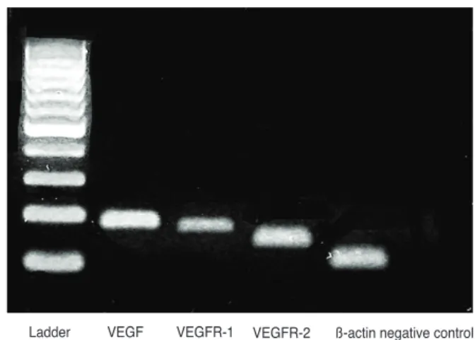
Identification of VEGF, VEGF receptor (VEGFR)-1 and VEGFR-2 expression in differentiated RGC-5 cell lines The expression of VEGF, VEGFR-1, and VEGFR-2 was assessed by real-time polymerase chain reaction (PCR) and Western blot (Table 1 and Fig. 1) after culturing RGC-5 cells differentiated using the above method.
cDNA synthesis
cDNA was synthesized from the extracted total RNA according to the manufacturer’s protocol using the Super- Script III First-Strand Synthesis System for real-time PCR (Gibco, Grand Island, NY, USA). Briefly, cultured RGC-5 cells were floated with trypsin and total RNA was extract- ed using RNeasy mini kit (Qiagen, Valencia, CA, USA). A combination of 1 μL of extracted total RNA, 1 μL of 50 μM oligo (dT), 1 μL of 10 mM deoxynucleoside triphos- phate (dNTP) mix and diethyl pyrocarbonate (DEP- C)-treated water for a total volume of 10 μL was incubated at 65°C for 5 minutes. The mixture was then put on ice for 1 minute to stop the reaction. Next, 2 μL of 10 × reverse transcription (RT) buffer, 4 μL of 25 mM MgCl2, 2 μL of 0.1 M dithiothreitol (DTT), 1 μL of RNaseOUT (40 U/μL) and 1 μL of SuperScript III RT (200 U/μL) was added for a total volume of 20 μL. Thereafter, the cells were incubated at 50°C for 50 minutes and incubated at 85°C for 5 minutes to stop the reaction, and then the tubes were placed on ice.
Table 1. Primer sequences for real-time PCR
Gene name Sequence
VEGF-A (172 bp) Forward: 5’-gcccatgaagtggtgaagtt-3’
Reverse: 5’-actccagggcttcatcattg-3’
VEGFR-1 (157 bp) Forward: 5’-tttatcagcgtgaagcatcg-3’
Reverse: 5’-ccgaatagcgagcagatttc-3’
VEGFR-2 (129 bp) Forward: 5’-acagttcccagagtggttgg-3’
Reverse: 5’-gtcactgacagaggcgatga-3’
PCR = polymerase chain reaction; VEGF = vascular endothelial growth factor; VEGFR = VEGF receptor.
Fig. 1. Expression of vascular endothelial growth factor (VEGF), VEGF receptor (VEGFR)-1 and VEGFR-2 assessed by real-time polymerase chain reaction. VEGF, VEGFR-1, and VEGFR-2 are all expressed in differentiated retinal ganglion cell-5 cells.
Ladder VEGF VEGFR-1 VEGFR-2 ß-actin negative control
The reaction mixture was collected by light centrifugation and 1 μL of RNase H was added and incubated at 37°C for 20 minutes to remove residual RNA. The resulting cDNA was stored at –20°C until further processing.
Real-time PCR
Real-time PCR was performed using the QuantiTect SYBR Green PCR kit (Qiagen). A mixture of 100 ng of cDNA, 2 μL of 20 μM primers, 25 μL of master mix, and DEPC-treated water was added to each sample to make a 50 μL mixture. The primer sequences are listed in Table 1.
Each primer product was identified via melting curve analysis. The temperature conditions of the amplification process are as follows. Cycle 1, 95°C for 3 minutes; cycle 2 (50 cycles; step 1, 95°C for 10 seconds; step 2, 55°C for 45 seconds); cycle 3, 95°C for 1 minute; cycle 4, 55°C for 1 minute; cycle 5 (80 cycles; step 1, 55°C for 10 seconds; step 2, increase set point temperature after 2nd cycle by 0.5°C).
Each mRNA level was calculated using the 2-∆Ct method based on the Ct value using β-actin as the reference house- keeping gene.
Western blot
Sodium dodecyl sulfate polyacrylamide gel electropho- resis was performed with 10%–15% sodium dodecyl sul- fate-polyacrylamide gel and 50 μg of cell lysate was loaded in each well. After electrophoresis, the cells were elec- tro-transferred to Immobilon-P transfer membrane (Milli- pore, Billerica, MA, USA) under 200 mA for 2 hours.
Three percent of BSA (AMRESCO, Solon, OH, USA) was used to inhibit nonspecific binding. After that, the primary antibody was added, reacted at room temperature for 1 hour, washed and secondary antibody was used. One of the ECL peroxidase-labeled anti-rabbit (Amersham, Pisca- taway, NJ, USA) or ECL peroxidase-labeled anti-mouse (Amersham) was selected for the primary antibody host, and then sensitized using Amersham ECL Western Blot- ting Detection Reagents (Amersham).
Induction of oxidative stress and evaluation of cell survival
The differentiated RGC-5 cells were treated with H2O2
(800 μM) for 24 hours to induce oxidative stress. Cell via-
bility was assessed using lactate dehydrogenase (LDH) as- say. The amount of LDH was measured via colorimetric assay (Calbiochem-Novabiochem, San Diego, CA, USA), and whole cells were frozen. In comparison with the amount of LDH measured after death, the degree of de- struction was evaluated. Serial dilutions of commercially available bevacizumab (Avastin; Genentech, San Francis- co, CA, USA) were performed to obtain treatment concen- trations of 0.01, 0.1, and 1 mg/mL.
Statistical analysis
Data are expressed as the mean ± standard deviation of at least three different experiments performed from sepa- rate cell preparations, and at least triplicate determinations were performed for each experiment. Statistical tests to determine the difference between groups were performed by Mann-Whitney U-test using SPSS ver. 18.0 (SPSS Inc., Chicago, IL, USA). A p-value of less than 0.05 was consid- ered statistically significant.
Results
Effect of bevacizumab on the survival of RGC-5 cell line under oxidative stress
Oxidative stress was induced in RGC-5 cells treated for 24 hours using 800 μM H2O2. At that time, the RGC-5 cells were simultaneously treated with the anti-VEGF anti- body, bevacizumab (0, 0.01, 0.1, and 1 mg), and the effect of this treatment on the survival was evaluated by LDH assay. We confirmed that the cell damage increased as the concentration of the bevacizumab increased. In the ab- sence of oxidative stress, RGC-5 cytotoxicity increased following treatment with 1 mg bevacizumab, which was higher than oxidative stress-induced cytotoxicity (Fig. 2).
Effect of bevacizumab on the expression of VEGF, VEGFR-1 and VEGFR-2 in RGC-5 Cells
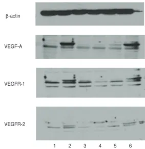
The expression of VEGF, VEGFR-1, and VEGFR-2 in bevacizumab-treated RGC-5 cells was confirmed by West- ern blot analysis. In addition, the expression of VEGF, VEGFR-1, and VEGFR-2 increased as the concentration of bevacizumab increased, and the expression of VEGF,
VEGFR-1, and VEGFR-2 increased in RGC-5 cells treated by H2O2 alone (Fig. 3).
Discussion
A number of recent studies have shown that bevacizum- ab injection therapy in the vitreous cavity has a remark- able therapeutic effect in many retinal vascular diseases including exudative age-related macular degeneration, dia- betic retinopathy, and neovascular glaucoma [12,13]. How- ever, VEGF plays an important role in the neuroprotection, development and maturation of the nerve tissues of the retina [7]. Normally, VEGF-mediated angiogenesis in the human body occurs to protect cells in a hypoxic environ- ment. The administration of bevacizumab to inhibit angiogenesis may cause unintended cytotoxicity and isch- emic damage [9]. Furthermore, the use of bevacizumab in patients with optic nerve weakness, such as glaucoma, is likely to cause damage to the retinal ganglion cells, which play an important pathophysiological role in glaucoma. In glaucoma, there is reduction of the retinal nerve fiber layer thickness and loss of retinal ganglion cells [19], and retinal ganglion cell damage may cause an irreversible field defect or visual loss.
Although intraocular pressure reduction still remains the mainstay of glaucoma therapy, recent studies have sug- gested that intraocular pressure reduction alone cannot
prevent irreversible damage to retinal ganglion cells. Neu- roprotection is a strategy for glaucoma treatment, and a number of studies have addressed the factors affecting the survival and death of the retinal ganglion cells [20,21].
Foxton et al. [15] observed that VEGF-A stimulates the survival of retinal ganglion cells in the glaucoma experi- mental model, and VEGF-A blockade significantly exacer- bates neuronal cell death. Saint-Geniez et al. [8] reported a reduction in retinal ganglion cell thickness when VEGF expression was suppressed.
In this study, we investigated the effects of bevacizumab concentration on RGC-5 cell survival under oxidative stress. We found that cytotoxicity increased when bevaci- zumab was applied to the differentiated RGC-5 cell line, and cell damage increased with increasing bevacizumab concentration. In addition, even in the absence of oxidative stress, when bevacizumab was applied to the RGC-5 cell line, the cytotoxicity of the cell line increased, and was greater than that from oxidative stress (Fig. 2). Our results confirm that bevacizumab, which is widely used clinically, would inhibit retinal ganglion cell survival. In addition, we confirmed that VEGF, VEGFR-1, and VEGFR-2 were all produced in the RGC-5 cells used in this experiment.
When bevacizumab and 800 μM H2O2 were simultaneous- Fig. 3. Western blot analysis of vascular endothelial growth fac- tor (VEGF), VEGF receptor (VEGFR)-1 and VEGFR-2 in retinal ganglion cell (RGC)-5 cells treated with bevacizumab. The ex- pression of VEGF, VEGFR-1, and VEGFR-2 increased with the increase in bevacizumab concentration, and the expression of VEGF, VEGFR-1, and VEGFR-2 was increased in RGC-5 cells treated with H2O2. 1, control; 2, bevacizumab 1 mg; 3, H2O2 800 µM; 4, bevacizumab 0.01 mg + H2O2 800 µM; 5, bevacizumab 0.1 mg + H2O2 800 µM; 6, bevacizumab 1 mg + H2O2 800 µM.
β-actin
VEGF-A
VEGFR-1
VEGFR-2
1 2 3 4 5 6
Fig. 2. Effects of bevacizumab on the survival of retinal ganglion cell (RGC)-5 cells under oxidative stress with 800 μM H2O2 for 24 hours. As the concentration of bevacizumab increases, the RGC-5 death rate increases. With the presence of bevacizumab, cell death increased without oxidative stress. Values are presented as mean ± standard deviation (*values are significantly different from the control; p < 0.05 by Mann-Whitney U-test).
% cytotoxicity
Concentration 0 Bevacizumab
(1 mg) H2O2
(800 μM) H2O2 (800 μM) + bevacizumab
(0.01 mg)
H2O2 (800 μM) + bevacizumab (0.1 mg)
H2O2 (800 μM) + bevacizumab (1 mg) 100
50
0
⁎
⁎ ⁎
⁎
ly treated with RGC-5, cell damage and the expression of VEGF in RGC-5 itself was also increased according to the increased concentration of bevacizumab.
This study shows that a high concentration of bevaci- zumab increases cell death in retinal ganglion cells. How- ever, since this experiment was not performed in vivo, we were unable to show different mechanisms and patterns in the human body due to interactions with other cells under oxidative stress. In the present study, we used the RGC-5 cell line, which has been reported to be cross-contaminat- ed with a mouse fibroblast cell line [22]. Furthermore, the RGC-5 cell line may not accurately represent the charac- teristics of all retinal ganglion cells. Therefore, additional experiments and studies will be needed to determine whether other animal models and primary retinal ganglion cells will produce similar results in the future.
This study is expected to provide a basis for the safe and effective use of bevacizumab in various ophthalmic dis- eases including glaucoma. In the future, bevacizumab should be used at the minimum concentration that does not cause cytotoxicity. In addition, intravitreal injection of bevacizumab for the treatment of retinal vascular disease often needs to be repeated rather than administered as a single treatment. Therefore, studies on the benefits and risks of long-term treatment according to the frequency of injection are needed to prevent potential adverse effects of indiscriminate bevacizumab treatment. In particular, con- sidering the fact that bevacizumab treatment may have a negative effect on eyes with optic nerve damage such as glaucoma, treatment should be performed according to in- dividual characteristics and more attention should be fo- cused on these conditions. In addition, long-term large- scale clinical trials and animal models are needed in the future to evaluate the efficacy and safety of bevacizumab therapy.
Conflict of Interest
No potential conflict of interest relevant to this article was reported.
Acknowledgements
This study was supported by a faculty research grant of
Yonsei University College of Medicine for 2008 (research project No. 6-2008-0201).
References
1. Wise GN. Retinal neovascularization. Trans Am Ophthal- mol Soc 1956;54:729-826.
2. Adamis AP, Miller JW, Bernal MT, et al. Increased vascu- lar endothelial growth factor levels in the vitreous of eyes with proliferative diabetic retinopathy. Am J Ophthalmol 1994;118:445-50.
3. Miller JW, Adamis AP, Shima DT, et al. Vascular endothe- lial growth factor/vascular permeability factor is temporal- ly and spatially correlated with ocular angiogenesis in a primate model. Am J Pathol 1994;145:574-84.
4. Drobek-Slowik M, Karczewicz D, Safranow K. The poten- tial role of oxidative stress in the pathogenesis of the age-related macular degeneration (AMD). Postepy Hig Med Dosw (Online) 2007;61:28-37.
5. Beatty S, Koh H, Phil M, et al. The role of oxidative stress in the pathogenesis of age-related macular degeneration.
Surv Ophthalmol 2000;45:115-34.
6. Esser S, Wolburg K, Wolburg H, et al. Vascular endothelial growth factor induces endothelial fenestrations in vitro. J Cell Biol 1998;140:947-59.
7. Wang Y, Mao XO, Xie L, et al. Vascular endothelial growth factor overexpression delays neurodegeneration and pro- longs survival in amyotrophic lateral sclerosis mice. J Neu- rosci 2007;27:304-7.
8. Saint-Geniez M, Maharaj AS, Walshe TE, et al. Endoge- nous VEGF is required for visual function: evidence for a survival role on muller cells and photoreceptors. PLoS One 2008;3:e3554.
9. Storkebaum E, Lambrechts D, Carmeliet P. VEGF: once re- garded as a specific angiogenic factor, now implicated in neuroprotection. Bioessays 2004;26:943-54.
10. Aiello LP, Avery RL, Arrigg PG, et al. Vascular endothelial growth factor in ocular fluid of patients with diabetic reti- nopathy and other retinal disorders. N Engl J Med 1994;331:1480-7.
11. Ferrara N, Mass RD, Campa C, Kim R. Targeting VEGF-A to treat cancer and age-related macular degeneration. Annu Rev Med 2007;58:491-504.
12. Ozaki H, Seo MS, Ozaki K, et al. Blockade of vascular en- dothelial cell growth factor receptor signaling is sufficient
to completely prevent retinal neovascularization. Am J Pathol 2000;156:697-707.
13. Gragoudas ES, Adamis AP, Cunningham ET Jr, et al.
Pegaptanib for neovascular age-related macular degenera- tion. N Engl J Med 2004;351:2805-16.
14. van der Reis MI, La Heij EC, De Jong-Hesse Y, et al. A systematic review of the adverse events of intravitreal an- ti-vascular endothelial growth factor injections. Retina 2011;31:1449-69.
15. Foxton RH, Finkelstein A, Vijay S, et al. VEGF-A is neces- sary and sufficient for retinal neuroprotection in models of experimental glaucoma. Am J Pathol 2013;182:1379-90.
16. Charles I, Khalyfa A, Kumar DM, et al. Serum deprivation induces apoptotic cell death of transformed rat retinal gan- glion cells via mitochondrial signaling pathways. Invest Ophthalmol Vis Sci 2005;46:1330-8.
17. Maher P, Hanneken A. The molecular basis of oxidative
stress-induced cell death in an immortalized retinal gangli- on cell line. Invest Ophthalmol Vis Sci 2005;46:749-57.
18. Na KD, Kang SY, Seong GJ, et al. Ischemic precondition- ing and the role of protein kinase C in cultured retinal gan- glion cell line. J Korean Ophthalmol Soc 2008;49:979-86.
19. Na JH, Lee K, Lee JR, et al. Detection of macular ganglion cell loss in preperimetric glaucoma patients with localized retinal nerve fibre defects by spectral-domain optical co- herence tomography. Clin Exp Ophthalmol 2013;41:870-80.
20. Chen SD, Wang L, Zhang XL. Neuroprotection in glauco- ma: present and future. Chin Med J (Engl) 2013;126:1567- 77.
21. Weinreb RN. Glaucoma neuroprotection: what is it? Why is it needed? Can J Ophthalmol 2007;42:396-8.
22. Krishnamoorthy RR, Clark AF, Daudt D, et al. A forensic path to RGC-5 cell line identification: lessons learned. In- vest Ophthalmol Vis Sci 2013;54:5712-9.