© 2019 The Korean Ophthalmological Society
This is an Open Access article distributed under the terms of the Creative Commons Attribution Non-Commercial License (http://creativecommons.org/licenses /by-nc/3.0/) which permits unrestricted non-commercial use, distribution, and reproduction in any medium, provided the original work is properly cited.
Original Article
Switching to Aflibercept in Diabetic Macular Edema after
Unsatisfactory Response to Other Anti-vascular Endothelial Growth Factor Drugs
Walid S. Ibrahim1, Zeiad H. Eldaly1, Mohamed G. Saleh1, Mahmoud F. Rateb1, Ahmed H. Aldoghaimy2
1Department of Ophthalmology, Assiut University, Assiut, Egypt
2Department of Ophthalmology, South Valley University, Qena, Egypt
Purpose: To evaluate the efficacy of switching to aflibercept in diabetic macular edema (DME) with suboptimal response to previous anti-vascular endothelial growth factor (anti-VEGF) injections.
Methods: A prospective interventional case series study recruited patients from a single center diagnosed with DME with suboptimal response to anti-VEGF injections. Three consecutive monthly injections of afliber- cept were performed. The primary outcome measure was mean change in visual acuity after switching to aflibercept.
Results: Forty-two patients (42 eyes) were included. Baseline logarithm of the minimum angle of resolution (logMAR) visual acuity was 0.87 ± 0.23 and improved significantly to 0.62 ± 0.29, 0.56 ± 0.34, and 0.46 ± 0.35 at 1, 2, and 3 months, respectively, after the first injection. Mean baseline retinal thickness was 451.57
± 107.09 μm and decreased significantly at 1, 2, and 3 months after switching to aflibercept (346.52 ± 79.03, 328.24 ± 81.98, and 313.71 ± 85.79 μm, respectively). Both visual improvement and mean change in retinal thickness were significant in patients with pre-aflibercept best-corrected visual acuity less than 1.0 logMAR but were not significant in patients with best-corrected visual acuity more than 1.0 logMAR.
Conclusions: Switching to aflibercept in DME patients with an unsatisfactory response to previous anti-VEGF injections provided acceptable short-term visual and retinal architectural improvement.
Key Words: Aflibercept, Diabetic retinopathy, Macular edema
Diabetic retinopathy is the leading cause of visual im- pairment in the working-age demographic in developed countries [1]. This problem is likely to increase in the fu- ture due to a growing diabetic population, especially in the
Middle East [2]. The Early Treatment Diabetic Retinopathy Study was a milestone study that defined laser photocoagu- lation as the benchmark treatment for preventing visual loss from diabetic macular edema (DME) [3].
Several studies have demonstrated the safety and effica- cy of different anti-vascular endothelial growth factor (an- ti-VEGF) drugs, namely ranibizumab [4] and bevacizumab [5], in the management of DME. Aflibercept gained US Food and Drug Administration approval to treat DME af- ter the phase 3 trials VIVID and VISTA and provided sig-
Received: April 2, 2018 Accepted: August 8, 2018
Corresponding Author: Zeiad H. Eldaly, MD. Department of Ophthal- mology, Assiut University Hospital, 6th floor, Assiut 71516, Egypt. Tel:
20-88-230-9970, Fax: 20-88-241-3626, E-mail: [email protected]
nificant visual and morphological improvement in patients suffering from DME [6,7]. In light of protocol T of the DRCR.net [8], many retinal specialists are now considering the use of aflibercept in DME cases, especially those with poor presenting vision. A growing body of research is in- vestigating different treatment protocols for management of resistant DME. Switching from bevacizumab or ranibi- zumab to aflibercept is one promising strategy for address- ing this challenging situation [9-11]. This positive effect could be explained by the different molecular structure [12], greater binding affinity [13], and/or longer intra-vitreal bio-availability of aflibercept [14].
This study aims to evaluate the short-term visual and retinal morphological changes and safety of switching to aflibercept injections in DME refractory to other an- ti-VEGF drugs.
Materials and Methods
Design
A prospective interventional case series study was con- ducted from October 2015 to November 2016 after approv- al of the Ethical Committee of the Faculty of Medicine, Assiut University, Egypt (264-15). All procedures were carried out under the tenets of the Helsinki Declaration.
Written consent was provided by all participants after dis- cussing the procedure, alternative treatment plans, fol- low-up schedules, and possible benefits and risks.
Patient selection
Patients suffering from previously diagnosed macular edema secondary to type 1 or 2 diabetes mellitus were in- cluded. For transition to aflibercept 2 mg/0.05 mL, a diag- nosis of resistant DME was required. Patients fulfilling one or more of the following criteria were considered to have resistant DME after at least 3 consecutive monthly bevaci- zumab 1.25 mg or ranibizumab 0.5 mg injections in the previous 6 months: central macular thickness greater than 300 μm by spectral-domain optical coherence tomography (SD-OCT), reduction of retinal thickness by less than 10%
of baseline retinal thickness, or suboptimal visual improve- ment (failure to gain at least 3 lines on the Snellen chart).
Exclusion criteria were unwillingness to participate, signif-
icant cataract or corneal opacity, DME associated with pro- liferative diabetic retinopathy, history of laser treatment in the previous 6 months (either focal or macular grid laser photocoagulation), history of steroid injection in the previ- ous 6 months (peri-ocular or intra-vitreal injection/im- plant), co-existing retinal pathology (e.g., retinal vascular occlusion, age-related macular degeneration), history of cataract surgery in the previous 12 months, associated optic nerve disorders (e.g., ischemic optic neuropathy), glyco- sylated hemoglobin higher than 8% at the time of partici- pation, ischemic heart disease, or previously complicated intra-vitreal injection of anti-VEGF. Participants were ex- cluded from the study if they had fewer than 3 consecutive aflibercept intra-vitreal injections.
Baseline evaluation
All participants underwent thorough ophthalmic and systemic evaluation. Ophthalmic evaluation included de- tailed history-taking, best-corrected visual acuity (BCVA) by Snellen chart (converted subsequently to equivalent logarithm of the minimum angle of resolution [logMAR]
values for statistical analysis), slit-lamp examination, and intraocular pressure (IOP) measurement before papillary dilatation (Goldmann applanation tonometer), followed by dilated fundus examination. Fundus fluorescein angiogra- phy (Topcon fundus camera; Topcon, Tokyo, Japan) and SD-OCT (Heidelberg Engineering, Heidelberg, Germany) were performed on every participant at initial evaluation.
In SD-OCT assessment, a 30 degree by 30 degree macu- lar grid scan acquisition was carried out with 64 horizontal lines, spaced 50 μm and averaged to 30 frames. Early Treatment Diabetic Retinopathy Study macular thickness map was then measured automatically with Heidelberg software. Poor OCT images (signal strength value below 10) were excluded. Retinal thickness was measured as the perpendicular line between the internal limiting membrane and Bruch’s membrane. Central 1 mm macular thickness was recorded with additional manual adjustment for mea- surement lines. Analysis and interpretation of OCT images were performed by an experienced ophthalmologist (ZE).
Aflibercept injection procedure
All participants received pre-injection prophylactic topi- cal gatifloxacin 0.3% eye drops and topical anesthetic ben-
oxinate 0.4% eye drops. After digital ocular massage to lower the IOP, the intra-vitreal injections were carried out under strict aseptic conditions in the operating room. The injection was performed in the temporal sclera 3.5 mm from the limbus after sweeping conjunctiva with a 5% Po- vidone Iodine-soaked sterile sponge. Assessment of pupil- lary response, detection of hand movement, and fundus ex- amination (if needed) were carried out to exclude possible post-injection IOP spikes. Intra-vitreal injection of afliber- cept was performed by a single experienced ophthalmolo- gist (WI).
Post-injection treatment and follow-up
All patients received topical gatifloxacin 0.3% eye drops post-injection for 5 days. Follow-up visits were scheduled at 1, 2, and 3 months after the initial injection. BCVA, slit- lamp examination, IOP measurement, and follow-up SD- OCT were performed at each injection.
Retinal morphological changes by SD-OCT
DME edema was stratified into spongiform macular edema, cystoid macular edema with or without the pres- ence of subretinal fluid, and/or vitreo-macular interface abnormality (VMI). Subretinal fluid was defined as accu- mulation of fluid between the neuro-sensory retina and retinal pigment epithelium. VMI abnormality was tagged by the presence of high reflective epi-retinal membrane (ERM), antero-posterior vitreo-macular traction, or thick- ened posterior hyaloid. Retinal morphological analysis was performed at baseline and at 3 months post-injection by SD-OCT and was interpreted by an experienced ophthal- mologist (ZE).
Study outcome measures
The primary outcome measure was mean change in vi- sual acuity after 3 consecutive monthly injections of af- libercept. Secondary outcome measures were mean change in retinal thickness, correlation between baseline visual acuity/baseline retinal thickness and final 3-month visual acuity/retinal thickness, and retinal architectural changes after switching to aflibercept.
Statistical analysis
Quantitative data were presented as mean ± standard de- viation. For comparison between mean BCVA and CMT change pre- and post-aflibercept injection, we used paired sample t-test. Comparison of means among subgroups was carried out by Mann-Whitney test. We utilized Pearson correlation coefficient for correlation between mean post-injection BCVA and central macular thickness (CMT).
Statistical significance was considered significant if p-val- ue < 0.05. Statistical analysis was carried out using IBM SPSS Statistics ver. 20.0 (IBM Corp., Armonk, NY, USA).
Results
Baseline evaluation
An interventional case series study recruited 66 consecu- tive participants. Fourteen participants were excluded be- cause they had received fewer than 3 consecutive intra-vit- real injections of a single anti-VEGF agent (10 patients) or 3 non-consecutive intra-vitreal injections (4 patients). Ten additional participants were excluded because they re- ceived only 2 intra-vitreal aflibercept injections and refused to have the third injection. A total of 42 patients (42 eyes) met the inclusion criteria and were included in the study.
Mean age of the included patients was 60.04 ± 6.89 years (range, 49 to 71 years). Twenty-six males and 16 females were included in the study. Table 1 demonstrates patient characteristics and baseline assessment.
Retinal thickness changes after switching to aflibercept Mean pre-aflibercept injection CMT was 451.57 ± 107.09 μm, which decreased significantly at 1, 2, and 3 months af- ter switching to aflibercept (346.52 ± 79.03, 328.24 ± 81.98, and 313.71 ± 85.79, respectively, p < 0.001) (Table 2). The patients were was subdivided into two groups based on baseline CMT: less than or equal to 450 μm and more than 450 μm. At the 3-month follow-up, mean reduction of CMT was 126.55 ± 34.71 and 150.30 ± 118.06 μm, respectively, in the two groups (p = 0.44, Mann-Whitney test).
Visual acuity changes after switching to aflibercept At baseline, mean logMAR visual acuity was 0.87 ± 0.23, which improved significantly to 0.62 ± 0.29 after a single aflibercept 2.0 injection (p = 0.04). Visual acuity continued to improve significantly after the second (0.56 ± 0.34, p = 0.02) and the third aflibercept injections (0.46 ± 0.35, p = 0.03) (Table 3). Furthermore, mean logMAR vi- sual acuity at 3-month follow-up was 0.25 ± 0.17 (equiva- lent to 6 / 12 on the Snellen chart) in patients with CMT less than or equal to 450 μm (22 eyes) and 0.68 ± 0.36 (equivalent to 6 / 30 on the Snellen chart) in patients with
CMT more than 450 μm (20 eyes) (p = 0.03, Mann-Whit- ney test). On the other hand, patients who presented with logMAR visual acuity greater than or equal to 1.0 (14 eyes) had a final logMAR visual acuity of 0.8 ± 0.33 (equivalent to 6 / 38 on the Snellen chart), while patients who present- ed with logMAR visual acuity less than 1.0 (28 eyes) had a final logMAR visual acuity of 0.28 ± 0.21 (equivalent to 6 / 12 on the Snellen chart).
Relationships of baseline visual acuity and retinal thickness changes to final outcome
Both baseline visual acuity and retinal thickness had a strong and significant relationship to final logMAR visual acuity and retinal thickness at 3 months (Pearson correla- tion coefficient 0.64, p < 0.001 and 0.66, p < 0.001, respec- tively).
Retinal architectural changes after shifting to afliber- cept
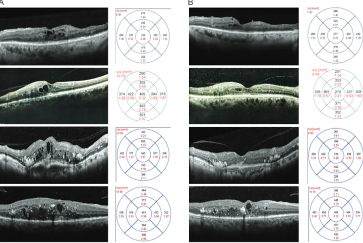
In SD-OCT follow-up scans, most of the eyes that pre- sented with spongiform macular edema, intra-retinal cysts, and subretinal fluid at the time of shifting to aflibercept showed improvement by the third month (Fig. 1A, 1B).
Progression of VMI abnormality was not found in any participant, and 1 eye developed ERM by the third month.
Adverse effects of aflibercept injection
No serious systemic adverse events (e.g., cerebro-vascu- lar stroke, myocardial infarction) were recorded during the study. Only four cases of subconjunctival hemorrhage were reported, with no other serious ocular adverse events (e.g., endophthalmitis, vitreous hemorrhage, retinal detach- ment).
Discussion
DME is one of the major causes of visual impairment in diabetic patients, especially in the working age group [1].
Despite the evolution of multiple treatment modalities for DME since implementation of the macular laser, it is not uncommon to experience DME that has failed to respond adequately to one of the treatment options [3]. There is on- Table 1. Baseline patient characteristics
Characteristics Value
Age (yr) 60.23 ± 6.89
Male : female 26 : 16
Diabetes mellitus duration (yr) 14.80 ± 2.48 Glycosylated hemoglobin (%) 7.32 ± 0.55 Lens status (phakic : pseudophakic) 30 : 12
BCVA (logMAR) 0.87 ± 0.23
Central macular thickness at 1 mm (μm) 451.57 ± 107.09
Number of injections 6.33 ± 1.15
Diabetic retinopathy stage (eyes)
Mild NPDR 5
Moderate NPDR 25
Severe NPDR 12
Anti-VEGF
Bevacizumab 1.25 mg only 14 (33.3)
Ranibizumab 0.5 mg only 16 (38.1)
Ranibizumab then shifted to bevacizumab 6 (14.3) Bevacizumab then shifted to ranibizumab 6 (14.3) Previous laser
Focal macular laser 12 (28.6)
Macular grid laser 6 (14.3)
Previous triamcinolone injection
IVTA 4 (9.5)
Peri-ocular TA 6 (14.3)
Values are presented as mean ± standard deviation, number, or number (%).
BCVA = best-corrected visual acuity; logMAR = logarithm of minimal angle of resolution; NPDR = non-proliferative diabetic retinopathy; VEGF = vascular endothelial growth factor; IVTA
= intra-vitreal triamcinolone acetonide; TA = triamcinolone ace- tonide.
going debate about the definition of unsatisfactory treat- ment in DME. Some authors advocate that unsatisfactory response in DME is diagnosed when reduction of retinal thickness is suboptimal; others define it as inadequate vi- sual improvement, while others may combine several pa- rameters [11,15]. Suboptimal response in DME could be at- tributed to many postulated mechanisms such as tachyphylaxis [16,17] or tolerance (due to receptor dysregu- lation or neutralizing antibody formation against the an- ti-VEGF agent) [18-20]. Many inflammatory mediators have been implemented in the development and progres- sion of diabetic retinopathy [21]. High VEGF level in the vitreous of diabetic patients could play a role in the patho- genesis and treatment response of DME [22].
There are many strategies in the management of resis- tant DME, such as switching to another anti-VEGF [23], switching to sustained-release steroid implants [24], com- bining treatments [25], or surgical intervention [26]. Con-
tinuation of anti-VEGF in the absence of satisfactory re- sponse was also suggested based on a proposed category of “late responders” [27]. In these eyes, it takes time and multiple injections to challenge high VEGF levels in the retina and/or vitreous, indicating that continuation of the original anti-VEGF may be the solution. The active afliber- cept molecule acts differently than those of bevacizumab and ranibizumab. It is a fusion protein with an intermedi- ate molecular weight of 115 KDa, which is between those of bevacizumab (149 KDa) and ranibizumab (48 KDa).
The active aflibercept molecule interacts not only with VEGF, but also with placental-derived growth factor, wid- ening the spectrum of its action [12].
The role of poor glycemic control in development and progression of diabetic retinopathy and DME cannot be overlooked [28]. Therefore, we included patients with gly- cosylated hemoglobin less than 8% in a trial to eliminate that possible confounding factor. We also included patients Table 2. Retinal thickness changes after shifting to aflibercept from baseline to 3 months after injection
Baseline 1-mon post-injection 2-mon post-injection 3-mon post-injection
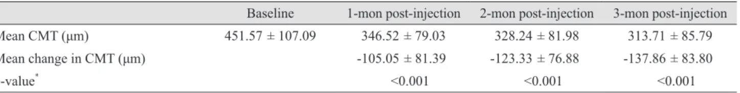
Mean CMT (μm) 451.57 ± 107.09 346.52 ± 79.03 328.24 ± 81.98 313.71 ± 85.79
Mean change in CMT (μm) -105.05 ± 81.39 -123.33 ± 76.88 -137.86 ± 83.80
p-value* <0.001 <0.001 <0.001
Values are presented as mean ± standard deviation.
CMT = central macular thickness.
*Paired sample t-test.
Table 3. Visual acuity change from baseline to 3 months after changing to aflibercept
Baseline 1 mon 2 mon 3 mon Previous
anti-VEGF 1.0 logMAR
or more (14 eyes)
No. of eyes 14 8 8 6 6.57 ± 1.39
(5–9 injections)
Mean BCVA 1.13 ± 0.16 0.88 ± 0.16 0.86 ± 0.27 0.79 ± 0.33
p-value* 0.07
Mean CMT 535.57 ± 142.33 417.29 ± 86.44 408.28 ± 94.63 398.57 ± 103.33
p-value* 0.04
Less than 1.0 logMAR (28 eyes)
No. of eyes 28 34 34 36 6.21 ± 1.05
(5–8 injections)
Mean BCVA 0.73 ± 0.10 0.49 ± 0.24 0.41 ± 0.26 0.29 ± 0.21
p-value* <0.001
Mean CMT 409.57 ± 50.95 311.14 ± 45.99 288.21 ± 32.12 271.29 ± 24.08
p-value* <0.001
Values are presented as mean ± standard deviation.
VEGF = vascular endothelial growth factor; logMAR = logarithm of minimal angle of resolution; BCVA = best-corrected visual acuity;
CMT = central macular thickness.
*Paired sample t-test.
who had undergone at least 3 consecutive intravitreal in- jections of anti-VEGF, as many studies recommend 3 con- secutive loading doses.
Our study revealed significant visual improvement after aflibercept injection along with significant reduction in ret- inal thickness. Most of the visual improvement and retinal thickness reduction was obtained after the first aflibercept injection. This does not support or invalidate certain pos- tulated theories mentioned before, as early response could be attributed to tachyphylaxis to drugs other than afliber- cept, new interactions between VEGF and fusion protein (aflibercept), or targeting of multiple inflammatory media- tors. Further molecular and clinical studies are needed to justify one theory over the other.
In DRCR.net protocol T, aflibercept was superior to oth- er anti-VEGF in treatment-naive eyes when baseline visual acuity was 6 / 15 or worse, though the difference was not clinically or statistically significant [29]. However, afliber- cept did not behave in a similar fashion in previously treat-
ed eyes. Despite significant reduction in central retinal thickness, eyes with pre-aflibercept retinal thickness great- er than 450 μm did not show much improvement compared to patients who presented with retinal thickness less than 450 μm. Also, patients with visual acuity better than 1.0 logMAR had a better response than patients who present- ed with vision worse than 1.0 logMAR. This could merely reflect the importance of baseline visual acuity as a signif- icant predictor of final visual outcome. It is noteworthy that the number of injections did not differ between the 2 groups (6.57 vs. 6.21 injections in patients with baseline vi- sual acuity <1.0 and >1.0 logMAR, respectively). As long as similar injections are given alongside a DME treatment plan over months, it is wise to receive anti-VEGF treat- ment early and regularly. Early intervention will maintain good vision rather than resorting to need for anti-VEGF treatment later when baseline vision is worse and the ex- pected final visual improvement will be unsatisfactory, both to physician and patient.
Fig. 1. (A) Retinal architectural changes between baseline and (B) 3 months after switching to aflibercept injection. Though variable morphological changes were observed, overall anatomical improvement was achieved.
a B
Many predictors for DME treatment response have been analyzed in an attempt to identify patients who would achieve satisfactory results and who would not. Many studies have concluded that visual acuity at the time of presentation is an important predictor of final visual out- come [30,31]. This hypothesis holds not only in treat- ment-naive eyes, but also in previously injected eyes. We reported that patients with good pre-aflibercept visual acu- ity will gain more visual improvement than those with poor pre-aflibercept visual acuity (1.13 to 0.79 logMAR when baseline vision was <1.0 logMAR in comparison with 0.73 to 0.29 logMAR when baseline vision was >1.0 logMAR).
Retinal structural improvement under aflibercept treat- ment was obvious in the current study but did not neces- sarily reflect visual gain. In the era of OCT, different reti- nal structural clues could be linked to resistant DME, such as intra-retinal high reflective foci. Also, other retinal ar- chitectural parameters could be associated with subopti- mal visual improvement, such as ISOS junction integrity [10], outer retinal layers thickness [32], disorganization of inner retinal layers [33], and inconsistent OCT angiogra- phy findings [34]. However, the present study did not ana- lyze the relationships between functional changes and prognostic OCT parameters. There are variable results from different studies about the interactions between an- ti-VEGF agents and VMIs [35,36]. In the current study, ERM developed in one eye with no obvious changes in other eyes with VMI after switching to aflibercept injec- tion. In addition, cases with VMI at baseline evaluation showed a variable response to injection-shift. A larger se- ries of cases is needed to verify the potential effect of VMI on treatment response in resistant cases.
Cumulative results of different studies regarding switch- ing to aflibercept in resistant cases of DME are expected to determine potential benefits of the wider spectrum of ac- tion of aflibercept in comparison to other anti-VEGFs. A randomized study with longer follow-up is needed to guar- antee the reproducibility of the current study findings.
Suboptimal response to anti-VEGF injection in DME is a challenging situation in diabetic retinopathy manage- ment. Switching to aflibercept after previous anti-VEGF injections provided acceptable anatomical and functional improvement and should be considered as a promising strategy for resolving this issue.
Conflict of Interest
No potential conflict of interest relevant to this article was reported.
References
1. Ting DS, Cheung GC, Wong TY. Diabetic retinopathy:
global prevalence, major risk factors, screening practices and public health challenges: a review. Clin Exp Ophthal- mol 2016;44:260-77.
2. Yau JW, Rogers SL, Kawasaki R, et al. Global prevalence and major risk factors of diabetic retinopathy. Diabetes Care 2012;35:556-64.
3. Photocoagulation for diabetic macular edema. Early Treat- ment Diabetic Retinopathy Study report number 1. Early Treatment Diabetic Retinopathy Study research group.
Arch Ophthalmol 1985;103:1796-806.
4. Nguyen QD, Brown DM, Marcus DM, et al. Ranibizumab for diabetic macular edema: results from 2 phase III ran- domized trials: RISE and RIDE. Ophthalmolog y 2012;119:789-801.
5. Rajendram R, Fraser-Bell S, Kaines A, et al. A 2-year pro- spective randomized controlled trial of intravitreal bevaci- zumab or laser therapy (BOLT) in the management of dia- betic macular edema: 24-month data: report 3. Arch Ophthalmol 2012;130:972-9.
6. Brown DM, Schmidt-Erfurth U, Do DV, et al. Intravitreal aflibercept for diabetic macular edema: 100-week results from the VISTA and VIVID studies. Ophthalmology 2015;122:2044-52.
7. Do DV, Nguyen QD, Boyer D, et al. One-year outcomes of the da Vinci Study of VEGF Trap-Eye in eyes with diabet- ic macular edema. Ophthalmology 2012;119:1658-65.
8. Diabetic Retinopathy Clinical Research Network, Wells JA, Glassman AR, et al. Aflibercept, bevacizumab, or ran- ibizumab for diabetic macular edema. N Engl J Med 2015;372:1193-203.
9. Rahimy E, Shahlaee A, Khan MA, et al. Conversion to af- libercept after prior anti-VEGF therapy for persistent dia- betic macular edema. Am J Ophthalmol 2016;164:118-27.
10. Bahrami B, Hong T, Zhu M, et al. Switching therapy from bevacizumab to aflibercept for the management of per- sistent diabetic macular edema. Graefes Arch Clin Exp Ophthalmol 2017;255:1133-40.
11. Chen YY, Chang PY, Wang JK. Intravitreal aflibercept for patients with diabetic macular edema refractory to bevaci- zumab or ranibizumab: analysis of response to aflibercept.
Asia Pac J Ophthalmol (Phila) 2017;6:250-5.
12. Papadopoulos N, Martin J, Ruan Q, et al. Binding and neu- tralization of vascular endothelial growth factor (VEGF) and related ligands by VEGF Trap, ranibizumab and beva- cizumab. Angiogenesis 2012;15:171-85.
13. Moradi A, Sepah YJ, Sadiq MA, et al. Vascular endothelial growth factor trap-eye (Aflibercept) for the management of diabetic macular edema. World J Diabetes 2013;4:303-9.
14. Stewart MW, Rosenfeld PJ, Penha FM, et al. Pharmacoki- netic rationale for dosing every 2 weeks versus 4 weeks with intravitreal ranibizumab, bevacizumab, and afliber- cept (vascular endothelial growth factor Trap-eye). Retina 2012;32:434-57.
15. Pacella F, Romano MR, Turchetti P, et al. An eigh- teen-month follow-up study on the effects of Intravitreal Dexamethasone Implant in diabetic macular edema refrac- tory to anti-VEGF therapy. Int J Ophthalmol 2016;9:1427- 32.
16. Schaal S, Kaplan HJ, Tezel TH. Is there tachyphylaxis to intravitreal anti-vascular endothelial growth factor phar- macotherapy in age-related macular degeneration? Oph- thalmology 2008;115:2199-205.
17. Gokce G, Durukan AH, Koylu MT, Kucukevcilioglu M.
Efficacy of aflibercept on exudative age-related macular degeneration in patients exhibiting complete ranibizumab resistance and tachyphylaxis. Arq Bras Oftalmol 2016;79:384-9.
18. Arjamaa O, Minn H. Resistance, not tachyphylaxis or tol- erance. Br J Ophthalmol 2012;96:1153-4.
19. Binder S. Loss of reactivity in intravitreal anti-VEGF ther- apy: tachyphylaxis or tolerance? Br J Ophthalmol 2012;96:1-2.
20. Forooghian F, Chew EY, Meyerle CB, et al. Investigation of the role of neutralizing antibodies against bevacizumab as mediators of tachyphylaxis. Acta Ophthalmol 2011;89:e206- 7.
21. Praidou A, Androudi S, Brazitikos P, et al. Angiogenic growth factors and their inhibitors in diabetic retinopathy.
Curr Diabetes Rev 2010;6:304-12.
22. Krizova L, Kalousova M, Kubena AA, et al. Correlation of vitreous vascular endothelial growth factor and uric acid concentration using optical coherence tomography in dia- betic macular edema. J Ophthalmol 2015;2015:478509.
23. Ferris FL 3rd, Maguire MG, Glassman AR, et al. Evaluat- ing effects of switching anti-vascular endothelial growth factor drugs for age-related macular degeneration and dia- betic macular edema. JAMA Ophthalmol 2016 Dec 22.
https://doi.org/10.1001/jamaophthalmol.2016.4820.
24. Khan Z, Kuriakose RK, Khan M, et al. Efficacy of the in- travitreal sustained-release dexamethasone implant for dia- betic macular edema refractory to anti-vascular endothelial growth factor therapy: meta-analysis and clinical implica- tions. Ophthalmic Surg Lasers Imaging Retina 2017;48:160- 6.
25. Maturi RK, Bleau L, Saunders J, et al. A 12-month, sin- gle-masked, randomized controlled study of eyes with per- sistent diabetic macular edema after multiple anti-vegf in- jections to assess the efficacy of the dexamethasone-delayed delivery system as an adjunct to bevacizumab compared with continued bevacizumab monotherapy. Retina 2015;35:1604-14.
26. Ghassemi F, Bazvand F, Roohipoor R, et al. Outcomes of vitrectomy, membranectomy and internal limiting mem- brane peeling in patients with refractory diabetic macular edema and non-tractional epiretinal membrane. J Curr Ophthalmol 2016;28:199-205.
27. Pieramici DJ, Wang PW, Ding B, Gune S. Visual and ana- tomic outcomes in patients with diabetic macular edema with limited initial anatomic response to ranibizumab in RIDE and RISE. Ophthalmology 2016;123:1345-50.
28. Prabhu M, Kakhandaki A, Chandra KR, Dinesh MB. A hospital based study regarding awareness of association between glycosylated haemoglobin and severity of diabetic retinopathy in type 2 diabetic individuals. J Clin Diagn Res 2016;10:NC01-4.
29. Wells JA, Glassman AR, Ayala AR, et al. Aflibercept, bev- acizumab, or ranibizumab for diabetic macular edema:
two-year results from a comparative effectiveness random- ized clinical trial. Ophthalmology 2016;123:1351-9.
30. Dugel PU, Hillenkamp J, Sivaprasad S, et al. Baseline visual acuity strongly predicts visual acuity gain in patients with diabetic macular edema following anti-vascular endothelial growth factor treatment across trials. Clin Ophthalmol 2016;10:1103-10.
31. Wells JA, Glassman AR, Jampol LM, et al. Association of baseline visual acuity and retinal thickness with 1-year ef- ficacy of aflibercept, bevacizumab, and ranibizumab for diabetic macular edema. JAMA Ophthalmol 2016;134:127- 34.
32. Eliwa TF, Hussein MA, Zaki MA, Raslan OA. Outer reti- nal layer thickness as good visual predictor in patients with diabetic macular edema. Retina 2018;38:805-811.
33. Sun JK, Radwan SH, Soliman AZ, et al. Neural retinal dis- organization as a robust marker of visual acuity in current and resolved diabetic macular edema. Diabetes 2015;64:2560- 70.
34. Lee J, Moon BG, Cho AR, Yoon YH. Optical coherence tomography angiography of DME and its association with anti-VEGF treatment response. Ophthalmology 2016;123:2368-
75.
35. Wong Y, Steel DH, Habib MS, et al. Vitreoretinal interface abnormalities in patients treatedwith ranibizumab for dia- betic macular oedema. Graefes Arch Clin Exp Ophthalmol 2017;255:733-42.
36. Sadiq MA, Soliman MK, Sarwar S, et al. Effect of vitreo- macular adhesion on treatment outcomes in the ranibizum- ab for edema of the macula in diabetes (READ-3) study.
Ophthalmology 2016;123:324-9.