© 2014 The Korean Ophthalmological Society
This is an Open Access article distributed under the terms of the Creative Commons Attribution Non-Commercial License (http://creativecommons.org/licenses /by-nc/3.0/) which permits unrestricted non-commercial use, distribution, and reproduction in any medium, provided the original work is properly cited.
Original Article
Serum Concentration of Vascular Endothelial Growth Factor after Bilateral Intravitreal Injection of Bevacizumab
Donghyun Wang, Kyung Seek Choi, Sung Jin Lee
Department of Ophthalmology, Soonchunhyang University College of Medicine, Seoul, Korea
Purpose: This study compared serum vascular endothelial growth factor (VEGF) concentration between pa- tients given the bilateral and unilateral intravitreal injections of bevacizumab.
Methods: In a prospective manner, serum VEGF levels in treatment-naive patients with age-related macular de- generation who underwent bilateral or unilateral intravitreal injections of bevacizumab were investigated. After informed consent, peripheral blood was collected from in patients who underwent bilateral or unilateral intrav- itreal injection of bevacizumab before and 1 month after the injection. Serum VEGF levels were measured by enzyme-linked immunosorbent assay after centrifugation. In addition, best-corrected visual acuity (BCVA) and central retinal thickness (CRT) before and 1 month after the injection were compared between each group.
Results: Twenty patients received bilateral injections (40 eyes) and 20 patients received unilateral injections.
The VEGF concentrations (pg/mL) before the bilateral injection were 235.75 ± 183.16 and 252.53 ± 233.52 for the unilateral injection. They were significantly reduced to 153.88 ± 113.26 and 189.42 ± 251.72 after 1 month, respectively (p = 0.037 and 0.019), which are showing no significant difference between the two groups (p = 0.771). And there were no significant intergroup difference in pre- and postoperative BCVA and CRT.
Conclusions: The bilateral simultaneous intravitreal injection of bevacizumab did not differ greatly from unilater- al intravitreal injection in the influence on serum VEGF levels and the therapeutic outcome.
Key Words: Bevacizumab, Intravitreal injections, Vascular endothelial growth factor
Vascular endothelial growth factor (VEGF) is an im- portant mediator of ocular angiogenesis and thus is import- ant in the etiology of various ophthalmic diseases including retinal and choroidal neovascularization. Intravitreal injec- tion of anti-VEGFs is the standard treatment for wet age-related macular degeneration (AMD). One major prob- lem with this treatment is the typical need for frequent in- jections. The interval between injections varies depending
upon the severity of disease and the physician; however, all patients are burdened with frequent hospital visits [1-6].
AMD tends to occur in both eyes. The incidence rate of bilateral AMD has been estimated to be at least 30% and 45% in Asian and European countries, respectively [7].
Also, 26% of patients diagnosed with unilateral AMD were reported to develope choroidal neovascularization in the opposite eye within five years [8]. Thus, patients under- going anti-VEGF treatment in both eyes should visit the hospital once every 1 to 2 weeks, which imposes economic and time burdens on patients. In addition, there is a need to inject anti-VEGF into both eyes at the same time on each treatment day.
Recently, some hospitals have been performing bilateral
Received: May 1, 2013 Accepted: July 29, 2013
Corresponding Author: Kyung Seek Choi, MD. Department of Ophthal- mology, Soonchunhyang University College of Medicine, #59 Daesag- wan-ro, Yongsan-gu, Seoul 140-743, Korea. Tel: 82-2-709-9354, Fax: 82-2- 710-3196, E-mail: [email protected]
simultaneous intravitreal injections to reduce costs and save time. A series of studies have reported that there is no sig- nificant difference in the occurrence rate of complications between bilateral simultaneous intravitreal injections and unilateral intravitreal injections [9-11], and some ophthal- mologists perform these injections in an office setting [11,12].
Bevacizumab (Avastin; Genentech, San Francisco, CA, USA) is a widely used anti-VEGF drug that consists of the full-length antibody. Researchers have found that unilater- ally-injected bevacizumab can reach the contralateral eye via systemic circulation [13-17]. However, the effect of bi- laterally-injected bevacizumab on the systemic circulation is not well known.
To compare the systemic effects of bilateral and unilat- eral intravitreal injections of bevacizumab, we investigated the serum concentration of VEGF from the peripheral blood of patients undergoing bilateral or unilateral intrav- itreal injections of bevacizumab.
Materials and Methods
This study was approved by the institutional review board of the Soonchunhyang University Hospital, and we adhered to the tenets of the Declaration of Helsinki. In- formed consent was obtained from all patients before in- travitreal injections and blood collections. In a prospective manner, we investigated preoperative and postoperative serum VEGF levels in treatment-naive AMD patients who underwent bilateral or unilateral intravitreal bevacizumab injections at the Soonchunhyang University Hospital be- tween May 2008 and December 2010. After informed con- sent was obtained, peripheral blood was collected before and one month after the injection. The collected blood was centrifuged at an authorized institute, and serum VEGF levels were measured by enzyme-linked immunosorbent assay according to the manufacturer’s protocol. Patients with systemic malignancy, infection, inflammatory disor- der, or other ophthalmologic diseases and who had previ- ous eye surgery were excluded. Patients who could not be followed for three months were also excluded.
Every patient underwent a complete ophthalmologic ex- amination including best-corrected visual acuity (BCVA) and a central retinal thickness (CRT) measurement using spectral optical coherence tomography (Heidelberg Engi- neering, Dossenheim, Germany) before and one month af-
ter the procedure. Every time a patient visited the hospital, ophthalmic complications such as anterior chamber in- flammation, endophthalmitis, retinal break, and retinal de- tachment were checked. Also, the patient was questioned about the occurrence of systemic complications such as heart disease, cerebrovascular disease, and infectious disease.
Intravitreal injection methods
Intravitreal injections were performed by two operators (KSC and SJL) under the same conditions for all patients.
Both eyes were anesthetized with the instillation of 0.5%
proparacaine hydrochloride (Alcaine; Alcon, Fort Worth, TX, USA). The eyelids and eyelashes on both sides were prepared with 10% povidone-iodine, and 5% povidone-io- dine was instilled onto the conjunctival surface. The oper- ators performed all procedures using aseptic techniques.
After the eyelid speculum and sterile drape were placed appositely, prepared bevacizumab (1.25 mg/0.05 mL) was injected using a 30-gauge needle at the superotemporal quadrant 3 to 3.5 mm posterior to the limbus. The bevaci- zumab ampule was opened just before the procedure, and the solution was dispensed into 1-mL syringes and was kept in aseptic conditions. Any remaining drug was dis- posed. The procedure on the opposite eye was performed after resterilization and with new gloves, drapes, surgical instruments, and syringes. Every patient was instructed to apply levofloxacin (Cravit; Santen, Osaka, Japan) into their eyes four times a day for one week post-injection.
Statistical analysis
Statistical data were analyzed using SPSS ver. 15.0 (SPSS Inc., Chicago, IL, USA). Mean changes in serum VEGF lev- el, BCVA, and CRT were analyzed by the Wilcoxon signed- ranks test and intergroup differences by the Mann-Whitney test.
Results
Twenty patients had bilateral intravitreal injections of bevacizumab (40 eyes) for bilateral wet AMD. The median age was 64 years (range, 34 to 92 years). There were 11 males and nine females. Over the same period, 20 patients underwent unilateral bevacizumab injections (nine males and 11 females). The median age was 63 years (range, 21 to
86 years). Between the two groups, there was no signifi- cant difference in the prevalence of diabetes (50% and 35%, p = 0.337) or hypertension (40% and 45%, p = 0.749).
There were no patients with renal disease. At the preopera- tive examination, BCVA, CRT, and serum VEGF levels did not show statistical intergroup differences (Table 1).
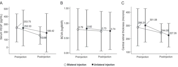
The serum VEGF level before and one month after the bilateral injection was 235.75 ± 183.16 pg/mL and 153.88 ± 113.26 pg/mL, respectively, representing a significant de- crease after the procedure (p = 0.037). Similarly, in the unilateral group, the serum VEGF level significantly de- creased after the procedure (252.53 ± 233.52 pg/mL to 189.42 ± 251.72 pg/mL, p = 0.019). However, there was no significant intergroup difference (p = 0.771).
The mean preoperative BCVA before the bilateral and unilateral injections was 0.79 ± 0.70 and 0.82 ± 0.62, respec- tively. The BCVA one month after the bilateral and unilater- al injections was 0.73 ± 0.67 and 0.74 ± 0.63, respectively.
There were statistically significant improvements in BCVA in both groups (bilateral injection group, p = 0.008; unilater- al injection group, p = 0.007). The mean preoperative CRT (µm) was 286.31 ± 140.80 in the bilateral group and 301.08 ± 127.58 in the unilateral group. CRT improved to 244.00 ± 69.73 and 227.35 ± 89.94 in the bilateral and unilateral groups, respectively, which were both statistically significant (p ≤ 0.001, 0.001, respectively). However, there were no sig- nificant intergroup differences in the pre- and postoperative BCVA and CRT values (Table 2 and Fig. 1).
A case of sudden intraocular pressure (IOP) elevation occurred in a 60-year-old woman who underwent bilateral intravitreal injection for wet AMD. The attack occurred in the right eye one day after the injection. The patient com- plained of a sudden headache with decreased visual acuity, and the IOP increased to 50 mmHg in the right eye. Slit lamp examination showed a shallow anterior chamber with diffuse corneal edema. Gonioscopy revealed a closed ante- rior chamber angle in 360 degrees. The patient had a histo- ry of an acute glaucoma attack in the opposite eye 20 years prior and had been treated with laser iridotomy. The medi- cal record showed bilateral shallow anterior chambers pri- or to the intravitreal injection. After laser iridotomy was performed on the right eye, the IOP was maintained at a steady-state for more than one year, during which the pa- tient received three additional bilateral simultaneous intra- vitreal injections. Serious complication events such as an- terior chamber inflammation, endophthalmitis, retinal
break, retinal detachment, cerebrovascular disease, and myocardial infarction did not occur in either group.
Discussion
With the increasing use of bevacizumab for VEGF-me- diated eye diseases, it is important to know the adverse systemic effects of intravitreal injection. In the CATT (Comparison of Age-related Macular Degeneration Treat- ments Trials) trial, acute myocardial infarction occurred in 0.7% of patients injected with 1.25 mg of bevacizumab monthly and in 0.3% of patients injected with bevacizum- ab as needed. Also, stroke events occurred in 0.7% and 0.7% of patients injected monthly and as needed, respec- tively, and venous thrombotic events occurred in 1.4% and 0.3% of the patients, respectively. These results were not statistically significant, but it remains unknown whether an increase in the systemic dose due to bilateral injections causes an increased risk [18].
The pharmacokinetics of bevacizumab have been deter- mined in other studies. In human nonvitrectomized eyes, the aqueous half-life of 1.5 mg intravitreally injected beva- cizumab is 8 to 10 days [19,20]. Recent studies have found that bevacizumab can flow into the serum via systemic circulation. In a study conducted on newborn infants, bev- acizumab concentration in the serum increased with time after injection into the vitreous cavity. Also, bevacizumab concentration reached its peak during the second week af- ter injection, which was inversely proportional to the VEGF level [21]. In another recent study in adult patients with diabetes, 24 hours after intravitreal injection of beva- cizumab, serum levels of VEGF were lower than basal lev- els, and the maximal reduction of serum VEGF was noted on the seventh postoperative day. Then, 28 days after the injection, VEGF levels increased to levels similar to those before the treatment [22]. The VEGF-lowering effect of bevacizumab has been known to diminish in the first few weeks after the injection. However, some studies have re- ported significantly decreased levels of blood VEGF even one month or more after intravitreal injection of bevaci- zumab [23,24]. In our study, it was impossible to obtain blood samples in the early postoperative period for ethical and technical reasons, and thus the data may be insuffi- cient to estimate the pharmacokinetics of intravitreally-in- jected bevacizumab. However, our results were in accor-
dance with previous studies showing that the effect of intravitreally-injected bevacizumab can last for one month.
Some researchers have reported a bilateral response after unilateral injection of bevacizumab [13-17]. Considering the fact that unilaterally-injected bevacizumab can reach the contralateral eye, it can be deduced that, after bilateral injection, a synergistic therapeutic effect can occur. How- ever, there were no significant intergroup differences in the preoperative and postoperative BCVA and CRT levels in our study.
In this study, there was no significant difference in the
preoperative and postoperative serum VEGF levels be- tween bilateral and unilateral intravitreal injections. These results suggest that bilateral injections have similar sys- temic effects compared to unilateral injections. However, it would take many more patients in order to assess the inci- dence of systemic complications. The reason why signifi- cant changes in serum level were not observed, even though more medicine was injected into the vitreous cavity, may be due to the relatively large distribution volume of the systemic circulation. The effect of bilateral injections in children or increasing the drug dose is still unknown. Ad- Table 1. Baseline characteristics
Bilateral injection Unilateral injection p-value
No. of patients 20 (40 eyes) 20 -
Age
Mean 64 63 0.828
Range 34-92 21-86 -
Gender
Male 11 (55%) 9 (45%) 0.527
Female 9 (45%) 11 (55%) -
Systemic diseases
Diabetes 10 (50%) 7 (35%) 0.337
Hypertension 8 (40%) 9 (45%) 0.749
Renal disease 0 0 NA
Malignant diseases/tumors 0 0 NA
Inflammatory/infectious disorders 0 0 NA
NA = not applicable.
Table 2. Mean serum concentration of VEGF and therapeutic outcome after bilateral intravitreal injection of bevacizumab Bilateral injection
(n = 20, 40 eyes) Unilateral injection
(n = 20) p-value*
Serum VEGF concentration (pg/mL)
Preoperative 235.75 ± 183.16 252.53 ± 233.52 0.62
Postoperative 1 month 153.88 ± 113.26 189.42 ± 251.72 0.77
Mean change -99.87 ± 146.75 -63.11 ± 29.51 0.53
p-value 0.04† 0.02†
BCVA (logMAR)
Preoperative 0.79 ± 0.70 0.82 ± 0.62 0.77
Postoperative 1 month 0.73 ± 0.67 0.74 ± 0.63 0.92
Mean change -0.06 ± 0.22 -0.08 ± 0.24 0.55
p-value‡ 0.008† 0.007†
CRT (µm)
Preoperative 286.31 ± 140.80 301.08 ± 127.58 0.51
Postoperative 1 month 244.00 ± 69.73 227.35 ± 89.94 0.37
Mean change -56.81 ± 92.53 -64.24 ± 102.32 0.74
p-value‡ <0.001† 0.001†
VEGF = vascular endothelial growth factor; BCVA = best-corrected visual acuity; logMAR = logarithm of the minimum angle of resolution; CRT = central retinal thickness.
*Mann-Whitney test; †p < 0.05; ‡Wilcoxon signed ranks test.
ditional causes for no changes in serum level could also be the blood sampling period of one month after the injection, which is a limitation of this study. Since the serum half- life of bevacizumab is known to be less than one week [22,24], the difference in serum VEGF level between the two groups one month after the injection would have been less than that in the early postoperative period. Future studies that include multiple blood tests from the early postoperative period will help more clearly determine the serum drug levels.
During this study, one serious complication, a sudden el- evation of IOP requiring emergency treatment, occurred.
Ordinarily, the IOP increases temporarily immediately af- ter the intravitreal injection of an anti-VEGF but decreases naturally with time [25,26]. However, recent studies have shown that IOP elevations can occur over a prolonged peri- od. An increase in vitreous volume, an inflammatory reac- tion of the aqueous humor or meshwork, direct damage to outflow tracks, and meshwork obstruction caused by high-molecular weight medicines such as bevacizumab are cited as the causes of the IOP increase [27-29]. It is rare for acute angle-closure glaucoma to occur after bilateral si- multaneous intravitreal injection, with only one case being reported to date [30]. The case in this study occurred in a patient who had a previous angle-closure glaucoma attack in the opposite eye. The vitreous humor increased in volume after the intravitreal injection and narrowed the anterior chamber angle. This may have created conditions suscepti-
ble to angle-closure attack. Accordingly, in patients who have a history of glaucoma or a shallow anterior chamber angle, intravitreal injection requires meticulous attention.
In recent years, researchers have reported on the safety of bilateral simultaneous intravitreal injections of anti-VEGFs.
Several studies have revealed that the complication rate af- ter bilateral intravitreal injections was not higher than that of unilateral injection [9-12,31]. However, there is still con- troversy on bilateral intravitreal injections of anti-VEGFs due to possible complications. Arteriothrombotic or venous thrombotic events, hypertension, and death are considered to be serious complications. For patients having cardiovas- cular risk factors, intravitreal injections require meticulous attention. If the patient has a history of a major systemic events such as stroke, cardiac arrest, or uncontrolled hy- pertension within the previous three months, intravitreal anti-VEGF injection should be avoided [32-35].
Our study has several limitations including a nonran- domized uncontrolled study, a small number of subjects, and short-term follow-up. In addition, postoperative blood tests were performed only once at one month post-injec- tion. Since it was shown in a newborn study that an in- crease in the serum concentration of bevacizumab after being injected into the vitreous cavity reaches its peak during the second week [21], our results may not demon- strate the lowest level of serum VEGF.
To our knowledge, there have been no studies comparing the serum VEGF levels in bilateral and unilateral intravit-
0 100
253.75 252.53
189.42 0.79 0.82 0.73 0.74
301.08 286.31
244.00 227.35 153.88
Preinjection
Serum VEGF (pg/mL)
Postinjection 200
300 400 500
Preinjection
BCVA (logMAR)
Postinjection 1.50
1.00
0.50
0.00
Preinjection
Central retinal thickness (microns)
Postinjection 400
300
200
100
Bilateral injection Unilateral injection
A B C
Fig. 1. Mean serum concentration of vascular endothelial growth factor (VEGF), best-corrected visual acuity (BCVA), and central retinal thickness before and 1 month after intravitreal injection of bevacizumab for wet age-related macular degeneration. logMAR = logarithm of the minimum angle of resolution.
real injections. In addition, BCVA and CRT did not differ between patients undergoing bilateral and unilateral intra- vitreal injections. Our results are encouraging; however, in order to understand the exact pharmacokinetic features and the impact of bilateral intravitreal injection on the sys- temic circulation, larger future studies are needed.
In conclusion, bilateral and unilateral intravitreal injec- tions of bevacizumab do not show significant differences with regard to postoperative serum VEGF level and thera- peutic outcome. This study has again demonstrated the ef- fectiveness of bilateral simultaneous intravitreal injection.
In-depth study of bilateral injection of bevacizumab with a large number of patients is still required.
Conflict of Interest
No potential conflict of interest relevant to this article was reported.
Acknowledgements
This work was supported by the Soonchunhyang Uni- versity Research Fund.
References
1. Lalwani GA, Rosenfeld PJ, Fung AE, et al. A variable-dos- ing regimen with intravitreal ranibizumab for neovascular age-related macular degeneration: year 2 of the PrONTO Study. Am J Ophthalmol 2009;148:43-58.
2. Engelbert M, Zweifel SA, Freund KB. “Treat and extend”
dosing of intravitreal antivascular endothelial growth fac- tor therapy for type 3 neovascularization/retinal angioma- tous proliferation. Retina 2009;29:1424-31.
3. Regillo CD, Brown DM, Abraham P, et al. Randomized, double-masked, sham-controlled trial of ranibizumab for neovascular age-related macular degeneration: PIER Study year 1. Am J Ophthalmol 2008;145:239-48.
4. Rosenfeld PJ, Rich RM, Lalwani GA. Ranibizumab: phase III clinical trial results. Ophthalmol Clin North Am 2006;19:
361-72.
5. Rosenfeld PJ, Brown DM, Heier JS, et al. Ranibizumab for neovascular age-related macular degeneration. N Engl J
Med 2006;355:1419-31.
6. Brown DM, Kaiser PK, Michels M, et al. Ranibizumab versus verteporfin for neovascular age-related macular de- generation. N Engl J Med 2006;355:1432-44.
7. Kawasaki R, Wang JJ, Amirul FM, et al. Is bilateral age-re- lated macular degeneration less common in Asians than Caucasians? Ophthalmic Epidemiol 2011;18:253-8.
8. Macular Photocoagulation Study Group. Five-year fol- low-up of fellow eyes of patients with age-related macular degeneration and unilateral extrafoveal choroidal neovas- cularization. Arch Ophthalmol 1993;111:1189-99.
9. Mahajan VB, Elkins KA, Russell SR, et al. Bilateral intra- vitreal injection of antivascular endothelial growth factor therapy. Retina 2011;31:31-5.
10. Davis RP, Schefler AC, Murray TG. Concomitant bilateral intravitreal anti-VEGF injections for the treatment of exu- dative age-related macular degeneration. Clin Ophthalmol 2010;4:703-7.
11. Lima LH, Zweifel SA, Engelbert M, et al. Evaluation of safety for bilateral same-day intravitreal injections of anti- vascular endothelial growth factor therapy. Retina 2009;29:
1213-7.
12. Bakri SJ, Risco M, Edwards AO, Pulido JS. Bilateral si- multaneous intravitreal injections in the office setting. Am J Ophthalmol 2009;148:66-9.
13. Hosseini H, Lotfi M, Esfahani MH, et al. Effect of intravit- real bevacizumab on retrobulbar blood flow in injected and uninjected fellow eyes of patients with neovascular age-re- lated macular degeneration. Retina 2012;32:967-71.
14. Matsuyama K, Ogata N, Matsuoka M, et al. Effects of in- travitreally injected bevacizumab on vascular endothelial growth factor in fellow eyes. J Ocul Pharmacol Ther 2011;
27:379-83.
15. Al-Dhibi H, Khan AO. Bilateral response following unilat- eral intravitreal bevacizumab injection in a child with uve- itic cystoid macular edema. J AAPOS 2009;13:400-2.
16. Wu Z, Sadda SR. Effects on the contralateral eye after in- travitreal bevacizumab and ranibizumab injections: a case report. Ann Acad Med Singapore 2008;37:591-3.
17. Avery RL, Pearlman J, Pieramici DJ, et al. Intravitreal bev- acizumab (Avastin) in the treatment of proliferative diabet- ic retinopathy. Ophthalmology 2006;113:1695.
18. CATT Research Group, Martin DF, Maguire MG, et al.
Ranibizumab and bevacizumab for neovascular age-related macular degeneration. N Engl J Med 2011;364:1897-908.
19. Meyer CH, Krohne TU, Holz FG. Intraocular pharmacoki-
netics after a single intravitreal injection of 1.5 mg versus 3.0 mg of bevacizumab in humans. Retina 2011;31:1877-84.
20. Krohne TU, Eter N, Holz FG, Meyer CH. Intraocular phar- macokinetics of bevacizumab after a single intravitreal in- jection in humans. Am J Ophthalmol 2008;146:508-12.
21. Sato T, Wada K, Arahori H, et al. Serum concentrations of bevacizumab (avastin) and vascular endothelial growth factor in infants with retinopathy of prematurity. Am J Ophthalmol 2012;153:327-33.
22. Davidovic SP, Nikolic SV, Curic NJ, et al. Changes of se- rum VEGF concentration after intravitreal injection of Avastin in treatment of diabetic retinopathy. Eur J Oph- thalmol 2012;22:792-8.
23. Ma Y, Zhang Y, Zhao T, Jiang YR. Vascular endothelial growth factor in plasma and vitreous fluid of patients with proliferative diabetic retinopathy patients after intravitreal injection of bevacizumab. Am J Ophthalmol 2012;153:307-13.
24. Matsuyama K, Ogata N, Matsuoka M, et al. Plasma levels of vascular endothelial growth factor and pigment epitheli- um-derived factor before and after intravitreal injection of bevacizumab. Br J Ophthalmol 2010;94:1215-8.
25. Gismondi M, Salati C, Salvetat ML, et al. Short-term effect of intravitreal injection of Ranibizumab (Lucentis) on in- traocular pressure. J Glaucoma 2009;18:658-61.
26. Falkenstein IA, Cheng L, Freeman WR. Changes of intra- ocular pressure after intravitreal injection of bevacizumab (avastin). Retina 2007;27:1044-7.
27. Choi DY, Ortube MC, McCannel CA, et al. Sustained ele- vated intraocular pressures after intravitreal injection of bevacizumab, ranibizumab, and pegaptanib. Retina 2011;31:
1028-35.
28. Adelman RA, Zheng Q, Mayer HR. Persistent ocular hy- pertension following intravitreal bevacizumab and ranibi- zumab injections. J Ocul Pharmacol Ther 2010;26:105-10.
29. Jalil A, Fenerty C, Charles S. Intravitreal bevacizumab (Avastin) causing acute glaucoma: an unreported complica- tion. Eye (Lond) 2007;21:1541.
30. Semoun O, Blumen-Ohana E, de Preobrajensky N, Nord- mann JP. Acute angle-closure glaucoma complicating an in- travitreal injection of bevacizumab. J Fr Ophtalmol 2009;
32:58.
31. Woo SJ, Han JM, Ahn J, et al. Bilateral same-day intravit- real injections using a single vial and molecular bacterial screening for safety surveillance. Retina 2012;32:667-71.
32. Rasier R, Artunay O, Yuzbasioglu E, et al. The effect of in- travitreal bevacizumab (avastin) administration on system- ic hypertension. Eye (Lond) 2009;23:1714-8.
33. Chung YR, Lee K, Cho EH, Lew HM. Blood pressure changes after intravitreal bevacizumab in patients grouped by ocular pathology. Eye (Lond) 2010;24:1320-4.
34. Comparison of Age-related Macular Degeneration Treat- ments Trials (CATT) Research Group, Martin DF, Maguire MG, et al. Ranibizumab and bevacizumab for treatment of neovascular age-related macular degeneration: two-year re- sults. Ophthalmology 2012;119:1388-98.
35. IVAN Study Investigators, Chakravarthy U, Harding SP, et al.
Ranibizumab versus bevacizumab to treat neovascular age-re- lated macular degeneration: one-year findings from the IVAN randomized trial. Ophthalmology 2012;119:1399-411.