Effect of Intravitreal Bevacizumab on Vascular Endothelial Growth Factor Expression in Patients with Proliferative
Diabetic Retinopathy
Eun Jee Chung,
1Shin Jeong Kang,
2Ja Seung Koo,
3Yoon Jung Choi,
4Hans E. Grossniklaus,
2,5and Hyoung Jun Koh
6Departments of 1Ophthalmology and 4Pathology, NHIC Ilsan Hospital, Goyang, Korea;
Departments of 2Ophthalmology and 5Pathology, Emory Eye Center, Emory University School of Medicine, Atlanta, Georgia, USA;
3Department of Pathology and 6The Institute of Vision Research, Department of Ophthalmology, Yonsei University College of Medicine, Seoul, Korea.
Received: April 22, 2010 Revised: May 27, 2010 Accepted: May 27, 2010
Corresponding author: Dr. Hyoung Jun Koh, Department of Ophthalmology,
Yonsei University College of Medicine, 250 Seongsan-ro, Seodaemun-gu, Seoul 120-752, Korea.
Tel: 82-2-2228-3570, Fax: 82-2-312-0541 E-mail: [email protected]
∙ The authors have no financial conflicts of interest.
© Copyright:
Yonsei University College of Medicine 2011 This is an Open Access article distributed under the terms of the Creative Commons Attribution Non- Commercial License (http://creativecommons.org/
licenses/by-nc/3.0) which permits unrestricted non- commercial use, distribution, and reproduction in any medium, provided the original work is properly cited.
Purpose: To investigate the effect of bevacizumab (Avastin; Genentech, San Francis- co, CA, USA) on vascular endothelial growth factor (VEGF) expression and inflam- mation in fibrovascular membranes in patients with proliferative diabetic retinopathy (PDR). Materials and Methods: Fibrovascular membranes from 19 eyes of 18 pa- tients with PDR were studied using immunohistochemistry and analyzed in the fol- lowing 3 groups; group 1: 4 inactive PDR eyes, group 2: 10 active PDR eyes treated preoperatively with adjunctive intravitreal bevacizumab, group 3: five active PDR eyes not treated preoperatively with bevacizumab. Immunohistochemical staining for VEGF, CD31 and CD68 were done. Results: The immunoreactivity to VEGF and CD 31-positive blood vessels was significantly higher in membranes from group 3 than group 1 (p = 0.007 for VEGF, 0.013 for CD 31-positive vessels). Intravitreal bev- acizumab caused a reduction in VEGF expression and vascular densities in 4 out of 10 (40%) excised membranes from eyes with PDR. However, six membranes (60%) in group 2 still demonstrated relatively strong VEGF expression and high vascular density. Infiltration of macrophages was observed in 16 out of the 19 membranes, and the density of macrophages was increased in group 2 compared with group 1 (p = 0.043). Conclusion: Intravitreal bevacizumab injections caused some reduction in VEGF expression and vascular densities in a limited number of active PDR patients.
A single intravitreal bevacizumab injection may not be enough to induce complete blockage of VEGF and pathologic neovascularization in active PDR patients. Repeat- ed injections, panretinal photocoagulation and/or PPV may be necessary following in- travitreal bevacizumab to reinforce the anti-VEGF effect of the drug.
Key Words: Intravitreal bevacizumab, proliferative diabetic retinopathy, vascular endothelial growth factor
INTRODUCTION
Proliferative diabetic retinopathy (PDR) is characterized by a pathological growth
tween 1 August 2006 and 28 February 2007. The study fol- lowed the tenets of Declaration of Helsinki and was ap- proved by the local Institutional Review Board. Signed and written informed consent was obtained from each patient.
The surgically removed fibrovascular membranes were classified into those with inactive (group 1) and active neo- vascularization. Neovascularization was considered to be active if the new vessels were perfused and had multi- branched preretinal capillaries, and considered to be inac- tive if a previously documented active proliferation had re- gressed, or if gliotic vessels or fibrosis were prominent. Of 15 active PDR eyes, 10 eyes had received an intravitreal in- jection of 1.25 mg bevacizumab one week before planned pars plana vitrectomy (PPV) (group 2), and 5 eyes had re- ceived PPV only (group 3).
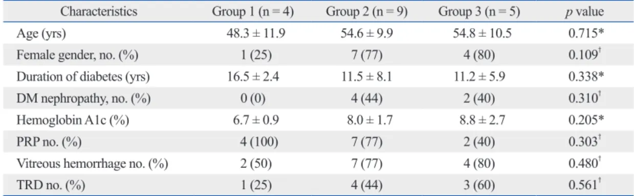
Ophthalmic examinations, including visual acuity and in- traocular pressure measurement, slit lamp examination, di- lated fundus examination, fluorescein angiography and col- or fundus photography, were performed at baseline. The patients’ clinical data are summarized in Table 1.
Tissue processing
The surgically excised proliferative fibrovascular mem- branes were fixated in 10% formalin solution in the operat- ing room and embedded in paraffin. Sections, 4 μm thick, were deparaffinized and prepared for immunohistochemi- cal study and hematoxylin-eosin (H&E) staining.
Immunohistochemical staining
Immunohistochemical analysis was performed using the in- direct immunoperoxidase staining technique. Endogenous peroxidase was blocked by methanol/hydrogen peroxide.
The poly-clonal rabbit anti-VEGF antibody (Santa Cruz, CA, USA) was diluted (1 : 300) in a 0.05-mol/L concentra- tion of tris (hydroxymethyl) aminomethane-buffered saline (pH 7.5) plus 1% bovine serum albumin and applied for 60 minutes. After washing, the slides were incubated with bio- tinylated goat antirabbit IgG for 30 minutes, washed and ex- posed to avidin-peroxidase complex. Three-amino-9-ethyl- carbazole (AEC) was used as the chromogen. The slides were counter-stained with Mayer hematoxylin. Observa- tions of the immunostaining were made under a light mi- croscope. When the specimens were too small to provide sufficient sections for all the immunostaining procedures, they were discarded from the study. Omission of the prima- ry antibody in a matched serial epiretinal membrane sec- tion, which lacks the specific staining of the respective pri- of new blood vessels that often results in catastrophic loss
of vision due to vitreous hemorrhage and/or tractional reti- nal detachment (TRD).
Vascular endothelial growth factor (VEGF) is an angio- genic mitogen, and the involvement of VEGF in PDR has been suggested by studies that have demonstrated that the vitreous humor from eyes with PDR contains a large amount of VEGF,1,2 and that VEGF is expressed in fibro- vascular tissues obtained from PDR patients.3-5 The discov- ery and cloning of VEGF in 1989 and subsequent develop- ment of antibodies to it allowed for the identification of VEGF’s key role in the development of retinal neovascular- ization in PDR.6,7 In addition to the key role of VEGF in the development of PDR, there is evidence that chronic inflam- mation may also be involved, as several studies have shown elevated levels of inflammatory markers in patients with diabetic retinopathy.8-10
Bevacizumab (Avastin, Genentech, San Francisco, CA, USA) is a humanized recombinant antibody that binds to most isoforms of VEGF.7 Recent reports have shown that intravitreal bevacizumab is well-tolerated in the short term for treatment of PDR.11-13 Beneficial effects of intravitreal bevacizumab as a preoperative adjunct for TRD in PDR has been reported.14,15 Previous studies have also demon- strated that intravitreal injection of bevacizumab in PDR patients significantly reduces the level of VEGF in aqueous humor.16-18 Although a reduction of the VEGF level in aque- ous humor and the regression of fibrovascular proliferation have been observed after clinical treatment intravitreal bev- acizumab, the effect of bevacizumab on retinal neovascular membranes is unknown.
In this study, we investigated the clinicohistopathology of surgically excised fibrovascular membranes from PDR pa- tients treated with an intravitreal bevacizumab injection pri- or to surgery. The expression of VEGF was determined by immunostaining, and the level of vascularization was eval- uated by immunodetection of the endothelial marker CD31.
The inflammatory infiltration was evaluated using an anti- body against CD68 as a marker for macrophages.
MATERIALS AND METHODS
Patients
Fibrovascular membranes were obtained from 19 eyes of 18 patients during vitrectomy at Yonsei University Eye &
Ear, Nose, and Throat Hospital vitreoretinal service, be-
Nemenyi-Damico-Wolfe-Dunn test. Statistical analyses were performed with the R (2.8.1) statistics program. The level of statistical significance was set at p < 0.05, and the results are expressed as mean ± standard deviation.
RESULTS
Histopathological examinations
The specimens from group 1 had sparse vascularized tissue with abundant extracellular matrix and fibrosis. The active neovascular membranes from group 3 were composed of highly vascularized fibrovascular tissue, and the vessels were surrounded by a loosely coherent extracellular matrix.
Four out of 10 specimens from group 2 demonstrated re- gressed vascular channels, with the remaining six speci- mens showing active PDR characteristics including highly vascularized tissue. The histopathological findings correlat- ed well with the clinical findings in the four eyes with re- gressed active PDR, with the membranes composed of large caliber vessels and fibroglial tissue (Fig. 1).
Immunohistochemistry
The results of immunohistochemical staining are summa- rized in Table 2. Immunoreactivity to VEGF was detected in the endothelial cells of newly formed vessels in the ex- cised fibrovascular membranes. The immunoreactivity to VEGF was 0.5 ± 0.6 in group 1, 2.0 ± 0.9 in group 2 and 2.6
± 0.6 in group 3 (Fig. 2). The numbers of CD31-positive blood vessels were 1.3 ± 2.5 in group 1, 11.6 ± 8.4 in group 2 and 17.0 ± 10.4 in group 3 (Fig. 3). The immunoreactivity to VEGF and the number of CD31-positive blood vessels were significantly higher in membranes from group 3 than mary antibody, was used as a negative control.
To study endothelial cell and macrophage distribution, monoclonal mouse antibodies against CD31 (1 : 100 dilu- tion; DakoCytomation, Glostrup, Denmark) and CD68 (1 : 100 dilution; DakoCytomation, Glostrup, Denmark) were used to visualize endothelial cells and macrophages, re- spectively. Counting of CD31 and 68-positive cells and im- munoreactivity scoring for VEGF were performed by an independent observer (JSK) with no knowledge of the pa- tients’ clinical information.
The immunoreactivity for VEGF was analyzed and semi- quantitatively scored on a scale from 0 to 3 depending on the staining intensity as follows: 0) no detectable staining in the specimen; 1) weak; 2) intermediate; 3) strong.
Immunohistochemical staining of vascular densities for CD31, which stains vascular endothelial cells, was con- ducted.19 The number of CD31-positive vessels were count- ed at ×400 power magnification covering the whole stromal area of the membrane and expressed as the average of the three highest readings. Any endothelial cells or clusters that were stained brown and clearly separated from the connec- tive tissue elements were considered as single countable microvessels. The number of CD68-positive cells was counted at ×400 power magnification covering the whole stromal area of the membrane and expressed as the average of the three highest readings.
Statistical analysis
The baseline demographic and clinical parameters were compared between treatment groups using a Kruskal-Wallis variance analysis for continuous variables and chi-squared tests for categorical variables. Comparisons of study end- points using comparisons between groups employed the
Table 1. Patient Demographic Data
Characteristics Group 1 (n = 4) Group 2 (n = 9) Group 3 (n = 5) p value
Age (yrs) 48.3 ± 11.9 54.6 ± 9.9 54.8 ± 10.5 0.715*
Female gender, no. (%) 1 (25) 7 (77) 4 (80) 0.109†
Duration of diabetes (yrs) 16.5 ± 2.4 11.5 ± 8.1 11.2 ± 5.9 0.338*
DM nephropathy, no. (%) 0 (0) 4 (44) 2 (40) 0.310†
Hemoglobin A1c (%) 6.7 ± 0.9 8.0 ± 1.7 8.8 ± 2.7 0.205*
PRP no. (%) 4 (100) 7 (77) 2 (40) 0.303†
Vitreous hemorrhage no. (%) 2 (50) 7 (77) 4 (80) 0.480†
TRD no. (%) 1 (25) 4 (44) 3 (60) 0.561†
n, number of patients; PDR, proliferative diabetic retinopathy; DM, diabetes mellitus; PRP, panretinal photocoagulation; TRD, tractional retinal detachment.
*Kruskal-Wallis variance analysis.
†Chi-square test.
expression, vascular density and macrophage infiltration were not significantly different between groups 2 and 3.
There were significant correlations between immunoreac- tivity to VEGF and the number of CD31-positive vessels (r
= 0.824, p < 0.001, Spearman correlation) and the number of CD68-positive macrophages (r = 0.485, p = 0.035).
DISCUSSION
We observed that immunoreactivity to VEGF was significant- ly stronger in membranes from active PDR compared to those from inactive PDR. The number of CD31-expressing blood vessels was also significantly higher in the active group. The those from group 1 (p = 0.007 for VEGF, 0.013 for CD
31-positive vessels, Nemenyi-Damico-Wolfe-Dunn test).
Intravitreal bevacizumab caused a reduction in VEGF ex- pression and vascular densities in four out of 10 eyes (40%) in group 2, and the histopathological findings in the excised membranes showed regressed PDR (Figs. 1B, 2B, and 3B).
However, six eyes (60%) in group 2 exhibited strong VEGF expression and high vascular densities even after the intravitreal bevacizumab injections (Figs. 1C, 2C, and 3C).
Infiltration of CD68-positive macrophages was observed in 16 out of 19 membranes. The number of CD68-positive macrophages was 1.5 ± 2.4 in group 1, 18.5 ± 8.4 in group 2 and 8.8 ± 5.7 in group 3 (Fig. 4). The number was signifi- cantly larger in group 2 than in group 1 (p = 0.043). VEGF
A B C D
Fig. 1. Representative fundus photographs and histopathologic findings. (A) Group 1. A fibrotic fibrovascular membrane can be seen in this fundus photo. This section of excised tissue shows sparsely vascularized fibrovascular tissue in H&E staining. (B) Group 2 with regression of active PDR. Regressed NVD, large caliber vessels and gliosis are evident. Sparsely vascularized tissue is present in H&E staining. (C) Group 2 with active PDR. (D) Group 3. Proliferative fibrovascu- lar membranes and preretinal hemorrhage can be seen. H&E staining shows highly vascularized tissue (original magnification × 400). PDR, proliferative diabetic retinopathy; NVD, neovascular at the disc.
A B C D
Fig. 2. Immunohistochemistry for vascular endothelial growth factor (VEGF) expression. (A) Group 1. Weak immunoreactivity to VEGF is observed in fibrovascular tissue. (B) Group 2 with regression of active proliferative diabetic retinopathy (PDR). The immunoreactivity to VEGF shows intermediate staining in vascular en- dothelial cells surrounding the vascular lumen. (C) Group 2 with active PDR (D) Group 3. Strong immunoreactivity to VEGF is shown (original magnification × 400).
Table 2. The Results of Immunohistochemical Staining
Primary antibody Group 1 (n = 4) Group 2 (n = 10) Group 3 (n = 5)
VEGF (SD) 0.5 (0.6) 2.0 (0.9) 2.6 (0.6)*
CD31-positve vessels (SD) 1.3 (2.5) 11.6 (8.4) 17.0 (10.4)*
CD68-positive cells (SD) 1.5 (2.4) 18.5 (8.4)* 8.8 (5.7)
n, number of eyes; VEGF, vascular endothelial growth factor; SD, standard deviation.
*indicates p < 0.05 compared to the group 1, Nemenyi-Damico-Wolfe-Dunn test.
Based on the observations of the current study and a recent report by Kubota, et al.20 vascular endothelial cells would not disappear in all patients in a short period after the injec- tion of bevacizumab. On the contrary, they showed signifi- cant decreases in VEGF expression in fibrovascular mem- branes after a bevacizumab injection, which is contradictory to our results.20 The tissue-expressed VEGF level may vary greatly among PDR patients, and a single bevacizumab in- jection would result in a different level of VEGF expression.
Further studies of larger groups are warranted to verify these findings. Some reports also demonstrated a clinically favor- able outcome associated with intravitreal bevacizumab in active PDR patients. However, even in those reports, partial regression of the active PDR was observed in 34 to 41% of the patients, indicating that a single bevacizumab injection may not be enough to cause complete regression of active PDR.13,21
VEGF is also a chemotactic factor for inflammatory cells, activating monocytes and macrophages, and induces the expression of ICAM-1 and E-selectin on vascular endo- thelial cells to facilitate adhesion and migration of inflam- matory cells.22-27 Since angiogenesis and inflammation are closely related, we hypothesized that VEGF inhibition by bevacizumab may influence new vessel formation as well as inflammatory activity in PDR membranes. In specimens treated with bevacizumab, the number of CD68-positive macrophages was comparable to that of active PDR cases without preoperative adjunctive therapy, and significantly results are consistent with previous reports that have demon-
strated increased VEGF levels in both vitreous humor and se- rum, and highly expressed VEGF in the endothelial cells of fi- brovascular membranes from eyes of PDR patients.1-5 There was a significant correlation between the immunoreactivity to VEGF and the number of CD31-positive vessels, supporting the theory that VEGF may play an important role in the pro- gression of neovascularization in PDR.1,2
In 40% of the eyes that were treated with intravitreal bev- acizumab injection and PPV, significant decreases in VEGF expression and vascular densities were observed. In those eyes, the decrease in immunoreactivity to VEGF and vas- cular densities correlated with the regression of NVD and large caliber vessels and the presence of fibroglial tissue comprising the NVE. However, relatively strong expres- sion of VEGF and high vascular densities were observed in the other 60% of membranes, even after preoperative ad- junct bevacizumab injection. In contrast with our observa- tions, previous studies have shown a significant reduction in VEGF level in both the vitreous and aqueous humor in active PDR patients after a single intravitreal bevacizumab injection.16-18 However, these studies reported levels of free VEGF in the vitreous or aqueous humor, unlike the tissue- expressed level as measured in the current study. Kubota, et al.19,20 also demonstrated that vascular endothelial cells were still present in the fibrovascular membranes from PDR patients and trabecular meshwork of neovascular glaucoma even after the intravitreal bevacizumab injection.
A B C D
Fig. 3. Immunohistochemistry for CD31. (A) Group 1. CD31-positive blood vessels are barely visible in fibrovascular tissue. (B) Group 2 with regression of active PDR. Vascular lumen with CD31-positive endothelial cells is visible. (C) Group 2 with active PDR. (D) Group 3. Highly vascularized tissue with the vascular lumen surrounded by CD31-positive cells (original magnification × 400).
A B C
Fig. 4. Immunohistochemistry for CD68. (A) Group 1. CD68-positive cells are barely visible in fibrovascular tissue. (B) Group 2. A significant increase in CD68- positive cells can be seen when compared with (A). (C) Group 3. Moderate increase in CD68-positive cells can be seen (original magnification × 400).
REFERENCES
1. Adamis AP, Miller JW, Bernal MT, D’Amico DJ, Folkman J, Yeo TK, et al. Increased vascular endothelial growth factor levels in the vitreous of eyes with proliferative diabetic retinopathy. Am J Ophthalmol 1994;118:445-50.
2. Aiello LP, Avery RL, Arrigg PG, Keyt BA, Jampel HD, Shah ST, et al. Vascular endothelial growth factor in ocular fluid of patients with diabetic retinopathy and other retinal disorders. N Engl J Med 1994;331:1480-7.
3. Boulton M, Foreman D, Williams G, McLeod D. VEGF localisa- tion in diabetic retinopathy. Br J Ophthalmol 1998;82:561-8.
4. Chen YS, Hackett SF, Schoenfeld CL, Vinores MA, Vinores SA, Campochiaro PA. Localisation of vascular endothelial growth fac- tor and its receptors to cells of vascular and avascular epiretinal membranes. Br J Ophthalmol 1997;81:919-26.
5. Matsuoka M, Ogata N, Minamino K, Matsumura M. Expression of pigment epithelium-derived factor and vascular endothelial growth factor in fibrovascular membranes from patients with pro- liferative diabetic retinopathy. Jpn J Ophthalmol 2006;50:116-20.
6. Adamis AP, Shima DT. The role of vascular endothelial growth factor in ocular health and disease. Retina 2005;25:111-8.
7. Ferrara N. Vascular endothelial growth factor: basic science and clinical progress. Endocr Rev 2004;25:581-611.
8. Bhavsar AR. Diabetic retinopathy: the latest in current manage- ment. Retina 2006;26:S71-9.
9. Meleth AD, Agrón E, Chan CC, Reed GF, Arora K, Byrnes G, et al. Serum inflammatory markers in diabetic retinopathy. Invest Ophthalmol Vis Sci 2005;46:4295-301.
10. Wellen KE, Hotamisligil GS. Inflammation, stress, and diabetes. J Clin Invest 2005;115:1111-9.
11. Minnella AM, Savastano CM, Ziccardi L, Scupola A, Falsini B, Balestrazzi E. Intravitreal bevacizumab (Avastin) in proliferative diabetic retinopathy. Acta Ophthalmol 2008;86:683-7.
12. Moradian S, Ahmadieh H, Malihi M, Soheilian M, Dehghan MH, Azarmina M. Intravitreal bevacizumab in active progressive pro- liferative diabetic retinopathy. Graefes Arch Clin Exp Ophthalmol 2008;246:1699-705.
13. Avery RL, Pearlman J, Pieramici DJ, Rabena MD, Castellarin AA, Nasir MA, et al. Intravitreal bevacizumab (Avastin) in the treatment of proliferative diabetic retinopathy. Ophthalmology 2006;113:1695.e1-15.
14. Oshima Y, Shima C, Wakabayashi T, Kusaka S, Shiraga F, Ohji M, et al. Microincision vitrectomy surgery and intravitreal bevaci- zumab as a surgical adjunct to treat diabetic traction retinal detach- ment. Ophthalmology 2009;116:927-38.
15. Chen E, Park CH. Use of intravitreal bevacizumab as a preopera- tive adjunct for tractional retinal detachment repair in severe pro- liferative diabetic retinopathy. Retina 2006;26:699-700.
16. Arimura N, Otsuka H, Yamakiri K, Sonoda Y, Nakao S, Noda Y, et al. Vitreous mediators after intravitreal bevacizumab or triam- cinolone acetonide in eyes with proliferative diabetic retinopathy.
Ophthalmology 2009;116:921-6.
17. Matsuyama K, Ogata N, Jo N, Shima C, Matsuoka M, Matsumura M. Levels of vascular endothelial growth factor and pigment epi- thelium-derived factor in eyes before and after intravitreal injec- tion of bevacizumab. Jpn J Ophthalmol 2009;53:243-8.
18. Sawada O, Kawamura H, Kakinoki M, Sawada T, Ohji M. Vascu- lar endothelial growth factor in aqueous humor before and after
higher than numbers observed in the inactive group. This may be a consequence of bevacizumab’s actions to in- crease inflammation in the process of new vessel regres- sion. Inhibition of VEGF has been shown to induce vascu- lar regression through endothelial cell apoptosis in neovascular vessels.28-30 Macrophages recognize and phagocytose apoptotic cells, and their depletion results in endothelial cell survival and persistence of functional capil- laries.31-33 During vascular regression, macrophages may be recruited in the process of endothelial cell apoptosis. Tatar, et al.34 also reported an unexpected increase in inflammato- ry activity in human choroidal neovascular tissues after in- travitreal bevacizumab. Our data corroborate this finding and further indicate that intravitreal bevacizumab injection may stimulate an inflammatory cellular reaction shortly af- ter the drug application.
Our sample population is small and the results are limit- ed. The results of this study might also be influenced by a selection bias because the membranes were obtained from the PDR patients without randomization. Further investiga- tions involving a larger group with appropriate randomiza- tion and a control group will be necessary to confirm our preliminary findings. In addition, the effects of intravitreal bevacizumab on VEGF expression were studied only in the fibrovascular membranes in this study. Since intravitreal bevacizumab injections may also affect VEGF expression in the neurosensory retina, including retinal vasculature, and induce possible vascular regression and ischemia as shown in previous case reports,35,36 further studies should include the effect of bevacizumab on the neurosensory reti- na, especially with vascular perfusion.
In summary, we observed that intravitreal bevacizumab injections cause a significant reduction in VEGF expression and vascular density in a limited number of patients. A sin- gle intravitreal bevacizumab injection may be insufficient to induce a complete blockage of VEGF on fibrovascular membranes in active PDR patients. Repeated injections, panretinal photocoagulation and/or PPV may be necessary following intravitreal bevacizumab in order to reinforce its anti-VEGF effect and result in complete regression of ac- tive PDR to an inactive membrane.
ACKNOWLEDGEMENTS
This work was supported by the National Health Insurance Corporation Ilsan Hospital grant (2009-31).
flammatory cytokines. Circ Res 1998;82:845-51.
27. Sunderkötter C, Steinbrink K, Goebeler M, Bhardwaj R, Sorg C.
Macrophages and angiogenesis. J Leukoc Biol 1994;55:410-22.
28. Alon T, Hemo I, Itin A, Pe’er J, Stone J, Keshet E. Vascular endo- thelial growth factor acts as a survival factor for newly formed ret- inal vessels and has implications for retinopathy of prematurity.
Nat Med 1995;1:1024-8.
29. Baffert F, Le T, Sennino B, Thurston G, Kuo CJ, Hu-Lowe D, et al. Cellular changes in normal blood capillaries undergoing regres- sion after inhibition of VEGF signaling. Am J Physiol Heart Circ Physiol 2006;290:H547-59.
30. Kamba T, Tam BY, Hashizume H, Haskell A, Sennino B, Mancu- so MR, et al. VEGF-dependent plasticity of fenestrated capillaries in the normal adult microvasculature. Am J Physiol Heart Circ Physiol 2006;290:H560-76.
31. Diez-Roux G, Argilla M, Makarenkova H, Ko K, Lang RA. Mac- rophages kill capillary cells in G1 phase of the cell cycle during programmed vascular regression. Development 1999;126:2141-7.
32. Diez-Roux G, Lang RA. Macrophages induce apoptosis in normal cells in vivo. Development 1997;124:3633-8.
33. Lang RA. Apoptosis in mammalian eye development: lens mor- phogenesis, vascular regression and immune privilege. Cell Death Differ 1997;4:12-20.
34. Tatar O, Yoeruek E, Szurman P, Bartz-Schmidt KU; Tübingen Be- vacizumab Study Group, Adam A, et al. Effect of bevacizumab on inflammation and proliferation in human choroidal neovascular- ization. Arch Ophthalmol 2008;126:782-90.
35. Chen E, Hsu J, Park CH. Acute visual acuity loss following intra- vitreal bevacizumab for diabetic macular edema. Ophthalmic Surg Lasers Imaging 2009;40:68-70.
36. Shimura M, Yasuda K. Macular ischaemia after intravitreal beva- cizumab injection in patients with central retinal vein occlusion and a history of diabetes and vascular disease. Br J Ophthalmol 2010;94:381-3.
intravitreal injection of bevacizumab in eyes with diabetic retinop- athy. Arch Ophthalmol 2007;125:1363-6.
19. Kubota T, Aoki R, Harada Y, Tou N, Kohno Y, Tawara A, et al.
Trabecular meshwork in neovascular glaucoma eyes after the in- travitreal injection of bevacizumab. Br J Ophthalmol 2009;93:
557-8.
20. Kubota T, Morita H, Tou N, Nitta N, Tawara A, Satoh H, et al.
Histology of fibrovascular membranes of proliferative diabetic retinopathy after intravitreal injection of bevacizumab. Retina 2010;30:468-72.
21. Arevalo JF, Wu L, Sanchez JG, Maia M, Saravia MJ, Fernandez CF, et al. Intravitreal bevacizumab (Avastin) for proliferative dia- betic retinopathy: 6-months follow-up. Eye (Lond) 2009;23:117- 23.
22. Barleon B, Sozzani S, Zhou D, Weich HA, Mantovani A, Marmé D. Migration of human monocytes in response to vascular endo- thelial growth factor (VEGF) is mediated via the VEGF receptor flt-1. Blood 1996;87:3336-43.
23. Clauss M, Gerlach M, Gerlach H, Brett J, Wang F, Familletti PC, et al. Vascular permeability factor: a tumor-derived polypeptide that induces endothelial cell and monocyte procoagulant activity, and promotes monocyte migration. J Exp Med 1990;172:1535-45.
24. Joussen AM, Poulaki V, Qin W, Kirchhof B, Mitsiades N, Wie- gand SJ, et al. Retinal vascular endothelial growth factor induces intercellular adhesion molecule-1 and endothelial nitric oxide syn- thase expression and initiates early diabetic retinal leukocyte adhe- sion in vivo. Am J Pathol 2002;160:501-9.
25. Kim I, Moon SO, Kim SH, Kim HJ, Koh YS, Koh GY. Vascular endothelial growth factor expression of intercellular adhesion mole- cule 1 (ICAM-1), vascular cell adhesion molecule 1 (VCAM-1), and E-selectin through nuclear factor-kappa B activation in endo- thelial cells. J Biol Chem 2001;276:7614-20.
26. Lee WS, Jain MK, Arkonac BM, Zhang D, Shaw SY, Kashiki S, et al. Thy-1, a novel marker for angiogenesis upregulated by in-