© 2018 The Korean Ophthalmological Society
This is an Open Access article distributed under the terms of the Creative Commons Attribution Non-Commercial License (http://creativecommons.org/licenses /by-nc/3.0/) which permits unrestricted non-commercial use, distribution, and reproduction in any medium, provided the original work is properly cited.
Original Article
Retinopathy of prematurity (ROP) is a proliferative retinal vascular disease exclusive to premature infants, and a major cause of childhood blindness [1,2]. Blood vessel growth in immature retinas of premature infants is disrupted due to
Received: January 22, 2018 Accepted: March 19, 2018
Corresponding Author: Min Kim, MD. Department of Ophthalmology, Institute of Vision Research, Gangnam Severance Hospital, Yonsei Univer-sity College of Medicine, #211 Eonju-ro, Gangnam-gu, Seoul 06273, Korea. Tel: 82-2-2019-3440, Fax: 82-2-3463-1049, E-mail: [email protected]
Anti-Vascular Endothelial Growth Factor Treatment of Retinopathy
of Prematurity: Efficacy, Safety, and Anatomical Outcomes
Hyun Goo Kang1, Eun Young Choi1, Suk Ho Byeon2, Sung Soo Kim2,
Hyoung Jun Koh2, Sung Chul Lee2, Min Kim1
1Department of Ophthalmology, Institute of Vision Research, Gangnam Severance Hospital, Yonsei University College of Medicine, Seoul, Korea 2Department of Ophthalmology, Institute of Vision Research, Severance Hospital, Yonsei University College of Medicine, Seoul, Korea
Purpose: To investigate the efficacy, safety, and anatomical outcomes associated with intravitreal anti-vascular endothelial growth factor (VEGF) treatment of retinopathy of prematurity (ROP).
Methods: We performed a retrospective review of intravitreal anti-VEGF (bevacizumab or ranibizumab) treat-ment of 153 eyes (83 infants) diagnosed with ROP at two tertiary hospitals from June 2011 to January 2017. The primary outcome was the rate of recurrence requiring additional treatment; secondary outcomes included incidence of major complications and final refractive error.
Results: A total of 101 eyes were treated with bevacizumab, and 52 with ranibizumab. The bevacizumab and ra-nibizumab groups were characterized by mean birthweights of 941.8 ± 296.1 and 1,257.7 ± 514.5 g, gestational ages at birth of 26.9 ± 1.9 and 28.1 ± 3.2 weeks, and postmenstrual ages at treatment of 40.4 ± 2.4 and 39.2 ± 2.3 weeks, respectively. The two groups differed significantly in birthweights and gestational ages at birth, but not in postmenstrual ages at treatment. The mean follow-up duration was 30.9 ± 18.4 months for the bev-acizumab group, and 13.9 ± 12.5 months for ranibizumab. More cases were classified as zone 1 ROP in the ranibizumab group (44.2% vs. 11.9%, p < 0.001). Major surgical interventions included scleral encircling and vitrectomy (one and two eyes, respectively, both in the bevacizumab group). Retinal detachment was noted in one eye treated with bevacizumab. There was no significant difference in the most recent spherical equiv-alence for the two groups (+0.10 ± 3.66 and +0.22 ± 3.00 diopters for bevacizumab and ranibizumab, respec-tively). Univariable analysis revealed that only ROP stage influenced the occurrence of major complications (odds ratio, 9.046; p = 0.012).
Conclusions: Intravitreal anti-VEGF treatment of ROP with both bevacizumab and ranibizumab achieved stable retinal vascularization with a low rate of complications and recurrence. Ranibizumab achieved similar anatomi-cal outcomes as bevacizumab, without additional risk for major complications.
lung immaturity and respiratory distress syndrome [2,3]. Exposure to relative hyperoxia and subsequent hypoxia in-duces release of vascular endothelial growth factor (VEGF), causing abnormal blood vessel growth in the reti-na, leading to retinal detachment and severe visual disabili-ties [4,5]. ROP contributes to 3% to 10% of childhood blindness worldwide [6-9]. A survey of the Korean Neona-tal Network database found that the toNeona-tal incidence rate of ROP was 34.1% among very-low-birth-weight infants, with 33.7% of these infants requiring treatment [10].
Following landmark studies such as the 2011 Bevacizum-ab Eliminates the Angiogenic Threat (BEAT)-ROP study [11], there has been widespread use of anti-VEGF injections as the primary treatment for zone 1 and posterior zone 2 ROP. The benefits of intravitreal anti-VEGF therapy in-clude increased peripheral vascularization with resulting improvement in the visual field, less macular dragging, less pathological myopia, and the ease of the procedure without need for general anesthesia [12]. However, several studies have shown systemic suppression of VEGF and other growth factors after intravitreal anti-VEGF injection in neonatal infants [13,14], raising concerns about neurological or developmental delay [15]. There have also been isolated reports of retinal detachment [16-18] and endophthalmitis [19] following intravitreal injections.
Bevacizumab has been more widely reported than ran-ibizumab for treating ROP [11,20], but the use of ranibi-zumab has increased recently due to its theoretically safer systemic profile [21,22]. Bevacizumab is a humanized monoclonal murine antibody that binds all VEGF isoforms; ranibizumab is a humanized recombinant antibody frag-ment derived from bevacizumab, with approximately ten-fold greater binding affinity [23]. Due to the smaller molec-ular weight of ranibizumab (48 vs. 149 kDa) and its shorter intravitreal half-life (26% that of bevacizumab) [24], ran-ibizumab is expected to clear faster from systemic circula-tion, potentially reducing systemic exposure and the risk of neurologic developmental defects [25].
In this study we compared the efficacy, anatomical out-comes, and rates of complications of anti-VEGF therapy for ROP using bevacizumab and ranibizumab.
Materials and Methods
Study designThis was a retrospective study conducted in two tertiary referral-based hospitals, Severance Hospital and Gangnam Severance Hospital, affiliated with Yonsei University Col-lege of Medicine. Patients diagnosed with ROP who received intravitreal anti-VEGF treatment from June 2011 to January 2017 were included, and their medical records were re-viewed. All infants with ROP who required treatment were hospitalized in neonatal intensive care units. This study was conducted with the approval of our institutional review board (3-2018-0050) and adhered to the principles of the Declaration of Helsinki. The legal guardian of each patient signed a consent form before any examination or treatment. Screening and treatment
Infants were screened for ROP if their gestational age at birth (GA) was <32 weeks and their birth weight (BW) was <1,500 g, or if they had an unstable clinical course as deter-mined by the primary neonatologist. All examinations were performed by qualified ophthalmologists using the 2005 International Classification of Retinopathy of Prema-turity [26]. Indications for treatment were infants who met criteria for type 1 ROP as defined in the ETROP (Early Treatment for Retinopathy of Prematurity) study [27], al-though earlier treatment was performed in some cases at the discretion of the primary ophthalmologist.
Intravitreal anti-VEGF—either bevacizumab (0.625 mg/0.025 mL; Avastin, Genentech, San Francisco, CA, USA) or ranibizumab (0.2 mg/0.02 mL; Lucentis, Novartis, Basel, Switzerland) [28,29]—was injected into each eye, with a gradual change in preference over the study period from bevacizumab to ranibizumab due to reports of safer systemic profiles for ranibizumab. The following tech-niques were used: topical anesthesia, sterile gloves, lid mar-gin cleaned with povidone-iodine swabs, patient’s face cov-ered with a surgical towel with eye holes, insertion of a lid speculum, instillation of topical povidone-iodine, indenta-tion of 0.5 to 1 mm posterior to the limbus with surgical calipers, injection of anti-VEGF with a sterile 30-gauge needle, removal of the needle with simultaneous compres-sion using a sterile cotton tip, instillation of topical tobra-mycin, and removal of the speculum. New equipment was
used for the fellow eye. Patients were reexamined the sub-sequent day and then weekly to monitor disease progres-sion. Additional treatments such as additional injections, laser treatment, or surgeries such as vitrectomy and scleral encircling were performed as needed.
Patients were divided into bevacizumab and ranibizum-ab groups. The primary outcomes were ROP recurrences requiring retreatment, refractive errors, and major compli-cations, defined as corneal opacity requiring transplanta-tion, lens opacity requiring cataract surgery, retinal or vitreous hemorrhage requiring vitrectomy, retinal detach-ment, optic atrophy and glaucoma surgery, as well as major systemic complications such as death.
Data analysis
Statistical analysis was performed using IBM SPSS Sta-tistics ver. 22.0 (IBM Corp., Armonk, NY, USA).
Kol-mogorov-Smirnov tests were used to analyze sample distri-butions. The independent t-test, the Mann-Whitney rank-sum test, the chi-square test, and the Fisher exact test were used. Logistic regression analyses were performed to assess the impact of patient and treatment factors on outcomes. A
p-value <0.05 was considered statistically significant.
Results
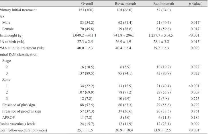
A total of 153 eyes from 83 infants were included in the study: 101 eyes treated with bevacizumab, and 52 with ra-nibizumab. The sex ratio, mean BW, GA, ROP classifica-tion, follow-up duraclassifica-tion, and other baseline characteristics are shown in Table 1. The mean GA was 27.3 weeks over-all, with a significant difference between the two groups (26.9 for bevacizumab, 28.1 for ranibizumab; p = 0.013). Accordingly, mean BW was higher in the ranibizumab
Table 1. Summary of ROP patients treated with intravitreal anti-vascular endothelial growth factor
Overall Bevacizumab Ranibizumab p-value*
Primary initial treatment 153 (100) 101 (66.0) 52 (34.0)
Sex
Male 83 (54.2) 62 (61.4) 21 (40.4) 0.017†
Female 70 (45.8) 39 (38.6) 31 (59.6) 0.017†
Birthweight (g) 1,049.2 ± 411.1 941.8 ± 296.1 1,257.7 ± 514.5 <0.001†
GA at birth (wk) 27.3 ± 2.5 26.9 ± 1.9 28.1 ± 3.2 0.013†
PMA at initial treatment (wk) 40.0 ± 2.3 40.4 ± 2.4 39.2 ± 2.3 0.090
Initial ROP classification Stage 2 16 (10.5) 6 (5.9) 10 (19.2) 0.022† 3 137 (89.5) 95 (94.1) 42 (80.8) 0.022† Zone 1 34 (22.2) 13 (12.9) 21 (40.4) <0.001† 2 107 (69.9) 78 (77.2) 29 (55.8) 0.009† 3 12 (7.8) 10 (9.9) 2 (3.8) 0.223
Presence of plus sign 88 (57.5) 66 (65.3) 29 (55.8) 0.292
Presence of pre-plus sign 57 (37.3) 37 (36.6) 20 (38.5) 0.861
APROP 11 (7.2) 5 (5.0) 6 (11.5) 0.186
Tunica vasculosis lentis 24 (15.7) 12 (11.9) 12 (23.1) 0.099
Total follow-up duration (mon) 25.1 ± 1.5 30.9 ± 18.4 13.9 ± 12.5 <0.001† Values are presented as number (%) or mean ± standard deviation.
ROP = retinopathy of prematurity; GA = gestational age; PMA = post-menstrual age; APROP = aggressive posterior retinopathy of prematurity.
group (1,257.7 vs. 941.8 g, p < 0.001). There was no differ-ence in mean postmenstrual age at primary treatment, which was 40.0 weeks overall. There was no difference in the proportion of eyes classified as type 1 ROP (62.4% for bevacizumab, 55.8% for ranibizumab; p = 0.487) or in ag-gressive posterior ROP (5.0% for bevacizumab, 11.5% for ranibizumab; p = 0.186). There were more eyes with zone 1 ROP in the ranibizumab group (40.4% vs. 12.9%, p < 0.001). There was a significantly longer mean follow-up period for the bevacizumab group (30.9 vs. 13.9 months, p < 0.001).
ROP recurrences requiring additional treatment are sum-marized in Table 2. A total of 15 (9.8%) eyes had recur-rences that required further intervention. Major interven-tions such as scleral encircling (one eye) or vitrectomy (two eyes) were required in a few cases in the bevacizumab
group. Further treatment with laser photocoagulation was needed in one case following bevacizumab treatment. More eyes required additional anti-VEGF therapy follow-ing treatment with ranibizumab than bevacizumab (13.5% vs. 4.0%, p = 0.037).
Major complications and anatomical outcomes are shown in Table 3. Retinal detachment and temporal macular drag-ging each occurred in one eye in the bevacizumab group. Spherical equivalence at the most recent visit was +0.11 ± 3.58 diopters (D) overall, with no significant difference be-tween the two groups (p = 0.922). There were more eyes with retinas fully vascularized to the ora serrata at follow-up in the bevacizumab grofollow-up than in the ranibizumab grofollow-up (100% vs. 85.0%, p < 0.001). The mean age at the most re-cent follow-up was 2.3 years old overall, and was higher in
Table 2. Retinopathy of prematurity recurrence requiring additional treatment after intravitreal anti-VEGF
Overall
(n = 153) Bevacizumab (n = 101) Ranibizumab (n = 52) p-value*
Additional laser 1 (0.7) 1 (1.0) 0 (0) 0.660
Additional anti-VEGF injection 11 (7.2) 4 (4.0) 7 (13.5) 0.037†
Scleral encircling/buckling 1 (0.7) 1 (1.0) 0 (0) 0.660
Vitrectomy 2 (1.3) 2 (2.0) 0 (0) 0.548
VEGF = vascular endothelial growth factor. *Bevacizumab vs. ranibizumab; †p < 0.05.
Values are presented as number (%) or mean ± standard deviation. *Bevacizumab vs. ranibizumab; †p < 0.05.
Overall
(n = 153) Bevacizumab (n = 101) Ranibizumab (n = 52) p-value* Complications
Retinal detachment 1 (0.7) 1 (1.0) 0 (0) 0.660
Vitreous hemorrhage 2 (1.4) 1 (1.0) 1 (1.9) 0.566
Macular dragging 1 (0.7) 0 (0) 1 (1.9) 0.340
Cataract 1 (0.7) 1 (1.0) 0 (0) 0.660
Pale disc without known neurologic defects 8 (1.4) 4 (4.0) 4 (7.7) 0.445
Glaucoma 0 (0) 0 (0) 0 (0)
-Known systemic complications (e.g., death) 0 (0) 0 (0) 0 (0)
-Strabismus operation 21 (13.7) 21 (20.8) 0 (0) <0.001†
Vascularization to ora serrata 130 (95.6) 96 (100) 34 (85.0) <0.001†
Age at most recent follow-up (yr) 2.3 ± 1.5 2.8 ± 1.5 1.3 ± 1.0 <0.001†
Final spherical equivalence (diopter) +0.11 ± 3.58 +0.10 ± 3.66 +0.22 ± 3.00 0.922
Table 3. Major complications and anatomical outcomes after anti-vascular endothelial growth factor injection for retinopathy of prematurity
the bevacizumab group (2.8 vs 1.3 years, p < 0.001).
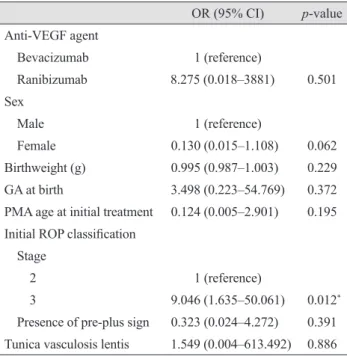
Univariable analysis of patient and treatment factors in-fluencing the incidence of major complications revealed that only ROP stage was a significant factor (Table 4), with ROP stage 3 having a much higher odds ratio than ROP stage 2 (odds ratio, 9.046; p = 0.012). No patient or treat-ment factors were associated with retinal detachtreat-ment (data not shown).
Discussion
Anti-VEGF injections promote retinal vascularization without permanent destruction of the peripheral retina, and are less time-consuming or risky than conventional laser therapy. However, many ophthalmologists still question the use of anti-VEGF as the primary treatment for ROP, due to concerns regarding long-term complications. From January 2013 to June 2014 in Korea, of 231 very-low-birth-weight infant eyes treated for ROP, 63.6% received only an opera-tion (laser, cryotherapy, or surgery), 16.9% received only
anti-VEGF treatment, and 19.5% received both an opera-tion and an anti-VEGF injecopera-tion [10]. We investigated the efficacy, anatomical outcomes, and complications associat-ed with intravitreal anti-VEGF ROP treatment using beva-cizumab and ranibizumab.
In our study, 22% of cases of zone 1 ROP and 40% of “pre-plus” retinas were treated with anti-VEGF. These high anti-VEGF treatment rates may be due to a recent change in our centers to aggressively treating ROP with anti-VEGF at earlier time points. ROP is a biphasic dis-ease, with a vaso-obliterative phase in which VEGF secre-tion is stimulated by relative hypoxia in the peripheral avascular retina, followed by a vasoproliferative phase [30,31]. A single burst of VEGF promotes vascular growth in ROP [32], which is different from other ocular neovas-cular diseases such as exudative neovasneovas-cular age-related macular degeneration, in which there is continual VEGF release. Delayed anti-VEGF treatment, administered when VEGF levels are decreasing, may not be as effective and may promote fibrosis [33,34]. Accordingly, in our study the overall rate of ROP recurrence requiring additional treat-ment was 9.8%, lower than in previous studies (13% to 35.4%) [20,22,28].
Significantly more eyes required additional anti-VEGF treatment in the ranibizumab group. This may be because ranibizumab has a shorter intravitreal half-life than bevaci-zumab due to its smaller molecular weight [24]. The ranibi-zumab group also had a greater proportion of zone 1 ROP, which requires more time for full vascularization following initial anti-VEGF treatment. During the extended period of vascular growth, elevation in VEGF levels may reactivate ROP, requiring additional anti-VEGF treatment [35].
In terms of safety, anti-VEGF injections appear to cause few cases of retinal detachment or macular temporal drag-ging, as only one case of each occurred (both in the beva-cizumab group). Furthermore, there was no significant dif-ference in incidence of major complications following ranibizumab and bevacizumab treatments (odds ratio, 8.275; p = 0.501). Though both anti-VEGF agents enter sys-temic circulation following intravitreal injection [36,37], ranibizumab appears to suppress systemic VEGF for less time than bevacizumab, and to clear more rapidly from systemic circulation [36,38]. Therefore, ranibizumab is the-oretically safer in its systemic profile, with less risk of neu-rologic deficits or developmental delays. In our analysis, more injections were required in the ranibizumab group,
Table 4. Univariable analysis of factors influencing occur-rence of major complications (retinal detachment, optic atro-phy, cataract surgery) after anti-VEGF injection for ROP
OR (95% CI) p-value Anti-VEGF agent Bevacizumab 1 (reference) Ranibizumab 8.275 (0.018–3881) 0.501 Sex Male 1 (reference) Female 0.130 (0.015–1.108) 0.062 Birthweight (g) 0.995 (0.987–1.003) 0.229 GA at birth 3.498 (0.223–54.769) 0.372 PMA age at initial treatment 0.124 (0.005–2.901) 0.195 Initial ROP classification
Stage
2 1 (reference)
3 9.046 (1.635–50.061) 0.012*
Presence of pre-plus sign 0.323 (0.024–4.272) 0.391 Tunica vasculosis lentis 1.549 (0.004–613.492) 0.886 VEGF = vascular endothelial growth factor; ROP = retinopathy of prematurity; OR = odds ratio; CI = confidence interval; GA = gestational age; PMA = post-menstrual age.
but there were cases in the bevacizumab group which re-quired major interventions such as scleral encircling/buck-ling or vitrectomy (the number of cases was too small for statistical comparison). There were no major systemic com-plications in either group.
Full retinal vascularization to the ora serrata by the most recent follow-up was achieved in more cases in the bevaci-zumab group than in ranibibevaci-zumab group (100% vs. 85.0%,
p < 0.001). This may be due to the shorter follow-up period
for the ranibizumab group (the mean age at the most recent follow-up was 2.3 years for bevacizumab, 1.3 years for ra-nibizumab; p < 0.001). There have been conflicting reports regarding the rate of retinal abnormalities and incomplete vascularization in long-term follow-up of ROP infants re-ceiving anti-VEGF. For example, Castellanos et al. [39] fol-lowed six eyes of ROP patients receiving ranibizumab for three years and found fully vascularized retinas with no fluorescein leakage on angiography in all eyes. Isaac et al. [40] found peripheral vascularization to the ora serrata in four of five eyes that received bevacizumab, with one pa-tient having too short a follow-up to determine vasculariza-tion potential properly. Huang et al. [22] noted incomplete vascularization without reactivation after ranibizumab treatment in 2.8% of 286 eyes. The BEAT-ROP study found the full vascularization rate to be 95.7% in 140 eyes. However, there have also been less favorable reports. Wu et al. [20] reported full vascularization in 88.0% of 162 beva-cizumab-treated eyes, and Chen et al. [28] reported full vascularization in only 46.4% of 151 ranibizumab-treated eyes. In our study, we noted a full vascularization rate of 95.6% of 136 eyes receiving anti-VEGF treatment, with the remaining eyes having too short a follow-up period to properly determine the vascularization potential.
Refractive error at the most recent follow-up revealed as-sociation with mild hyperopia, with similar values for ra-nibizumab and bevacizumab. These results are consistent with previous results, which showed less association with myopia in anti-VEGF–treated eyes [12,41].
This study has several limitations. First, it was a retro-spective study with a variable follow-up period and without appropriate controls; as such, significant differences in a few baseline characteristics between the treatment groups were noted. Second, the study population was limited to Asian patients in a tertiary hospital setting. Third, systemic complications may not have been properly observed or re-ported by the neonatologists. Last, we did not routinely
perform fluorescein angiography to document fully vascu-larized retinas; however, the primary retina specialist per-formed thorough fundus examinations at each follow-up visit. The strengths of this study include the large number of patients with a mean follow-up period >12 months, and all patients being treated at only two hospitals using the same treatment protocols.
In conclusion, we found that intravitreal anti-VEGF in-jections achieve stable retinal vascularization with a low rate of complications and recurrences requiring additional treatment. Ranibizumab appears to achieve similar ana-tomical outcomes as bevacizumab, without additional risk for major complications such as retinal detachment or sys-temic side effects.
Conflict of Interest
No potential conflict of interest relevant to this article was reported.
References
1. Blencowe H, Lawn JE, Vazquez T, et al. Preterm-associated visual impairment and estimates of retinopathy of prema-turity at regional and global levels for 2010. Pediatr Res 2013;74 Suppl 1:35-49.
2. Kushner BJ, Essner D, Cohen IJ, Flynn JT. Retrolental Fibroplasia. II. Pathologic correlation. Arch Ophthalmol 1977;95:29-38.
3. Tsui I, Chu A. Hot topics in retinopathy of prematurity. Pe-diatr Ann 2017;46:e415-22.
4. Casteels I, Cassiman C, Van Calster J, Allegaert K. Edu-cational paper: retinopathy of prematurity. Eur J Pediatr 2012;171:887-93.
5. Malcolm W. Beyond the NICU: comprehensive care of the high-risk infant. New York: McGraw Hill Professional; 2014. p. 347-65.
6. Darlow BA. Retinopathy of prematurity: new develop-ments bring concern and hope. J Paediatr Child Health 2015;51:765-70.
7. Goggin M, O'Keefe M. Childhood blindness in the Republic of Ireland: a national survey. Br J Ophthalmol 1991;75:425-9. 8. Hoogerwerf A, Schalij-Delfos NE, van Schooneveld MJ,
the last decade in the Central Netherlands. Neonatology 2010;98:137-42.
9. Isaza G, Arora S, Bal M, Chaudhary V. Incidence of reti-nopathy of prematurity and risk factors among premature infants at a neonatal intensive care unit in Canada. J Pedi-atr Ophthalmol Strabismus 2013;50:27-32.
10. Hwang JH, Lee EH, Kim EA. Retinopathy of prematurity among very-low-birth-weight infants in Korea: incidence, treatment, and risk factors. J Korean Med Sci 2015;30 Suppl 1:S88-94.
11. Mintz-Hittner HA, Kennedy KA, Chuang AZ; BEAT-ROP Cooperative Group. Efficacy of intravitreal bevacizumab for stage 3+ retinopathy of prematurity. N Engl J Med 2011;364:603-15.
12. Geloneck MM, Chuang AZ, Clark WL, et al. Refractive outcomes following bevacizumab monotherapy compared with conventional laser treatment: a randomized clinical trial. JAMA Ophthalmol 2014;132:1327-33.
13. Kong L, Bhatt AR, Demny AB, et al. Pharmacokinetics of bevacizumab and its effects on serum VEGF and IGF-1 in infants with retinopathy of prematurity. Invest Ophthalmol Vis Sci 2015;56:956-61.
14. Wu WC, Lien R, Liao PJ, et al. Serum levels of vascular endothelial growth factor and related factors after intravit-reous bevacizumab injection for retinopathy of prematurity. JAMA Ophthalmol 2015;133:391-7.
15. Morin J, Luu TM, Superstein R, et al. Neurodevelopmental outcomes following bevacizumab injections for retinopa-thy of prematurity. Pediatrics 2016 Mar 17. https://doi. org/10.1542/peds.2015-3218.
16. Jang SY, Choi KS, Lee SJ. Delayed-onset retinal detach-ment after an intravitreal injection of ranibizumab for zone 1 plus retinopathy of prematurity. J AAPOS 2010;14:457-9. 17. Yonekawa Y, Wu WC, Nitulescu CE, et al. Progressive
retinal detachment in infants with retinopathy of prematu-rity treated with intravitreal bevacizumab or ranibizumab. Retina 2018;38:1079-83.
18. Lee BJ, Kim JH, Heo H, Yu YS. Delayed onset atypical vitreoretinal traction band formation after an intravitreal injection of bevacizumab in stage 3 retinopathy of prematu-rity. Eye (Lond) 2012;26:903-9.
19. Wang J, Xiang D. Early clinical characteristics of bacterial endophthalmitis in retinopathy of prematurity after intra-vitreal bevacizumab injection: a case report. Exp Ther Med 2017;13:3563-6.
20. Wu WC, Kuo HK, Yeh PT, et al. An updated study of the
use of bevacizumab in the treatment of patients with pre-threshold retinopathy of prematurity in Taiwan. Am J Oph-thalmol 2013;155:150-8.
21. Chen SN, Lian I, Hwang YC, et al. Intravitreal anti-vascular endothelial growth factor treatment for retinopathy of prematurity: comparison between ranibizumab and bevaci-zumab. Retina 2015;35:667-74.
22. Huang Q, Zhang Q, Fei P, et al. Ranibizumab injection as primary treatment in patients with retinopathy of prematu-rity: anatomic outcomes and influencing Factors. Ophthal-mology 2017;124:1156-64.
23. Csaky K, Do DV. Safety implications of vascular endothe-lial growth factor blockade for subjects receiving intravit-real anti-vascular endothelial growth factor therapies. Am J Ophthalmol 2009;148:647-56.
24. Semeraro F, Morescalchi F, Duse S, et al. Aflibercept in wet AMD: specific role and optimal use. Drug Des Devel Ther 2013;7:711-22.
25. Ferrara N, Damico L, Shams N, et al. Development of ranibizumab, an anti-vascular endothelial growth factor antigen binding fragment, as therapy for neovascular age-related macular degeneration. Retina 2006;26:859-70. 26. International Committee for the Classification of
Reti-nopathy of Prematurity. The International Classification of Retinopathy of Prematurity revisited. Arch Ophthalmol 2005;123:991-9.
27. Good WV; Early Treatment for Retinopathy of Prematurity Cooperative Group. Final results of the Early Treatment for Retinopathy of Prematurity (ETROP) randomized trial. Trans Am Ophthalmol Soc 2004;102:233-48.
28. Chen Y, Fen J, Meng X. Effects of ranibizumab in zone I and zone II retinopathy of prematurity patients. Chinese J Ophthalmol 2015;31:6-9.
29. Darlow BA, Ells AL, Gilbert CE, et al. Are we there yet? Bevacizumab therapy for retinopathy of prematurity. Arch Dis Child Fetal Neonatal Ed 2013;98:F170-4.
30. Chen J, Smith LE. Retinopathy of prematurity. Angiogen-esis 2007;10:133-40.
31. Hartnett ME, Penn JS. Mechanisms and management of retinopathy of prematurity. N Engl J Med 2012;367:2515-26. 32. Micieli JA, Surkont M, Smith AF. A systematic analysis of
the off-label use of bevacizumab for severe retinopathy of prematurity. Am J Ophthalmol 2009;148:536-43.
33. Klaassen I, van Geest RJ, Kuiper EJ, et al. The role of CTGF in diabetic retinopathy. Exp Eye Res 2015;133:37-48. 34. Hartnett ME. Vascular endothelial growth factor
antago-nist therapy for retinopathy of prematurity. Clin Perinatol 2014;41:925-43.
35. Geisen P, Peterson LJ, Martiniuk D, et al. Neutralizing an-tibody to VEGF reduces intravitreous neovascularization and may not interfere with ongoing intraretinal vascular-ization in a rat model of retinopathy of prematurity. Mol Vis 2008;14:345-57.
36. Hong YR, Kim YH, Kim SY, et al. Plasma concentrations of vascular endothelial growth factor in retinopathy of pre-maturity after intravitreal bevacizumab injection. Retina 2015;35:1772-7.
37. Avery RL, Castellarin AA, Steinle NC, et al. Systemic phar-macokinetics following intravitreal injections of ranibizum-ab, bevacizumab or aflibercept in patients with neovascular AMD. Br J Ophthalmol 2014;98:1636-41.
38. Hoerster R, Muether P, Dahlke C, et al. Serum concen-trations of vascular endothelial growth factor in an infant treated with ranibizumab for retinopathy of prematurity. Acta Ophthalmol 2013;91:e74-5.
39. Castellanos MA, Schwartz S, Garcia-Aguirre G, Quiroz-Mercado H. Short-term outcome after intravitreal ranibizumab injections for the treatment of retinopathy of prematurity. Br J Ophthalmol 2013;97:816-9.
40. Isaac M, Mireskandari K, Tehrani N. Does bevacizumab al-ter vascularization potential in retinopathy of prematurity? Ophthalmology 2016;123:2042-3.
41. Hwang CK, Hubbard GB, Hutchinson AK, Lambert SR. Outcomes after intravitreal bevacizumab versus laser pho-tocoagulation for retinopathy of prematurity: a 5-year retro-spective analysis. Ophthalmology 2015;122:1008-15.