https://doi.org/10.20307/nps.2018.24.1.13
13
Estragole Exhibits Anti-inflammatory Activity with the Regulation of NF- κB and Nrf-2 Signaling Pathways in LPS-induced RAW 264.7 cells
Anupom Roy
1, Hee-Juhn Park
2, Hyun Ah Jung
3,*, and Jae Sue Choi
1,*
1
Department of Food and Life Science, Pukyong National University, Busan 608-737, Republic of Korea
2
Department of Pharmaceutical Engineering, Sangji University, Wonju 220-702, Republic of Korea
3
Department of Food Science and Human Nutrition, Chonbuk National University, Jeonju 561-756, Republic of Korea
Abstract − Estragole is a naturally occurring phenylpropanoid obtained from essential oils found in a broad diversity of plants. Although the phenylpropanoids show many biological activities, clear regulation of the inflammatory signaling pathways has not yet been determined. Here, we scrutinized the anti-inflammatory effect of estragole. The anti-inflammatory effect of estragole was determined through the inhibitory mechanisms of inducible nitric oxide synthase (iNOS), cyclooxygenase (COX-2), nuclear factor kappa B (NF-κB), and mitogen- activated protein kinases (MAPK) pathways and the activation of nuclear factor erythroid 2-related factor 2 (Nrf- 2)/heme oxygenase (HO)-1 pathways in lipopolysaccharide (LPS)-stimulated RAW 264.7 cells. Estragole significantly inhibited NO production, iNOS and COX-2 expression as well as LPS-induced NF-κB and MAPK activation. Furthermore, estragole suppressed LPS-induced intracellular ROS production but up-regulated the stress response gene HO-1 via the activation of transcription factor Nrf-2. These findings demonstrate that estragole inhibits the LPS-induced expression of inflammatory mediators via the down-regulation of iNOS, COX- 2, NF-κB, and MAPK pathways, as well as the up-regulation of the Nrf-2/HO-1 pathway, indicating that this phenylpropanoid has potential therapeutic and preventive applications in various inflammatory diseases.
Keywords − Essential oils, Estragole, Anti-inflammation, Nrf-2, HO-1, RAW 264.7 cells
Introduction
Inflammation is a complex physiological and patho- logical process activated in response to infection or tissue trauma. Inflammation can be caused by several elements such as microbial infections, chemicals, and immunological reactions. It is a protective response that affects immune cells, blood vessels, and molecular mediators. The treatment of inflammatory diseases focuses on the suppression of a few inflammatory mediators or signaling pathways including nitric oxide (NO), inducible nitric oxide synthase (iNOS), cyclooxygenase (COX-2), prostaglandin E
2(PGE
2), nuclear factor κB (NF-κB), mitogen-activated protein kinase (MAPK), reactive oxygen species (ROS), and pro- inflammatory cytokines (TNF- α, IL-6, and IL-1β).
1During inflammation, macrophages play essential roles,
producing inflammatory cytokines when activated by endotoxin. NO is a vital cellular signaling molecule involved in many physiological and pathological processes.
NO is produced as a metabolic by-product when L- arginine is converted into L-citrulline by the interference of nitric oxide synthase (NOS). There are three isoforms of NOS, neuronal NOS (nNOS), endothelial NOS (eNOS), and iNOS.
2During the inflammatory disease condition, iNOS produces an excessive amount of NO.
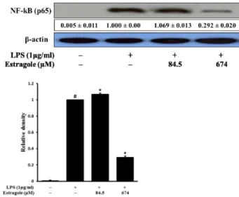
Eventually, NO and iNOS are the important targets for the treatment of inflammatory diseases. NF- κB is a transcription factor that plays a fundamental role in the inflammatory and acute response.
3Normally, NF- κB subunits are inactive and bound to I κB. Phosphorylation of IκB activates NF- κB to enter the nucleus and activate gene expression.
4NF- κB is one of the key regulators of proinflammatory gene expression and mediates the synthesis of cytokines such as TNF- α, IL-1β, IL-6, and IL-8.
5It also regulates the transcription of other inflam- matory mediators such as COX-2 or iNOS.
5Therefore, NF- κB is a vital target for the treatment of inflammatory diseases. MAPKs are protein chains that regulate NF- κB
*Author for correspondence
Jae Sue Choi, Department of Food and Life Science, Pukyong National University, Busan 608-737, the Republic of Korea.
Tel: +82-51-629-5845; E-mail: [email protected]
Hyun Ah Jung, Department of Food Science and Human Nutrition, Chonbuk National University, Jeonju 561-756, Republic of Korea.
Tel: +82-63-270-4882; E-mail: [email protected]
activation. These MAPKs include the extracellular signal- regulated kinases 1/2 (ERK 1/2), c-Jun amino-terminal kinases 1/2/3 (JNK 1/2/3), and p38 isoforms ( α, β, γ, and δ).
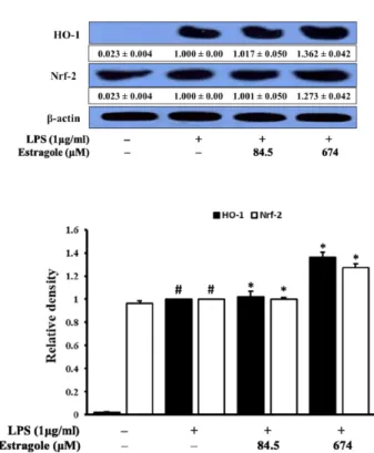
6Recently, HO-1 has been reported to implicate in inhibition of the excessive production of pro- inflammatory cytokines and ROS in LPS-stimulated RAW 264.7 cells.
7At the transcription level, HO-1 induction is regulated by a transcription factor called nuclear factor erythroid 2-related factor 2 (Nrf-2). Nrf-2 contributes to the anti-inflammatory process through the regulation of the gene expression of HO-1 and is a drug target for the treatment of inflammatory diseases.
8Essential oils are volatile, concentrated hydrophobic, natural, complex compounds extracted from aromatic plants and are characterized by their strong odor. Essential oils were first used in the Middle Ages by Arabs and are known for their antiseptic, i.e. bactericidal, fungicidal, and virucidal, and medicinal properties as well as their smell.
9Essential oils were used in embalmment, preservation of foods, and as antimicrobial, analgesic, sedative, anti- inflammatory, spasmolytic, and anesthetic compounds.
9Estragole is a phenylpropene and a colorless natural primary constituent of the essential oil of tarragon.
9In addition, we have previously reported that the Korean mint herb Agastache rugosa contained essential oil components of eugenol, methyl eugenol, and estragole through gas chromatographic analysis.
10Estragole was the major compound obtained from Agastache rugosa, with 148 ± 1.73 µg/g dry weight.
10In a preliminary study, we demonstrated that Agastache rugose extract showed anti- inflammatory activity through the inhibition of NO production in LPS-stimulated RAW 264.7 cells. Previous studies have shown that estragole has several biological activities including antioxidant,
11antimicrobials,
12anxi- olytics,
13induction of contraction of skeletal muscles,
14and anti-inflammatory.
15,16Although estragole shows many biological activities, clear regulation of the inflammatory signaling pathways has not yet been determined.
The aim of this study is to focus on the anti- inflammatory activity of estragole through the inhibition of iNOS, COX-2, NF- κB, and MAPKs expression and the induction of Nrf-2/HO-1 expression in LPS-stimulated RAW 264.7 cells.
Experimental
Chemicals and reagents − Estragole, LPS from Escherichia coli, Griess reagent, 3-[4,5-dimethylthiazol-2- yl]-2,5-di-phenyl tetrazolium bromide (MTT), dimethyl sulfoxide (DMSO), 2',7'-dichlorodihydrofluorescein diacetate
(DCFH-DA), 2-amino-5,6-dihydro-6-methyl-4H-1,3-thiazine hydrochloride (AMT), 6-hydroxy-2,5,7,8-tetramethylchro- man-2-carboxylic acid (Trolox), phenylmethylsulfonyl fluoride (PMSF), fetal bovine serum (FBS), and anti- biotics were purchased from Sigma-Aldrich Co. (St.
Louis, MO, USA), and Dulbecco’s modified Eagle’s medium (DMEM) was from Hyclone (Logan, UT, USA).
Various primary antibodies (iNOS, COX-2, NF- κB (p65), p-ERK, ERK, p-JNK, JNK, p-p38, p38, HO-1, Nrf-2, and β-actin) and secondary antibodies were obtained from Cell Signaling Technology Inc. (Beverly, MA, USA) and Santa Cruz Biotechnology, Inc. (Santa Cruz, CA, USA).
Polyvinylchloride fluoride (PVDF) membrane (Immobilon- P) was obtained from Millipore Co. (Billerica, MA, USA).
Super-signal
®West Pico Chemiluminescent Substrate was obtained from Pierce Biotechnology, Inc. (Rockford, IL, USA). All other chemicals and solvents were purchased from Sigma-Aldrich Co. unless stated otherwise.
Cell culture − Murine RAW 264.7 macrophage cells were obtained from the American Type Culture Collection (ATCC Rockville, MD, USA). RAW 264.7 cells were cultured in DMEM supplemented with 10% heat- inactivated FBS, 1% penicillin-streptomycin, and 0.1%
amphotericin B. The cells were incubated in a humidified atmosphere of 5% CO
2at 37
oC.
Cell viability − Cell viability was assessed using 3- [4,5-dimethylthiazol-2-yl]-2,5-di-phenyl tetrazolium bromide (MTT) assay. Briefly, RAW 264.7 cells were seeded into 96-well plates at a density of 1 × 10
4cells per well and incubated at 37
oC for 24 h. The cells were then treated with various sample concentrations. After incubation for an additional 24 h at 37
oC, 100 μL MTT (0.5 mg/mL in PBS) was added to each well, and the incubation continued for another 2 h. The resulting color was assayed at 540 nm using a microplate spectrophotometer (Molecular Devices, Sunnyvale, CA, USA).
NO production − The nitrite concentration in the medium was measured using Griess reagent as an indicator of NO production. Briefly, RAW 264.7 cells (2 × 10
4cells/well in a 24-well plate with 500 mL culture medium) were pretreated with various concentration of samples for 2 h and incubated for 18 h with LPS (1 μg/
mL). After incubation, the nitrate concentration of the supernatants (100 μL/well) was measured after adding 100 μL of Griess reagent. The absorbance values of the mixtures were determined using a microplate spectropho- tometer at 540 nm. The iNOS inhibitor AMT was used as a positive control.
Measurement of intracellular ROS − The intracellular
ROS scavenging activity of estragole was measured using
the DCFH-DA fluorescent probe. Cells plated on a black 96-well plate at a density of 1 × 10
4cells/well were co- treated with various concentrations of the above four compounds and LPS (1 µg/mL) for 2 h. Cells were treated with 20 µM DCFH-DA for 30 min at 37
oC. The fluorescence intensity was measured at an excitation wavelength of 485 nm and an emission wavelength of 528 nm using a fluorescence microplate reader (Dual Scanning SPECTRAmax, Molecular Devices, Sunnyvale, CA, USA).
Western blot analysis − Western blotting was used to measure the protein expression of iNOS, COX-2, MAPKs, HO-1, and Nrf-2. First, RAW 264.7 cells (5 × 10
4cells/mL) were cultured in 100-mm culture dishes in the presence or absence of LPS (1.0 µg/mL), with or without test samples for 18 h. Afterward, the cells were washed twice with ice-cold PBS and lysed with cell lysis buffer (50 mM Tris-HCl, 150 mM NaCl, 1% Nonidet P- 40, 1% Tween 20, 0.1% sodium dodecyl sulfate (SDS), 1 mM Na
3VO
4, 10 µg/mL leupeptin, 50 mM NaF, and 1 mM PMSF, pH 7.5) on ice for 30 min. Cell extracts were obtained at 14,000 × g at 4
oC for 20 min. The protein amount was determined by Protein Bradford assay. Cytosolic proteins were electrophoretically separated on SDS-PAGE and transferred to PVDF membranes. The membranes were immediately blocked with nonfat dry milk (50 g/L) in Tris-buffered saline containing 0.1%
Tween-20 (pH 7.4) (TBST) buffer at room temperature for 1 h. The membranes were then washed three times (10 min) in TBST buffer, incubated with primary antibody, and then diluted 1:1000 in nonfat dry milk (50 g/L) in TBST buffer at 4
oC overnight. After three washes with TBST buffer (10 min), the membranes were incubated with horseradish peroxidase (HRP)-conjugated secondary antibody diluted 1:2000 in nonfat dry milk (50 g/L) in TBST buffer at room temperature for 2 h. After washing three times in TBST buffer for 10 min, the antibody labeling was visualized with the Supersignal West Pico Chemiluminescent Substrate (Pierce, Rockford, IL, USA) according to the manufacturer’s instructions and was then exposed to X-ray film (GE Healthcare Ltd., Amersham, United Kingdom). Pre-stained blue markers were used for molecular weight determination. Bands were quantified by densitometry analysis using an ATTO CS analyzer.
Statistical analysis − Data are expressed as the mean ± standard deviation (SD) of at least three independent experiments unless otherwise indicated. Data were com- pared using one-way ANOVA. P values < 0.05, 0.01, and 0.001 were considered statistically significant. All analyses were performed using SPSS for windows, version 23
(SPSS Inc., Chicago, IL, USA).
Result and Discussion
Effect of estragole on RAW 264.7 cell viability − The effect of estragole was tested in the MTT cell viability assay using RAW 264.7 cells. Cell viability was tested to determine the appropriate concentration ranges of the selected compounds. Estragole did not show cytotoxicity to RAW 264.7 cells at doses of 84.5, 168.5, 337, and 674 µM (data not shown). These non-toxic concentrations were used for the following experiments.
Effect on NO production − Nitrite concentration was determined in the culture media using Griess reagent to evaluate the anti-inflammatory activity of estragole on NO production in LPS-stimulated RAW 264.7 cells.
Treatment of RAW 264.7 cells with the selected com- pound significantly suppressed LPS-induced NO produc- tion (Fig. 1). The results clearly indicated that estragole dos-dependently inhibited LPS-induced NO production in RAW 264.7 cells. AMT, a positive iNOS inhibitor, significantly inhibited LPS-induced NO production.
Effect on the production of iNOS and COX-2 − The expression of iNOS was determined by Western blot to evaluate the cause of decreased NO production. COX-2 expression was also determined to exhibit the inhibition Fig. 1. Inhibitory effect of estragole on the production of nitric oxide (NO) in LPS-stimulated RAW264.7 cells. Cells were pretreated with different concentrations of estragole for 2 h and stimulated with LPS (1 μg/mL) for 24 h. NO production was measured by Griess reaction. Data are presented as the mean ± standard deviation of three independent experiments.
###