• 검색 결과가 없습니다.

Anti-inflammatory Effect of Branches Extracts from Quercus mongolica in LPS-induced RAW264.7 Cells

N/A
N/A
Protected

Academic year: 2021

Share "Anti-inflammatory Effect of Branches Extracts from Quercus mongolica in LPS-induced RAW264.7 Cells"

Copied!
7
0
0

로드 중.... (전체 텍스트 보기)

전체 글

(1)

Introduction

Inflammation is protective response of tissues to harmful irritate, such as injury and microbes. Under pathophysiological conditions, acute or chronic inflammation may cause various inflammatory diseases such as diabetes, arthritis, septic shock and cancer. The macrophages produce preventative inter- mediates such as interleukin 6 (IL-6) interleukin 1β (IL-β), tumor necrosis factor α (TNF-α) and nitrogen oxides (NO), which play a pivotal role in the inflammatory process and also serve as a host defense mechanism. NO has been reported to activate nuclear factor kappa B (NF- κB) in peripheral blood monaryotic cells, which is a transcription factor in the expression of the inducible nitric oxide synthases (iNOS) gene in response to inflammation (Tripathi et al., 2007; Wang et al., 2018). Heme oxygenase 1 (HO-1) is induced by various stimuli, such as proinflammatory cytokines and heavy metals. Expression of HO-1 has been reported to provide an important protective response of cells against oxidative damage, because HO-1 expression may decrease the cellular

heme level and elevate the level of bilirubin, which is demonstrated a potent scavenging activity against reactive oxygen species (Doi et al., 1999; Fang et al., 2003).

Quercus mongolica (QM) is a species of oak native to Korea. QM has been reported to exert anti-oxidation, anti- microbial, and anti-allergic activities as an oriental traditional medicine. The components of leaves and bark of QM are known to be flavonoids, phenols, tannins and triterpenoid (Kong et al., 2001a; Kong et al., 2001b; Yeo et al., 2008; Bak et al., 2011; Yin et al., 2019) There are many research on leaves and barks of QM, but little is known about branches (QM-B). Therefore, we evaluates the anti-inflammatory effect of QM-B in LPS-induced RAW264.7 cells and elucidates the mechanism of NF- κB and mitogen activated protein kinase (MAPK) regulation and HO-1 expression as the molecular target.

Materials and methods

Materials

Dulbecco’s Modified Eagle medium (DMEM)/F-12 1:1 Modified medium (DMEM/F-12) was purchased from Lonza

Anti-inflammatory Effect of Branches Extracts from Quercus mongolica in LPS-induced RAW264.7 Cells

Hyun Ji Eo

1

, Youngki Park

2

, Jin Taek Kang

3

and Gwang Hun Park

4

*

1

Post-doc,

2

Senior Researcher and

4

Researcher, Forest Medicinal Resources Research Center, National Institute of Forest Science, Yeongju 36040, Korea

3

Senior Researcher, Division of Forest Industry Research, National Institute of Forest Science, Seoul 02455, Korea

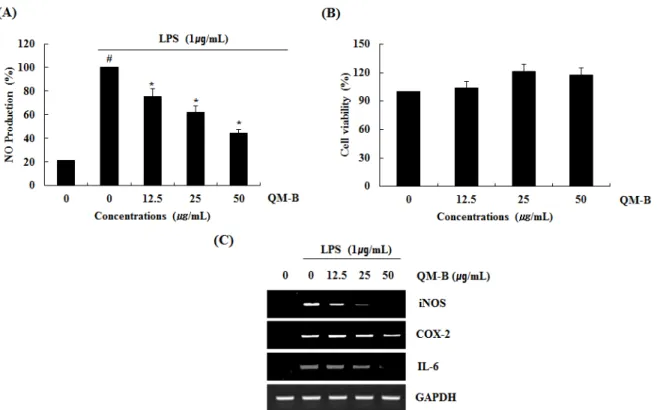
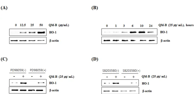
Abstract - Quercus mongolica (QM), which belongs to fagaceae, is one of the oak native to Korea. We evaluated the anti- inflammatory effect of branches extracted with 70% ethanol of QM (QM-B) and elucidated the potential signaling pathway in LPS-induced RAW264.7 cells. The QM-B showed anti-inflammatory activity through inhibition of NO production. The QM-B dose-dependently suppressed NO production by inhibiting iNOS, COX-2 and IL-6 expression in LPS-induced RAW264.7 cells. The QM-B inhibited the degradation and phosphorylation of IκB-α and NF-κB activation. The QM-B suppressed the phosphorylation of p38 and ERK1/2. Also, the QM-B increased HO-1 expression. These results suggested that QM-B may utilize anti-inflammatory activity by suppressing NF-κB and MAPK signaling pathway and inducing HO-1 expression indicated that the QM-B can be used as a natural anti-inflammatory drugs.

Key words – Anti-inflammatory effect, HO-1, MAPK, NF-κB, Quercus mongolica

*Corresponding author. E-mail : [email protected] Tel. +82-54-630-5638

ⓒ 2019 by The Plant Resources Society of Korea

Original Research Article

(2)

(Walkersville, MD, USA). lipopolysaccharide (LPS) and 3-(4,5-dimethylthiazol-2-yl)-2,5-diphenyltetrazolium bromide (MTT) were purchased from Sigma-Aldrich (St.

Louis, MO, USA). Antibodies against phospho-IκB-α, IκB- α, phospho-extracellular signal-regulated kinase1/2 (p-ERK 1/2), ERK1/2, phospho-p38 (p-p38), p38, HO-1 and β-actin were purchased from Cell Signaling technology (Bervely, MA, USA).

Sample preparation

The branches of Q. mongolica (QM-B, specimen number : FMCQm-P-20190521-005~009) were collected from Wolhak-ri, Buk-myeon, Inje-gun, Kangwon-do. 10g of dried QM-B were extracted with 200 mL of 70% ethanol for 72 hours under shaking at the room temperature. After 72 hours, the ethanol-soluble fraction was filtered and concentrated a rotary vacuum evaporator. The ethanol extracts from the QM-B were kept in a refrigerator until use.

Cell culture and treatment

RAW264.7 cells were purchased American Type Culture Collection (ACTT, Virginia, USA) and grown in Dulbecco's Modified Eagle (Lonza, Walkerscille, MD, USA) supple- mented with 10% fetal bovine serum (FBS), penicillin (100 units/mL) and streptomycin (100 ㎍/mL) under an atmos- phere of 5% CO

2

at 37 ℃. QM-B was dissolved in dimethyl sulfoxide (DMSO) and treated to cells. DMSO was used as a vehicle and the final DMSO concentration did not exceed 0.1% (v/v).

Cell viability assay

RAW264.7 cells viability were estimated by determining mitochondrial reductase function with the MTT assay.

Briefly, RAW264.7 cells (3 × 10

4

cells/well) were cultured in 12-well plate and incubated for 24 hours. The RAW264.7 cells were treated with QM-B at the indicated concentrations for 24 hours. Then, the cells were cultured with 200 μL of 1

㎎/mL MTT solution for an additional 2 hours. The resulting crystals were dissolved in DMSO. The formazan synthesised was dissolved in DMSO and the optical density was mea- sured at 570 ㎚ using UV/Visible spectrophotometer (Perkin Elmer, Nowolk, CT, USA).

Measurement of NO production

RAW264.7 cells were cultured in 12-well plate for overnight.

The cells were pretreated with QM-B at the indicated concentrations for 6 hours and then co-treated with LPS (1 ㎍ /mL) for 18 hours. NO levels were estimated by Griess assay.

Briefly, 50 μL of the cell culture supernatants were mixed with 50 μL of Griess reagent (Sigma Aldrich, St. Louis, MO, USA) and followed by reaction for 15 minutes at the room temperature. After 15 minutes, absorbance values were mea- sured using a UV/Visible spectrophotometer (Perkin Elmer, Norwolk, CT, USA) at 540 ㎚.

Western blot analysis

The cells were harvested in ice-cold 1XPBS, and lysed in 1X radioimmunoprecipitation assay (RIPA) buffer (Cell signaling, Bervely, MA, USA) supplemented with protease inhibitor cocktail and phosphatase inhibitor cocktail, and centrifuged at 15,000 rpm for 10 minutes at 4 ℃. The protein content of the cell lysates was then determined using bicinchoninic acid (BCA). Protein in each sample were resolved by 10% Sodium dodecyl sulphate polyacrylamide gel electrophoresis (SDS- PAGE), transferred to the nitrocellulose membranes (Bio- Rad, CA, USA). The membranes were blocked for non- specific binding with 5% skim milk in Tris-buffered saline containing 0.05% Tween 20 (TBS-T) for 1 hour at room temperature. The membrane was incubated with diluted primary antibodies for proper times and then washed three times with TBS-T and further incubated with secondary antibody for 1 hour at room temperature. The chemilumine- scence was detected with ECL Western blotting substrate and visualized in Chemi Doc MP Imaging system (Bio-rad, CA, USA).

Reverse transcriptase-polymerase chain reaction (RT-PCR)

Total RNA was prepared using a RNeasy Mini Kit (Qiagen,

Valencia, CA, USA) and total RNA (1 ㎍) was reverse-

transcribed using a Verso cDNA Kit (Thermo Scientific,

Pittsburgh, PA, USA) according to the manufacturer’s

protocol for cDNA synthesis. PCR was carried out using PCR

Master Mix Kit (Promega, Madison, WI, USA) with primers

for mouse iNOS, mouse COX-2, mouse IL-6 and mouse

GAPDH (Table 1.).

(3)

Statistical analysis

Data are expressed as mean ± standard deviation (SD). Each experiment was repeated at least three times. Statistical analysis was performed with one-way ANOVA followed by Dunnett's test. Differences with *P or #P < 0.05 were considered statistically significant.

Results and Discussion

The effect of QM-B on NO production and iNOS, COX-2 and IL-6 expression in LPS-induced RAW264.7 cells Nitric oxide synthases (NOS) synthesize the metastable free radical NO. NO is an important mediator and regulator of various processes in the immune, cardiovascular and nervous systems. Also, NO is implicated in pathophysiologic states as diverse as hypertension, septic shock, stroke, and neurodege- nerative diseases (Ignarro, 1990; Davies et al., 1997; Hou et al., 1999). During the inflammatory process, large amounts of the pro-inflammatory mediators, NO and prostaglandin E2 (PGE2) are generated by the iNOS and cyclooxygenase-2 (COX-2) (Kim et al., 2007; Wang et al., 2019).

To determine if the QM-B could suppress NO generation by LPS, RAW264.7 cells were pretreated with the QM-B for 6 hours and then co-treated with LPS (1 ㎍/mL) for the additional 18 hours. As shown in Fig. 1A, treatment of LPS without QM-B induced NO overproduction in LPS-induced RAW264.7 cells, while pretreatment of QM-B suppress LPS mediated NO overproduction. As shown in Fig. 1B, QM-B maintained the cell viability. Since NO production is regulated by iNOS, COX-2 and IL-6 expression, the effect of QM-B on iNOS, COX-2 and IL-6 expression was performed

by western blot and RT-PCR. As shown in Fig. 1C, the treatment of QM-B attenuated the mRNA expression of iNOS, COX-2 and IL-6. Thus, QM-B blocked the over- production of NO by suppressing LPS-induced iNOS, COX-2 and IL-6 over-expression in RAW264.7 cells.

The effect of QM-B on NF- κB signaling activation in LPS-induced RAW264.7 cells

NF-KB is one of the most key transcription factors and is found in cytokines, growth factors, chymokins, cell adhesives, and cell types that express some acute-phase proteins in healthy and diseased conditions. NF-KB plays a crucial role in the regulation of cell survival genes and is known to coordinate the expression of cytokine with proinflammatory enzymes such as iNOS, COX-2, IL-6 and TNF-α. NF-KB is located in the cytoplasm as an inactivated complex bound to IKB- α, which is phosphorylated and degradaded, and then dissociates to produce activated NF-KB (Kaibori et al., 1999; Kim et al., 2007; Li et al., 2017; Ran et al., 2018; Woo et al., 2018).

To elucidate the effect of QM-B on NF- κB signaling activation, we performed a western blot for degradation and phosphorylation of IκB-α in LPS-stimulated RAW264.7 cells. As shown in Fig. 2, LPS induced subsequent degra- dation and phosphorylation of I κB-α at 30 minutes after the stimulation. However, pretreatment of QM-B blocked LPS- induced IκB-α degradation in a dose-dependent manner.

The effect of QM-B on MAPK signaling activation in LPS-induced RAW264.7 cells

MAPK plays an important role in regulating cell growth and differentiation and controlling cellular responses to cytokines Table 1. The primer sequences used in this study

iNOS Forward 5′-GTGCTGCCTCTGGTCTTGCAAGC-3′

Reverse 5′-AGGGGCAGGCTGGGAATTCG-3′

COX-2 Forward 5′-GGAGAGACTATCAAGATAGTGATC-3′

Reverse 5′-ATGGTCAGTAGACTTTTACAGCTC-3′

IL-6 Forward 5′-GAGGATACCACTCCCAACAG-3′

Reverse 5′-TTCACAGAGGATACCACTCC-3′

GAPDH Forward 5′-CAGGAGCGAGACCCCACTAACAT-3′

Reverse 5′-GTCAGATCCACGACGGACACATT-3′

(4)

Fig. 1. The effect of QM-B on NO production and iNOS, COX-2 and IL-6 expression in LPS-stimulated RAW264.7 cells. (A) RAW264.7 cells were pre-treated with QM-B at the indicated concentrations for 6 hours and then co-treated with LPS (1 ㎍/mL) for the additional 18 hours. After treatment, NO production was measured using the media and Griess reagent. (B) RAW264.7 cells were treated with QM-B at the indicated concentrations for 24 hours. Cell viability was measured using MTT assay system and expressed as % cell viability. (C) For RT-PCR, RAW264.7 cells were pre-treated with QM-B at the indicated concentrations for 6 hours and then co-treated with LPS (1 ㎍/mL) for the additional 18 hours. Total RNA was isolated and RT-PCR was performed for iNOS and COX-2 and IL-6. Values given are the mean ± SD (n = 3). *P < 0.05 compared to LPS treatment without QM-B. GAPDH was used as an internal control for RT-PCR.

Fig. 2. The effect of QM-B on NF- κB signaling activation in LPS-stimulated RAW264.7 cells. (A) RAW264.7 cells were pretreated with QM-B for 6 hours and then co-treated with LPS (1 ㎍/mL) for 30 minutes. For Western blot analysis, the cell lysates were subjected to SDS-PAGE and the Western blot was performed using antibodies against p-I κB-α and IκB-α. β -actin was used as internal control.

Fig. 3. The effect of QM-B on MAPK signaling activation in LPS-stimulated RAW264.7 cells. RAW264.7 cells were pre- treated with QM-B for 6 hours and then co-treated with LPS (1

㎍/mL) for 30 minutes. For Western blot analysis, the cell

lysates were subjected to SDS-PAGE and the Western blot was

performed using antibodies against p-ERK1/2, p-p38 and

Total p-38 and Total ERK1/2 were used as internal control.

(5)

and stress. MAPK are involved in the LPS-induced iNOS expression signaling pathway (Kim et al., 2007). There is growing evidence that activation of MAPK by phosphory- lating ERK1/2, p38 and JNK mediates excessive production of the pro-inflammatory mediators, which results to the initiation and development of inflammatory disease (Park et al., 2018; Ran et al., 2018). Also, MAPK has considered as a typical molecular target for the development of anti- inflammatory agents. To further investigate whether decrease of NF-κB activation by QM-B treatment is associated with the regulation of p38 and ERK1/2 activation, we evaluated the effects of QM-B on phosphorylation of p38 and ERK1/2 in LPS-induced RAW264.7 cells. As shown in Fig. 3, Increase of p38 and ERK1/2 phosphorylation was observed in RAW264.7 cells by LPS. However, QM-B suppressed phosphorylation of p38 and ERK1/2, indicating that QM-B suppress the inflammatory response by inhibiting p38 and ERK1/2 activation in LPS-induced RAW264.7 cells.

The effect of QM-B on HO-1 activation in RAW264.7 cells HO-1 is an enzyme that promotes the oxidation degradation of heme, forming bilburdine, carbon monoxide, and free iron.

The evidence represented an important role of HO-1 in cell protection and other more diverse biological functions. The high expression of HO-1 occurs in various tumors, and HO-1 is known to play an important role in rapid tumor growth because of its anti-oxidant and anti-apoptotic effects.

Induction of HO-1 expression protects against the cytotoxicity of oxidative stress and apoptotic cell death (Eyssen- Hernandez et al., 1996; Doi et al., 1999; Jung et al., 2006;

Paine et al., 2010). In this study, our data showed that QM-B increased HO-1 expression (Fig. 4A and B), and inhibition of HO-1 by the treatment of PD98059 (ERK inhibitor) (Fig. 4C) or SB230580 (p38 inhibitor) (Fig. 4D). These findings indicate that HO-1 may be a potential target for the anti-inflammatory effect of QM-B.

Taken together, these results demonstrated that QM-B blocked NO production via inhibiting iNOS, COX-2 and IL-6 expression through suppressing the activation of NF-κB and MAPK signaling pathway. In addition, the induction of HO-1 expression by QM is involved in the anti-inflammatory effect of QM-B. These findings suggest that QM-B may have great potential for the development of anti-inflammatory drug to treat acute and chronic inflammatory disorders.

Fig. 4. Effect of QM-B on HO-1 expression in RAW264.7 cells. (A) RAW264.7 cells were treated with QM-B at the indicated

concentrations for 24 hours. (B) RAW264.7 cells were treated with 25 ㎍/mL of QM-B for the indicated times. For Western blot

analysis, the cell lysates were subjected to SDS-PAGE and the Western blot was performed using antibody against HO-1. (C)

RAW264.7 cells were treated with PD98059 (20 μM, ERK1/2 inhibitor) or SB203580 (20 μM, p38 inhibitor) for 2 hours, and then

co-treated with QM-B for 10 hours. For Western blot analysis, the cell lysates were subjected to SDS-PAGE and the Western blot was

performed using antibody against HO-1. β-actin was used as internal control for Western blot analysis.

(6)

Acknowledgments

This work was supported by the National Research Found- ation of Korea (NRF-2018M3C1B5052453), and by a grant from National Institute of Forest Science in 2019.

References

Bak, J.P., J.B. Kim, J.H. Park, Y.J. Yang, I.S. Kim, E.S. Choung and S.C. Kang. 2011. Screening and compound isolation from natural plants for anti-allergic activity. J. Korean Soc.

Appl. Biol. Chem. 54(3):367-375.

Davies, S.A., E.J. Stewart, G.R. Huesmaan, N.J. Skaer, S.H.

Maddrell, N.J. Tublitz and J.A. Dow. 1997. Neuropeptide stimulation of the nitric oxide signalling pathway in Drosophila melanogaster Malpighian tubules. Am. J.

Physiol. 273:823-827.

Doi, K., T. Akaike, S. Fujii, S. Tanaka, N. Ikebe, T. Beppu, S.

Shibahara, M. Ogawa and H. Maeda. 1999. Induction of haem oxygenase-1 nitric oxide and ischaemia in experi- mental solid tumours and implications for tumour growth.

Brit. J. Cancer 80(12):1945-1954.

Eyssen-Hernandez, R., A. Ladoux, C. Frelin. 1996. Differential regulation of cardiac heme oxygenase-1 and vascular endothelial growth factor mRNA expressions by hemin, heavy metals, heat shock and anoxia. FEBS Lett. 382(3):

229-233.

Fang, J., T. Sawa, T. Akaike, T. Akuta, S.K. Sahoo, G. Khaled, A. Hamada and H. Maeda. 2003. In vivo antitumor activity of pegylated zinc protoporphyrin: targeted inhibition of heme oxygenase in solid tumor. Cancer Res. 63(13):3567-3574.

Hou, Y.C., A. Janczuk and P.G. Wang. 1999. Current trends in the development of nitric oxide donors. Curr. Pharm. Design 5(6):417-441.

Ignarro, L.J. 1990. Nitric Oxide. A novel signal transduction mechanism for transcellular communication. Hypertension 16(5):477-483.

Jung, J.H., H.R. Kim, E.J. Kim, K.E. Hwang, S.Y. Kim, J.H.

Park, H.J. Kim, S.H. Yang and E.T. Jeong. 2006. The role of heme oxygenase-1 in lung cancer cells. Tuberc. Respir. Dis.

60(3):304-313.

Kaibori, M., K. Sakitani, M. Oda, Y. Kamiyama, Y. Masu, M.

Nishizawa, S. Ito and T. Okumura. 1999. Immunosuppressant FK506 inhibits inducible nitric oxide synthase gene

expression at a step of NF-kappaB activation in rat hepatocytes. J. Hepatol. 30(6):1138-1145.

Kim, J.B., A.R. Han, E.Y. Park, J.Y. Kim, W. Cho, J. Lee, E.K.

Seo and K.T. Lee. 2007. Inhibition of LPS-induced iNOS, COX-2 and cytokines expression by poncirin through the NF-kappaB inactivation in RAW 264.7 macrophage cells.

Biol. Pharm. Bull. 30(12):2345-2351.

Kong, Y.J., T.S. Kang, M.K. Lee, B.K. Park and D.H. Oh.

2001a. Antimicrobial and antioxidative activities of solvent fractions of Quercus mongolica leaf. J. Korean Soc. Food Sci. Nutr. 30(2):338-343.

Kong, Y.J., B.K. Park and D.H. Oh. 2001b. Antimicrobial activity of Quercus mongolica leaf ethanol extract and organic acids against food-borne microorganisms. Korean J.

Food Sci. Technol. 33(2):178-183.

Li, D., J. Chen, J. Ye, X. Zhai, J. Song, C. Jiang, J. Wang, H.

Zhang, X. Jia and F. Zhu. 2017. Anti-inflammatory effect of the six compounds isolated from Nauclea officinalis Pierrc ex Pitard, and molecular mechanism of strictosamide via suppressing the NF- κB and MAPK signaling pathway in LPS-induced RAW 264.7 macrophages. J. Ethnopharmacol.

20:66-74.

Paine, A., B. Eiz-Vesper, R. Blasczyk and S. Immenschuh.

2010. Signaling to heme oxygenase-1 and its anti-inflammatory therapeutic potential. Biochem. Pharmacol. 80(12):1895- 1903.

Park, S.B., H.M. Song, H.N. Kim, G.H. Park, H.J. Son, Y. Um, J.A. Park and J.B. Jeong. 2018. Anti-inflammatory effect of Biji (Soybean curd residue) on LPS-stimulated RAW264.7 cells. Korean J. Plant Res. 31(2):117-123.

Ran, X., Y. Li, G. Chen, S. Fu, D. He, B. Huang, L. Wei, Y. Lin, Y. Guo and G. Hu. 2018. Farrerol ameliorates TNBS- Induced colonic inflammation by inhibiting ERK1/2, JNK1/2, and NF-κB signaling pathway. Int. J. Mol. Sci.

19:E2037.

Tripathi, P., P. Tripathi, L.Kashyap and V. Singh. 2007. The role of nitric oxide in inflammatory reactions. FEMS Immunol. Med. Mic. 51(3):443-452.

Wang, C.Y., H.J. Jang, Y.K. Han, X.D. Su, S.W. Lee, M.C.

Rho, H.S Wang, S.Y. Yang and Y.H. Kim. 2018. Alkaloids from Tetrastigma hemsleyanum and their anti-inflammatory effects on LPS-Induced RAW264.7 cells. Molecules 23(6):E1445.

Wang, C.Y., S. Lee, H.J. Jang, X.D. Su, H.S. Wang, Y.H. Kim

and S.Y. Yang. 2019. Inhibition potential of phenolic

(7)

constituents from the aerial parts of Tetrastigma hemsleyanum against soluble epoxide hydrolase and nitric oxide synthase. J. Enzym. Inhib. Med. Ch. 34(1):753-760.

Woo, H.S., S.M. Lee, D.H. Jeong, M.S. Lee, Y.S. Kim and D.W. Kim. 2018. Anti-inflammatory activity of extracts of Hovenia dulcis on lipopolysaccharides-stimulated RAW- 264.7 cells. Korean J. Plant Res. 31(5):466-477.

Yeo, H.D., H.C. Lee, B. K. Lim, H.K. Kim, M.S. Choi and J.K.

Yang. 2008. Antifungal activity of the Quercus Mongolica extracts against Botrytis cinerea. J. Korean Wood Sci.

Technol. 36(1):88-101.

Yin, J., H.H. Kim, I.H. Hwang, D.H. Kim and M.W. Lee. 2019.

Anti-Inflammatory effects of phenolic compounds isolated from Quercus Mongolica Fisch. ex Ledeb. on UVB- Irradiated human skin cells. Molecules 24(17):E3094.

(Received 13 November/ 2019 ; Revised 23 November 2019 ; Accepted 25 November 2019)

수치

Fig. 1. The effect of QM-B on NO production and iNOS, COX-2 and IL-6 expression in LPS-stimulated RAW264.7 cells
Fig. 4. Effect of QM-B on HO-1 expression in RAW264.7 cells. (A) RAW264.7 cells were treated with QM-B at the indicated  concentrations for 24 hours

참조

관련 문서

thunbergii was determined by measuring expression of the LPS-induced inflammatory proteins, inducible nitric oxide synthase (iNOS), cyclooxygenase-2 (COX-2) and nuclear

Moreover, we demonstrated that LicE sup- pressed iNOS and COX-2 expression as well as the release of proinflammatory cytokines, possibly via the inhibition of LPS-induced

Furthermore, combined RA and luteolin more strongly suppressed NF-κB activation than RA or luteolin alone, by inhibiting the degradation of inhibitor of NF-κB (IκB)-α and

Effects of Chondrus nipponicus Yendo ethanol extract on LPS-induced iNOS, COX-2, and NF-κB p65 expression in RAW 264.7 cells.. The level of NF-κB p65 in nuclear protein

Effect of onion peel ethanol extract on LPS-induced iNOS, COX-2, and NF-κB p65 expression in LPS-induced RAW 246.7 cells.. The levels of iNOS and COX-2 in the cytosolic protein

AC-L dose-dependently inhibited NO production by suppressing iNOS, COX-2 and IL-β expression in LPS-stimulated RAW264.7 cells.. AC-L inhibited the degradation and phosphorylation of

Effect of tuna oil (TO) on western blot analysis of NF- κB p65, iNOS, and COX-2 protein expression on LPS-induced inflam- matory responses in RAW 264.7 cells, and (B-D) relative unit

Taken together, the results of the in vitro cell line assay for MTT, NO production, ROS generation; the Western blot analysis for the inhibition of iNOS, COX-2, NF- κB, and