Received on November 2, 2010. Revised on November 11, 2010. Accepted on November 15, 2010.
CC This is an open access article distributed under the terms of the Creative Commons Attribution Non-Commercial License (http://creativecommons.org/licenses/by-nc/3.0) which permits unrestricted non-commercial use, distribu- tion, and reproduction in any medium, provided the original work is properly cited.
*Corresponding Author. Tel: 82-51-510-2526; Fax: 82-51-513-9258; E-mail: [email protected] Keywords: Kalopanax pictus, NO, NF-κB, MAPK, HO-1, Nrf2
The Stem Bark of Kalopanax pictus Exhibits Anti-inflammatory Effect through Heme Oxygenase-1 Induction and NF-κB Suppression
Soo Young Bang1, Ga-Young Park1, Sun Young Park1, Ji-Hee Kim1, Yun Kyoung Lee1, Sang-Joon Lee2 and YoungHee Kim1*
1Department of Molecular Biology, and 2Department of Microbiology, College of Natural Sciences, Pusan National University, Busan 609-735, Korea
Backgroud: The stem bark of Kalopanax pictus (KP) has been used in traditional medicine to treat rheumatoidal ar- thritis, neurotic pain and diabetes mellitus in China and Korea. In this study, the mechanism responsible for anti-in- flammatory effects of KP was investigated. Methods: We ex- amined the effects of KP on NO production, nitric oxide syn- thase (iNOS) and HO-1 expression, NF-κB, Nrf2 and MAPK activation in mouse peritoneal macrophages. Results:
The aqueous extract of KP inhibited LPS-induced NO secre- tion as well as inducible iNOS expression, without affecting cell viability. KP suppressed LPS-induced NF-κB activation, phosphorylation and degradation of IκB-α, phosphor- ylation of extracellular signal-regulated kinase 1/2 (ERK1/2) and c-Jun N-terminal kinase (JNK). Furthermore, KP in- duced HO-1 expression and Nrf2 nuclear translocation.
Conclusion: These results suggest that KP has the inhibitory effects on LPS-induced NO production in macrophages through NF-κB suppression and HO-1 induction.
[Immune Network 2010;10(6):212-218]
INTRODUCTION
Kalopanax pictus (KP) NAKAI (Araliaceae) is distributed in Korea, Japan, and China. The stem bark of KP has been used in traditional medicine to treat rheumatoidal arthritis, neurotic pain and diabetes mellitus (1). The constituents such as sap- onins, polyacetylenes, phenylpropanoid glycosides, lignans,
and simple phenolic glycosides have been isolated from this plant (2). Kalopanaxsaponin A, the constituent of KP, has been reported to inhibit iNOS, COX-2 expression, and TNF-al- pha release in vitro (1). However, the underlying mechanism of KP function has remained to be characterized so far.
Nitric oxide (NO) is a free radical with multiple effects on various organ systems. The most prominent physiological ac- tions of NO as a biological mediator include cGMP-dependent vasodilation, neural communication, host defense, inflamma- tion, immune suppression and blood clotting (3). NO is pro- duced in physiological and pathophysiological conditions by NO synthase (NOS), and inducible NOS (iNOS) is induced by inflammatory cytokines and/or bacterial lipopolysaccharide (LPS) in various cell types including macrophages. A large amount of NO, particularly synthesized by iNOS, induces an inflammatory response to inhibit the growth of invading mi- croorganisms and tumor cells. This strong inflammatory re- sponse to foreign cells could also cause further damage for the neighboring cells and tissues of the host. Therefore iso- zyme specific inhibitors of NOS are essential for therapeutic purposes and drugs that specifically inhibit iNOS could be useful in treating diseases mediated by NO overproduction (4). A major transcription factor that regulates iNOS gene ex- pression is NF-κB. In unstimulated cells, NF-κB is present in the cytosol bound to the inhibitory protein I kappa B (IκB).
In response to stimulation such as LPS, IκBs are rapidly ubiquitinated and degraded by 26S proteasome complex. The
free NF-κB dimers translocate to the nucleus and stimulate target genes expression. The critical event which triggers the degradation of IκBs is their stimulus-dependent phosphor- ylation at two serine residues (Ser32 and 36) that are located within their conserved N-terminal regulatory region.
Heme oxygenase-1 (HO-1) is a ubiquitous stress-inducible enzyme that catalyzes the oxidative degradation of free heme.
HO-1 cleaves heme to form carbon monoxide (CO), iron and biliverdin (BV), the latter being subsequently converted into bilirubin (5). HO-1 expression is up-regulated in response to various inflammatory stimuli, and this is associated with re- duced inflammation. The anti-inflammatory effect of HO-1 is due to these by-products of HO-1 activity. CO is the most responsible for anti-inflammatory action and suppresses the production of tumor necrosis factor-α (TNF-α), interleukin-1β (IL-1β), and macrophage inflammatory protein-1, iNOS and cyclo-oxygenase-2 (COX-2) (6,7). BV has the anti-inflamma- tory effects on organ transplantation including decreased leu- kocyte infiltration, less T cell proliferation, and extended sur- vival of allogeneic heart transplants (8). It reduced production of the pro-inflammatory cytokines and enhanced the anti-in- flammatory cytokines in rat model (9). HO-1 gene expression is regulated by various stimuli and transcription factors.
Above all, nuclear factor E2-related factor 2 (Nrf2) mainly reg- ulates HO-1 gene expression via various intracellular signaling molecules including phosphatidylinositol 3-kinase (PI3K)/Akt pathway and mitogen-activated protein kinase (MAPK) path- way (10). In basal condition, cytoplasmic Nrf2 is associated with Kelch-like ECH associated protein 1 (Keap1) and de- graded via ubiquitination. Numerous prooxidant stimuli cause dissociation of Nrf2 from Keap1, which permits subsequent nuclear translocation of Nrf2. In nucleus, Nrf2 binds to anti- oxidant response element (ARE) or electrophile responsive el- ement (EpRE) of various antioxidant enzymes including HO-1, NAD(P)H:quinone oxidoreductase-1 (NQO1), and su- peroxide dismutase (SOD) (11).
In this study, we investigated the effects of KP on NF-κB activation and MAPKs activities which have been known to regulate NF-κB in mouse peritoneal macrophages. We also examined whether KP upregulate HO-1 expression which is responsible for anti-inflammatory effect.
MATERIALS AND METHODS Preparation of extract
The stem bark of KP was purchased from a local herb store,
Kwang Myoung Herb Medicine (Pusan, Korea) in April 2005.
The roots were identified and authenticated by Professor W.
S. Ko, College of Oriental Medicine, Dongeui University (Pusan, Korea). A voucher specimen (number KP-05-04) has been deposited at the Department of Molecular Biology, Pusan National University, Busan, Korea. The dry roots (300 g) were extracted with distilled water at 100oC for 4 h. The extract was filtered through 0.45μm filter and freeze-dried (yield, 27 g) and kept at 4oC. The dried extract was dissolved in phosphate buffered saline (PBS) and filtered through 0.22 μm filter before use.
Macrophage culture
C57BL/6 mice purchased from Dae Han Laboratory Animal Center (Korea) were used between 8 to 12 weeks of age (25
∼30 g). Thioglycollate (TG) broth (Brewer, DIFCO, Detroit, MI)-elicited macrophages were harvested 3 days after intra- peritoneal injection of 2.5 ml TG into mice and isolated as reported previously (12). Murine macrophage RAW 264.7 cells were maintained in Dulbecco’s modified Eagle’s medium supplemented with glutamine (1 mM) and 10% FBS.
Measurement of nitrite concentration
NO synthesis in cell cultures was measured by a microplate assay method. To measure nitrite, 100μl aliquots were re- moved from conditioned medium and incubated with an equal volume of the Griess reagent (1% sulfanilamide/0.1%
N-(1-naphthyl)-ethylenediamine dihydrochloride/2.5% H3PO4) at room temperature for 10 min. Nitrite concentration was de- termined by measuring the absorbance at 540 nm with a Vmax 96-well microplate spectrophotometer (Molecular De- vices, Menlo Park, CA). The sodium nitrite was used as a standard.
Cell viability assay
The cytotoxicity of KP was assessed using the microculture tetrazolium (MTT)-based colorimetric assay. The remaining cells after Griess reaction were used for MTT assay. 5μl of MTT solution (5 mg/ml) was added to each well (final con- centration is 62.5μg/ml). After incubation for 3 h at 37oC and 5% CO2, the supernatant was removed and the formed formazan crystals in viable cells were solubilized with 150μl of DMSO. The absorbance of each well was then read at 570 nm using microplate reader.
Preparation of nuclear extract and eletrophoretic mobility shift assay
Nuclear extracts were prepared as described previously (12).
Eletrophoretic mobility shift assay (EMSA) of nuclear extracts was performed according to the manufacturer’s instructions (Promega, Madison, WI) with some modifications. In brief, the probe consisted of a double-stranded oligonucleotide con- taining the consensus binding sequence for NF-κB (5’-AGTT- GAGGGGACTTTCCCAGGC-3’, Promega, Madison, WI) was end-labeled with [γ-32P]-ATP (3,000 Ci/mmol at 10μCi/ml) using T4 polynucleotide kinase. Nuclear extracts were in- cubated with gel shift binding buffer (10 mM HEPES, pH 7.9, 50 mM KCl, 0.2 mM EDTA, 2.5 mM DTT, 10% glycerol, 0.05% NP-40, 0.25 mg/ml poly dI/poly dC, protease inhibitor cocktail) for 10 min at room temperature and then the mix- ture was incubated with 32P-labeled probe for 20 min at room temperature. The incubation mixture was loaded onto non- denaturating gel and run in 0.25× Tris/borate/EDTA buffer.
Gels were dried and exposed to X-ray film at −70oC.
Western blot analysis
The cells were washed with phosphate buffered saline (PBS) three times and scraped off and lysed with lysis buffer (1%
Triton X-100, 1% deoxycholate). Protein concentration of ly- sates was determined and equal amounts of protein were sep- arated electrophoretically using 10% SDS-PAGE (sodium do- decyl sulfate-polyacrylamide gel electrophoresis), and then the gel was transferred to 0.45μm nitrocellulose paper. The blot was incubated with anti-iNOS, IκB-α, HO-1, Nrf2, β- tubulin antibody (Santa Cruz Biotechnology), anti-phospho- IκB-α, JNK, phospho-JNK, ERK1/2, phospho-ERK1/2 anti- body (Cell Signaling Technology), and secondary antibody and then detected by the enhanced chemiluminescence de- tection system according to the recommended procedure (Amersham Co.).
Statistical analysis
All results were expressed as means±SE. Each experiment was repeated at least three times. Statistical significances were compared between each treated group and analyzed by the paired Student’s t-test. Data with p<0.05 were considered statistically significant.
RESULTS
KP inhibits NO synthesis and iNOS expression in macro- phages
To investigate the anti-inflammatory effect of KP, we exam- ined the effect of KP on NO synthesis. TG-elicited mouse per- itoneal macrophages were incubated with KP for 1 h and stimulated with 0.1μg/ml LPS for 24 h. The amount of NO released into culture medium was measured by the method of Griess. Whereas LPS-treated cells produced a large amount of NO, KP suppressed NO release into culture supernatant in a dose-dependent manner (Fig. 1A). To determine whether the decreased nitric oxide synthesis is correlated with iNOS expression, we analyzed the amount of iNOS by Western blot analysis. TG-elicited mouse peritoneal macrophages were treated with KP as mentioned above. The level of iNOS was dramatically reduced by KP in a dose-dependent manner (Fig. 1B). On the other hand, cell viability was not affected by KP as measured by MTT assay (Fig. 1C). These results suggest that KP inhibits NO release by affecting the iNOS ex- pression level without affecting cell viability.
KP suppresses NF-κB activity and phosphorylation/
degradation of IκB-α in macrophages
Since iNOS promoter contains 2 binding sites for transcription factor NF-κB and NF-κB plays a critical role in LPS-induced iNOS expression, we investigated whether KP suppresses iNOS expression through the regulation of NF-κB activity.
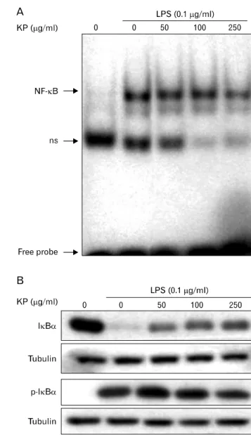
TG-elicited mouse peritoneal macrophages were incubated with KP for 1 hr and stimulated with 0.1μg/ml LPS for 30 minutes. A low level of NF-κB/DNA complex was observed in unstimulated macrophages, while a large quantity of NF-κB/
DNA complex was induced by LPS treatment. This increased NF-κB binding affinity was inhibited by KP in a dose-de- pendent manner (Fig. 2A). These results suggest that KP sup- presses iNOS expression though inhibition of NF-κB activity.
To analyze whether KP affect NF-κB signaling pathway, we investigated the effects of KP on upstream signaling path- way of NF-κB, that is, IκB-α degradation. TG-elicited mouse peritoneal macrophages were pretreated with KP for 1 h and stimulated with LPS (0.1μg/ml) for 15 min, and then the levels of IκB-α or phospho-IκB-α protein were exam- ined by Western blotting. As shown in Fig. 2B, IκB-α was degraded in response to LPS, but KP suppressed the degrada- tion of IκB-α in a dose-dependent manner. Incubation of macrophages with LPS for 15 min also caused significant
Figure 1. Effect of KP on the NO secretion and iNOS expression in TG-elicited mouse peritoneal macrophages. (A) Cells were incubated with various concentrations of KP for 1 h and then stimulated with 0.1μg/ml LPS for 24 h at 37oC. The amount of nitrite released was measured by the method of Griess. Values are means±S.E. of three independent experiments. *p<0.05 and †p<0.01 vs LPS-treated group. (B) Cells were treated with KP and/or LPS as mentioned above and equal cytosolic extracts were analyzed by Western blotting with anti-iNOS antibody. Western blot detection of β-tubulin was estimated protein-loading control for each lane. (C) Cells were incubated with various concentration of UR in presence of 0.1μg/ml of LPS for 24 h. Then cell viability was measured by MTT assay as described in Materials and Methods. Data represent the relative viability to control group and are expressed as the means±S.E. of three independent experiments.
Figure 2. Inhibitory effect of KP on NF-κB activity and phosphory- lation/degradation of IκB-α. (A) Effect of KP on DNA binding activity of NF-κB. TG-elicited mouse peritoneal macrophages were incubated with various concentrations of KP for 1 h, and then stimulated with LPS (0.1μg/ml) for 30 min. Nuclear proteins were extracted and assayed for NF-κB DNA binding affinity by EMSA as described in Materials and methods. ns, non-specific band. (B) Effect of KP on LPS-stimulated phosphorylation and degradation of IκB-α. Cells were incubated with various concentrations of KP for 1 hr, and then stimulated with 0.1μg/ml LPS for 15 min. Cells were harvested and equal cytosolic extracts were analyzed by Western blotting with anti- IκB-α and -phospho-IκB-α. Western blot detection of β-tubulin was estimated protein-loading control for each lane.
phosphorylation of IκB-α, but KP markedly inhibited the phosphorylation of IκB-α. These results indicate that KP blocks NF-κB translocation into nucleus through suppression of IκB-α phosphorylation and degradation.
Figure 3. Effect of KP on ERK1/2 and JNK activity in LPS-stimulated macrophages. TG-elicited mouse peritoneal macrophages were treated with indicated concentrations of KP for 1 h and stimulated with 0.1μ g/ml LPS for 15 min. Equal amount of cell extracts was analyzed by western blotting with anti-phospho-ERK1/2 or -phospho-JNK antibody.
Western blot detection of non-phosphorylated kinases was estimated protein-loading control for each lane.
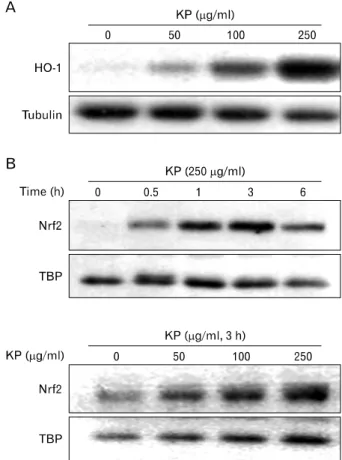
Figure 4. Effect of KP on HO-1 expression and nuclear translocation of in macrophages. (A) TG-elicited mouse peritoneal macrophages were incubated with indicated concentrations of KP for 24 h. HO-1 protein levels of cytosolic extracts were analyzed by Western blotting.
(B) Cells were incubated with indicated concentrations of KP for 3 h and the levels of Nrf2 in nuclear extracts were analyzed by Western blotting. Western blot detection of β-tubulin was estimated protein- loading control for each lane.
KP inhibits LPS-induced MAPK activities
To elucidate the molecular target of KP in further upstream signaling pathway, we examined the effect of KP on MAPKs activities which regulate the function of NF-κB (13). Since extracellular signal-regulated kinase1/2 (ERK1/2) and c-Jun N-terminal kinase (JNK) have been known to be phosphory- lated and activated in response to LPS, the activities were monitored by its phosphorylation. TG-elicited mouse peri- toneal macrophages were treated with indicated concen- trations of KP for 1 h and then stimulated with 0.1μg/ml LPS for 15 min. KP suppressed LPS-induced activation of ERK1/2 and JNK in a dose-dependent manner (Fig. 3), while the amount of kinases was unaffected by either LPS or KP treatment.
HO-1 expression is induced by KP in macrophages It is known that by-products of HO-1 have anti-inflammatory effect. To determine whether KP exhibit anti-inflammatory ef- fect through the induction of HO-1, HO-1 expression by KP was examined. TG-elicited mouse peritoneal macrophages were treated with KP for 24 h. KP induced HO-1 expression in a dose-dependent manner (Fig. 4A).
To determine whether KP-induced HO-1 expression is caused by Nrf2 transcription factor, we examined the effect of KP on Nrf2 nuclear translocation. TG-elicited mouse peri- toneal macrophages were treated with KP and cytosolic and nuclear extracts were separately harvested. The amount of
nuclear Nrf2 was increased by KP in dose-dependent and time-dependent manners, whereas the amount of cytosolic Nrf2 did not showed prominent changes (Fig. 4B).
DISCUSSION
The application of medicinal herbs dates back to the begin- ning of civilization, and interestingly, medicinal herbs are still routinely used by most of world’s population. Over time, many information has been accumulated by using these herbs in practice, and it is experientially proved that these herbs have the effect of pain alleviation, life extension and disease prevention. Of these, the stem bark of KP has been used for different remedies and expresses specific therapeutic effects
in China and Korea. The stem bark of KP is mainly used for treating neuralgia, rheumatic arthritis, lumbago, furuncle, car- buncle, wound, diarrhea and scabies (2). It has been reported that hederagenin monodesmosides and bisdesmosides iso- lated from the stem bark of KP have antimutagenic and cyto- toxic activity (14). Kalopanaxsaponin A and pictoside A from the stem bark of KP reduce vascular permeability (2). The other constituent of KP, α-hederin methyl ester, showed anti- carrageenan activity in rats and antiarthritic activity in rats and mice (15). Kalopanaxsaponin A was reported to have an- ti-rheumatoidal (16-19), anti-tumor (20), anti-diabetic (21), an- tifungal (22) activities. Kalopanaxsaponins A inhibits the pro- duction of NO, prostaglandin E2 (PGE2) and TNF-α in mac- rophage cell line RAW 264.7 (1). In this study, KP sig- nificantly inhibited LPS-induced NO production and iNOS ex- pression in mouse peritoneal macrophages (Fig. 1), which is consistent with the results of Kim et al. (1). Because KP did not show cytotoxic effects, inhibition of iNOS expression might be specific. Major transcriptional regulators of iNOS gene are the NF-κB family of transcription factors, which are also key regulators of a variety of genes involved in immune and inflammatory responses. Since dysregulation of NF-κB function is associated with inflammation, a development of drug that control NF-κB is one of promising candidates for the therapeutic strategy in the treatment of inflammatory disease. Our study showed that KP significantly inhibited NF- κB DNA binding activity in macrophages. Although it was reported that the combined extracts containing KP and other herb medicine suppress NF-κB DNA binding activity in RAW 264.7 cells (23), the effect of KP on NF-κB activity has re- mained to be clarified. Moreover, our study showed that KP suppressed not only the LPS-stimulated degradation but also phosphorylation of IκB-α. The phosphorylation of N-termi- nal regulatory serines on IκB-α and IκB-α is known to be catalyzed by IKK. Thus, KP could regulate the IKK activity. Since MAPKs have been shown to be involved in the LPS-mediated induction of many inflammatory genes includ- ing iNOS gene and are important for the activation of NF-κB (13), we investigated the effect of KP on ERK1/2 and JNK activation in LPS-stimulated mouse peritoneal macrophages.
KP greatly inhibited the LPS-induced ERK1/2 and JNK after LPS application. These results suggest that KP inhibits LPS-in- duced NF-κB activation by down-regulating ERK1/2 and JNK.
HO breaks down free heme made from intracellular heme- containing proteins. Two types of isozyme have been identi-
fied; HO-1 and 2. Whereas HO-2 is a constitutively expressed form, HO-1 is an inducible form that expressed by either in- flammatory or oxidative stress (5). Recently, many reports showed that HO-1 expression is associated with reduced inflammation. HO-1 plays a critical protective role during the development of intestinal inflammation (24,25). HO-1 attenu- ates metabolic syndrome in obese mice (26), LPS-induced lung injury (27), cardiovascular desease (28) and liver desease (29). HO-1-deficient mice develop increased end-organ dam- age and have increased mortality after LPS administration (30). Human HO-1 deficiency presents with severe hemolysis, asplenia, inflammation and nephritis (31). In addition, LPS-in- duced iNOS expression is reduced by HO-1 expression (24).
In this study, HO-1 expression was increased by KP, and nu- clear translocation of Nrf2, a major transcription factor of HO-1 expression, also induced by KP. These results suggest that KP exhibit anti-inflammatory effect by suppressing NO production via HO-1 induction.
In conclusion, we demonstrated that KP inhibited NO re- lease and iNOS expression in LPS-stimulated macrophages, and that these effects are mediated by inhibition of the activ- ity of IκB/NF-κB and induction of HO-1. Our results pro- vide a molecular basis for understanding the inhibitory effects of KP on endotoxin-mediated inflammation. As the effect of KP is mediated via inhibition of NF-κB and HO-1 induction, KP could be developed into potent anti-inflammatory drugs and be used in pathological processes in which NF-κB and HO-1 are involved.
ACKNOWLEDGEMENTS
This work was supported by the Korea Research Foundation Grant funded by Korea Government (MOEHRD, Basic Re- search Promotion Fund) (KRF-2005-204-C00050).
CONFLICTS OF INTEREST
The author have no financial conflict of interest.
REFERENCES
1. Kim YK, Kim RG, Park SJ, Ha JH, Choi JW, Park HJ, Lee KT: In vitro antiinflammatory activity of kalopanaxsaponin A isolated from Kalopanax pictus in murine macrophage RAW 264.7 cells. Biol Pharm Bull 25;472-476, 2002 2. Li DW, Lee EB, Kang SS, Hyun JE, Whang WK: Activity-
guided isolation of saponins from Kalopanax pictus with
anti-inflammatory activity. Chem Pharm Bull (Tokyo) 50;
900-903, 2002
3. Moncada S, Palmer RM, Higgs EA: Nitric oxide: physiology, pathophysiology, and pharmacology. Pharmacol Rev 43;109-142, 1991
4. Southan GJ, Szabó C: Selective pharmacological inhibition of distinct nitric oxide synthase isoforms. Biochem Pharma- col 51;383-394, 1996
5. Pae HO, Chung HT: Heme oxygenase-1: its therapeutic roles in inflammatory diseases. Immune Netw 9;12-19, 2009 6. Otterbein LE, Bach FH, Alam J, Soares M, Tao Lu H, Wysk M, Davis RJ, Flavell RA, Choi AM: Carbon monoxide has anti-inflammatory effects involving the mitogen-activated protein kinase pathway. Nat Med 6;422-428, 2000 7. Suh GY, Jin Y, Yi AK, Wang XM, Choi AM: CCAAT/en-
hancer-binding protein mediates carbon monoxide-induced suppression of cyclooxygenase-2. Am J Respir Cell Mol Biol 35;220-226, 2006
8. Yamashita K, McDaid J, Ollinger R, Tsui TY, Berberat PO, Usheva A, Csizmadia E, Smith RN, Soares MP, Bach FH:
Biliverdin, a natural product of heme catabolism, induces tolerance to cardiac allografts. FASEB J 18;765-767, 2004 9. Sarady-Andrews JK, Liu F, Gallo D, Nakao A, Overhaus M, Ollinger R, Choi AM, Otterbein LE: Biliverdin administration protects against endotoxin-induced acute lung injury in rats. Am J Physiol Lung Cell Mol Physiol 289;L1131-L1137, 2005
10. Paine A, Eiz-Vesper B, Blasczyk R, Immenschuh S: Signal- ing to heme oxygenase-1 and its anti-inflammatory ther- apeutic potential. Biochem Pharmacol 80;1895-1903, 2010 11. Kundu JK, Surh YJ: Nrf2-Keap1 signaling as a potential tar-
get for chemoprevention of inflammation-associated carci- nogenesis. Pharm Res 27;999-1013, 2010
12. Kim Y, Moon JS, Lee KS, Park SY, Cheong J, Kang HS, Lee HY, Kim HD: Ca2+/calmodulin-dependent protein phosphatase calcineurin mediates the expression of iNOS through IKK and NF-kappaB activity in LPS-stimulated mouse peritoneal macrophages and RAW 264.7 cells.
Biochem Biophys Res Commun 314;695-703, 2004 13. Bubici C, Papa S, Dean K, Franzoso G: Mutual cross-talk
between reactive oxygen species and nuclear factor-kappa B: molecular basis and biological significance. Oncogene 25;6731-6748, 2006
14. Lee KT, Sohn IC, Park HJ, Kim DW, Jung GO, Park KY:
Essential moiety for antimutagenic and cytotoxic activity of hederagenin monodesmosides and bisdesmosides isolated from the stem bark of Kalopanax pictus. Planta Med 66;
329-332, 2000
15. Li DW, Hyun JE, Jeong CS, Kim YS, Lee EB: Antiinflamma- tory activity of alpha-hederin methyl ester from the alkaline hydrolysate of the butanol fraction of Kalopanax pictus bark extract. Biol Pharm Bull 26;429-433, 2003
16. Choi J, Huh K, Kim SH, Lee KT, Kwon SH, Park HJ:
Toxicology of Kalopanax pictus extract and hematological effect of the isolated anti-rheumatoidal kalopanaxsaponin A on the Freunds complete adjuvant reagent-treated rat.
Arch Pharm Res 24;119-125, 2001
17. Choi J, Huh K, Kim SH, Lee KT, Lee HK, Park HJ:
Kalopanaxsaponin A from Kalopanax pictus, a potent anti- oxidant in the rheumatoidal rat treated with Freund's com-
plete adjuvant reagent. J Ethnopharmacol 79;113-118, 2002 18. Choi J, Huh K, Kim SH, Lee KT, Park HJ, Han YN:
Antinociceptive and anti-rheumatoidal effects of Kalopanax pictus extract and its saponin components in experimental animals. J Ethnopharmacol 79;199-204, 2002
19. Kim DH, Bae EA, Han MJ, Park HJ, Choi JW: Metabolism of kalopanaxsaponin K by human intestinal bacteria and antirheumatoid arthritis activity of their metabolites. Biol Pharm Bull 25;68-71, 2002
20. Park HJ, Kwon SH, Lee JH, Lee KH, Miyamoto K, Lee KT:
Kalopanaxsaponin A is a basic saponin structure for the an- ti-tumor activity of hederagenin monodesmosides. Planta Med 67;118-121, 2001
21. Park HJ, Kim DH, Choi JW, Park JH, Han YN: A potent anti-diabetic agent from Kalopanax pictus. Arch Pharm Res 21;24-29, 1998
22. Kim DW, Bang KH, Rhee YH, Lee KT, Park HJ: Growth inhibitory activities of kalopanaxsaponins A and I against human pathogenic fungi. Arch Pharm Res 21;688-691, 1998 23. Kim IT, Park YM, Shin KM, Ha J, Choi J, Jung HJ, Park
HJ, Lee KT: Anti-inflammatory and anti-nociceptive effects of the extract from Kalopanax pictus, Pueraria thunbergiana and Rhus verniciflua. J Ethnopharmacol 94;165-173, 2004 24. Vareille M, Rannou F, Thélier N, Glasser AL, de Sablet T,
Martin C, Gobert AP: Heme oxygenase-1 is a critical regu- lator of nitric oxide production in enterohemorrhagic Escherichia coli-infected human enterocytes. J Immunol 180;5720-5726, 2008
25. Takagi T, Naito Y, Uchiyama K, Yoshikawa T: The role of heme oxygenase and carbon monoxide in inflammatory bowel disease. Redox Rep 15;193-201, 2010
26. Burgess A, Li M, Vanella L, Kim DH, Rezzani R, Rodella L, Sodhi K, Canestraro M, Martasek P, Peterson SJ, Kappas A, Abraham NG: Adipocyte heme oxygenase-1 induction at- tenuates metabolic syndrome in both male and female obese mice. Hypertension 56;1124-1130, 2010
27. Yin H, Li X, Gong Q, Jin X, Gu H, Yuan B, Zhang B, Zheng F, Gong F, Zhu J: Heme oxygenase-1 upregulation im- proves lipopolysaccharide-induced acute lung injury involv- ing suppression of macrophage migration inhibitory factor.
Mol Immunol 47;2443-2449, 2010
28. Wu ML, Ho YC, Yet SF: A central role of heme oxygen- ase-1 in cardiovascular protection. Antioxid Redox Signal 2010 [Epub ahead of print]
29. Mandal P, Pritchard MT, Nagy LE: Anti-inflammatory path- ways and alcoholic liver disease: role of an adiponectin/in- terleukin-10/heme oxygenase-1 pathway. World J Gastro- enterol 16;1330-1336, 2010
30. Wiesel P, Patel AP, DiFonzo N, Marria PB, Sim CU, Pellacani A, Maemura K, LeBlanc BW, Marino K, Doerschuk CM, Yet SF, Lee ME, Perrella MA: Endotoxin-in- duced mortality is related to increased oxidative stress and end-organ dysfunction, not refractory hypotension, in heme oxygenase-1-deficient mice. Circulation 102;3015-3022, 2000
31. Radhakrishnan N, Yadav SP, Sachdeva A, Pruthi PK, Sawhney S, Piplani T, Wada T, Yachie A: Human heme oxygenase-1 deficiency presenting with hemolysis, neph- ritis, and asplenia. J Pediatr Hematol Oncol 33;74-78, 2011