서 론
최근 간세포암종(hepatocellular carcinoma, HCC)의 조기 및 전암 병변을 진단하고자 하는 노 력 및 영상 진단학의 발달로 만성 간질환에서, 특히 간경변에서 발생하는 비교적 큰 크기의 결절에 대 해 관심이 집중되고 있다. 이러한 결절이 발견되어 병리조직학적으로 살펴보면, 소간세포암종(small HCC)인 경우도 있으나, 그 밖에 결절내부에서 비 교적 정상크기의 문맥역(portal tract)들이 존재하며, 결절을 구성하는 간세포들에서 세포학적 및 구조적 비정형성이 관찰되지 않거나, 비정형성이 관찰되나 HCC의 진단에는 부족한 경우 등이 있다. 이러한 결절들은 그 동안의 추적 관찰을 통하여 간의 다른 부위에 HCC가 존재하거나, 또는 발생할 가능성이 높 음 이 알 려 졌 으 며 , 이 러 한 결 절 의 내 부 에 서
microscopic HCC의 발생이 관찰되는 경우도 있다1-
11).
이 병변들은 그 동안 다양한 이름으로 보고되었는 데‘선종성 과증식 (adenomatous hyperplasia, AH)’은 Edmondson이 한정된 성장능을 가진 병변 이란 의미로 처음 기술한 이래 주로 일본의 연구자 들 사이에 널리 사용되어왔으며1-3,7,15), 거대증식성 결절(macroregenerative nodule, MRN)6,8,10)은 구미 의 연구 논문에서 주로 쓰여 왔다. 그 밖에 border- line lesion13), hepatocellular pseudotumor12) 등의 이름으로도 불려져 왔다. 그러나 이러한 명칭들은 병변의 특성을 나타내는데 다소 부적절한 점이 있 어, 최근 working international party에서 이형성 결절 (dysplastic nodule)로 명명하기를 제안하였다
14).
이형성 결절의 정의
Dysplastic nodule (DN), which is nodular hepatocellular proliferation of at least 1mm in diameter, is detected most often through radiological and pathological observations in chronic advanced liver dis- eases. DNs characteristically contain portal tracts and they can be classified into low grade for mild atypia and high grade for at least moderate atypia that is insufficient for the diagnosis of malignancy.
DNs are supplied portal venous blood and arterial blood supply. In unpaired arteries, new angiogene- sis, shows stepwise increases in the following order of DN low grade, DN high grade, early hepatocel- lular carcinoma and hepatocellular carcinoma. There are convincing clinicopathological data to sup- port the premise that DNs are considered to be precancerous lesions and early stages of multistep pro- cesses of hepatocellular carcinogenesis. Extensive clinicopathological and molecular study can provide a better understanding of the characteristics of DNs and new therapeutic approaches to DNs.
Key words: Dysplastic nodule, Precancerous lesion, Hepatocarcinogenesis, 연세대학교 의과대학 병리학 교실
박 영 년
Fig. 2. Dysplastic nodule, low grade in a liver with cirrhosis due to chronic hepatitis C. The nodule (arrows) is not truley enca-psulated, but it is surrounded by fibrous tissue similar to that surrounding cirrhotic nodule. Fibrous island and septa within the nodule containing portal structures (arrow heads) are distributed throught the leison (A, x3; B, x40). Minimal dyplasia of hepatocyte in the nodule (C, x200)
Fig. 3. Dysplastic nodule, high grade showing diffuse fatty change (A, x3), por- tal tract (arrows)(B, x100), small liver cell dys- plasia (C, X200), unpaired arteries without accom- paning bile duct (arr- ows)(D, x200), Mallory bodies in the hepatocytes (E, x400). Early hepat- ocellular carcinoma show-ing microscopic foci of well diffe- renciated hep-atocellular carcinoma (arrows) in the dysplastic nodule, high grade (F, x40)
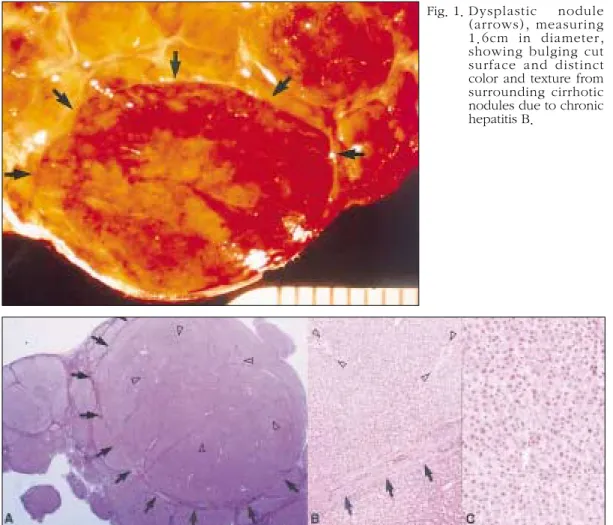
Fig. 1. Dysplastic nodule (arrows), measuring 1.6cm in diameter, showing bulging cut surface and distinct color and texture from surrounding cirrhotic nodules due to chronic hepatitis B.
1994년 world congress of gastroenterology에서 working international party의 토의를 거쳐 제정한 새로운 명칭인 이형성 결절은 현재로서는 양성인지 악성인지 확실히 결정하기 어려우나 앞으로 간세포 암종이 발생될 가능성이 높다는 생물학적 특성에 중점을 둔 명칭이다14). 이형성 결절 (dysplastic nodule)은 이형성을 동반한 장경 1mm 이상의 간 의 결절성 병변으로 확실한 악성의 조직학적 소견 은 없으며 대부분 간경변증에서 발생한다. 육안 소 견상 이형성 결절은 대부분 주위 간조직과 색, 성상 등이 틀리며 절단면이 불룩하게 나오는 특징을 보 인다 (Fig. 1). 이형성 병소 (dysplastic foci)는 장경 1mm 이하의 간세포 군집으로 이형성은 관찰되나 확실한 악성의 조직학적 소견은 없다.
그 동안 여러 논문에서 쓰여져 왔던 MRN 또는 AH는 장경 0.8cm9,15)또는 1.0cm 이상6,8,10) 의 결절 만을 지칭하였으나, 새로운 정의에서는 장경 1mm 이상은 이형성 결절로, 그 이하는 이형성 병소(dys- plastic foci)로 정의함으로써 이형성 결절을 진단시 병변의 크기는 중요성이 적어졌다14).
이형성 결절의 발생 빈도
이형성 결절의 발생빈도는 간경변 환자의 14- 25%으로, 이는 장경 0.8cm 또는 1cm이상의 결절 만을 연구대상으로한 보고들에 의한 것이다6-10).
이형성 결절은 대부분 간경변 환자에서 발생되나, 드물게는 간경변이 발생되지 않은 만성 간질환 환 자에서도 발생될 수 있다16). 이형성 결절이 발생된 경우 간경변 및 만성 간질환을 일으킨 원인은 다양 하며, 미국에서 간이식을 시행한 환자 155예를 대 상으로 한 연구 보고에 의하면, 그 중 44예 (28%)에 서 이형성결절이 발생되었으며, 원인별로는 C형 간 염에서 71%, B형 간염에서 67%, 알코올성 간질환 에서 33%, 자가면역성간염에서 20%, 원발성 경화 성 담관염에서 18%, 원발성 담즙성 간경변증에서 12%의 이형성 결절의 발생율을 보였으며, 원인을 알 수 없는 간경변증에서도 60%의 높은 발생율을 보였다10). 우리나라에서 저자가 경험한 이형성결절 은 B형 간염에 의한 간경변증 환자에서 발생된 것
이 대부분이었다.
이형성 결절의 분류 및 감별 진단
1) 이형성 결절의 분류
이형성 결절은 병리조직학적 특성에 따라 저등도 (low grade)와 고등도 (high grade)로 분류된다.
저등도 이형성 결절에서는 결절을 구성하는 간세포 의 이형성이 미약하며, 고등도 이형성 결절에서는 중등도 이상의 이형성이 관찰되나 악성 종양의 소 견은 없다 (Fig. 2 와 3).
이것을 이전의 분류와 비교하여 보면 대략 다음과 같이 연관지어 볼 수있다.
Type I MRN - ordinary AH - dysplastic nodule, low grade
Type II MRN - atypical AH - dysplastic nodule, high grade
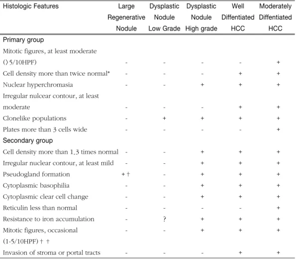
이형성 결절의 분류에 따른 병리조직학적 진단기 준은 학자들간에 다소 다를 수 있으나, 기본적인 진 단기준은 Table 1과 같다14).
2) 조기 간세포암종이란?
조기 간세포암종 (early HCC)은 이형성 결절에서 HCC가 발생된 것을 지칭하며, HCC의 다단계 발생 과정 중 이형성 결절과 HCC의 중간 단계로 생각된 다. 대부분 기존의 간엽 구조의 파괴없이 분화가 좋 은 HCC가 발생하며, 병변내에 문맥역이 관찰되는 것이 특징적이다5,17,18)(Fig 3).
연구보고자들에 따라서는 이형성 결절에서 육안 적으로는 관찰되지 않는 즉 microscopic HCC가 발 생된 경우를 early HCC으로, 이형성 결절에서 육안 적으로 인지할 수 있는 HCC가 발생되는 경우, 즉 macroscopic HCC 주위로 고등도 이형성결절가 관 찰되는 경우를 early advanced HCC (HCC with early component)로 분류하기도 한다19, 20).
이에 반하여 소간세포암종 (small HCC)은 이러한 조기 병변, 즉 이형성 결절의 소견없이 전체 병변이 HCC의 소견을 보이는 장경 2cm 이하의 결절을 지 칭한다.
이들 병변을 방사선학적 소견 또는 육안 소견만으 로 이형성 결절, early HCC 또는 small HCC인지 감 별하는 것은 대부분의 경우 불가능하며, 정확한 진 단을 위해서는 병리조직학적 검색이 필수적이다.
3) 거대 재생결절 (Large regenerative nodule)과의 감별
거대 재생결절이란 간경변의 재생결절들 중 장경 약 0.5cm 이상의 큰 크기의 결절을 지칭한다.
Kondo 등21)은 17명의 간경변 환자에서 발생한 거 대 재생결절들을 치료하지않고 13 - 52 개월간 추 적 관찰한 연구에서 4개의 결절은 관찰기간 중 소 실되어 더 이상 방사선학적 검사상 관찰이 되지 않
았으며, 나머지 결절들도 크기의 변화를 보이지 않 았다. 또한 생검 소견상 비정형성이 관찰되지 않아 이 결절들은 전암병변으로서의 특성이 없는 것으로 보고하였다.
거대 재생결절과 이형성 결절의 감별진단에 도움 이 되는 병리학적 소견은 Table 2와 같다22). 그러나 실제로는 거대 재생결절과 이형성이 미약한 이형성 결절 저등도는 감별이 어려운 경우가 많으며, 정확 한 감별 진단을 위해서는 분자병리학적 검색이 필 요하리라 생각된다. Hytiroglou 등10)은 155 예의 간 이식시 적출된 간 조직을 검색한 결과, 10예에서 무 수히 많은 결절의 발생이 관찰되었으며, 육안 및 현 미경적 소견이 이형성 결절 저등도와 유사하였다.
Table 1. Histological Criteria to Distinguish Hepatocellualr Nodules14
Histologic Features Large Dysplastic Dysplastic Well Moderately Regenerative Nodule Nodule Diffentiated Diffentiated
Nodule Low Grade High grade HCC HCC Primary group
Mitotic figures, at least moderate
(>5/10HPF) - - - - +
Cell density more than twice normal* - - - + +
Nuclear hyperchromasia - - + + +
Irregular nulcear contour, at least
moderate - - - + +
Clonelike populations - + + + +
Plates more than 3 cells wide - - - - +
Secondary group
Cell density more than 1.3 times normal - - + + +
Irregular nuclear contour, at least mild - - + + +
Pseudogland formation +† - + + +
Cytoplasmic basophilia - - + + +
Cytoplasmic clear cell change - - + + +
Reticulin less than normal - - - - +
Resistance to iron accumulation - ? + + +
Mitotic figures, occasional - - + + +
(1-5/10HPF)††
Invasion of stroma or portal tracts - - - + +
이 예들은 모두 간염으로 인한 괴사후 발생된 간경 변에서 발생되었으며, 발생 연령이 통상의 이형성 결절이 발생된 환자들에 비해 의의 있게 낮았다 (평 균연령 31세 vs. 48세). 또한 이 예들에서의 HCC의 발생 빈도는 10%로 전체 간경변 환자의 HCC발생 빈도인 8%와 비슷하였다. 이상의 소견으로 이들 병 변은 거대 재생결절일 가능성이 높을 것으로 추정 하였으며, 간염에 의한 괴사후 발생된 간경변증에 서 10개 이상의 결절의 발생이 관찰되는 경우는 이 형성 결절이라기 보다는 거대 재생결절일 가능성이 더 높음을 시사하였다.
이형성 결절의 전암병변으로서의 의의 및 증거
현재 이형성 결절, 특히 고등도 이형성 결절는 전 암병변으로 생각되며, 이를 지지하는 소견들은 다 음과 같다. 이형성 결절은 주로 HCC가 발생된 간 의 종괴 주변에서 발견되며, 이는 통계학적으로 의 의가 있다8, 10). 이형성 결절에서 한개 또는 서너개의 microscopic HCC의 발생이 관찰되는 경우가 있다1,3, 4, 6, 15, 16, 23)
. 분자병리학적 연구를 통해 이형성 결절 이 단일 클론의 증식임이 증명되었다24, 25). 이형성 결절 고등도에서는 비배수성의 발생이 증가된다26,
27). 이형성 결절의 세포증식능의 증가는 이형성 결 절 저등도, 고등도 및 HCC가 발생함에 따라 의의있 게 증가되나, 저등도 이형성 결절에서는 주위 간경 변 결절과 비슷한 정도의 세포증식능을 보인다28-30). 그 밖에 이형성 결절, 특히 고등도 이형성 결절에서
는 전암병변의 특성으로 알려진 다음과 같은 병리 조직학적 소견이 자주 관찰된다; 소간세포 이형성 (small liver cell dysplasia) 3, 10, 11, 15), Mallory 소체의
출현31, 32), 이형성 결절내 철분 축적 증가33), 철분이
과축적된 결절 내에서 철분의 축적이 없는 병소의
출현34,35), 이형성이 심해질수록 동양구조의 모세혈
관화 즉 혈관내피세포의 유창 감소, 기저막 축적, factor VIII 및 CD34 의 표현 등의 증가36-38), 지방변 성39).
저등도 이형성 결절에서는 위에 기술한 고등도 이형성 결절에서 자주 관찰되는 전암병변으로서의 병리학적 특성들 및 세포증식능의 증가, DNA의 비 배수성의 출현 등이 관찰되지 않아26, 30), 저등도 이 형성 결절의 전암병변으로서의 의의에 대한 의문점 이 제기될 수 있다. 그러나 이형성 결절만으로도 HCC의 발생과 의의있는 연관성을 보이며8, 10), 저등 도 이형성 결절에서 주위의 간경변 결절에 비해 의 의 있게 증가된 신생혈관의 증식을 보이는 점38)은 저등도 이형성 결절가 아직 병리조직학적 비정형성 은 보이지 않으나 전암병변일 가능성을 시사한다.
이상과 같이 이형성 결절, 특히 고등도 이형성 결 절과 HCC는 긴밀한 연관관계를 가지고 있어, 적어 도 이형성 결절의 존재는 앞으로 HCC가 발생될 가 능성이 높을 것을 예견하는 지표로 생각된다. 고등 도 이형성 결절을 절제한 후 3년간 추적 관찰한 연 구보고에 따르면 고등도 이형성 결절에서 micro- scopic HCC가 관찰된 경우에는 모든 예(3/3)에서, Table 2. Features Distinguishing Dysplastic Nodules from Regenerative Nodules22)
Dysplastic nodules
Clonelike features in the entire nodule or clone-like domains of cells within the nodule Unpaired arteries (i.e., without accompanying bile ducts)
Dysplasia, focal or diffuse, particularly of small cell type Compression of portal structures
Dysplastic nodules are usually few in number(<10) within a liver Large regenerative nodules
Sinusoidal dilatation
Often seen diffusely (uncountable numbers) in a cirrhotic, posthepatitic liver Absence of features of dysplastic nodules
microscopic HCC가 관찰되지 않는 경우에는 36%(4/11)에서 HCC가 발생되었으나, 저등도 이형 성 결절 10예에서는 HCC가 발생되지 않았다2). 또 한 방사선학적 검사에서 고등도 이형성 결절가 관 찰되어 연속 생검을 통해 추적 관찰한 보고에 의하 면 HCC가 발생되기까지의 최단 기간은 4개월이었 다3). 그러나 아직 모든 고등도 이형성 결절에서 장 차 악성 종양이 발생될 것인지 또는 HCC의 어느 정 도가 이형성 결절에서 발생된 것인지는 확실하지 않으며, 이들 병변의 생물학적 특성을 규명하기 위 해서는 분자병리학적 연구를 포함한 좀 더 많은 연 구가 필요하리라 생각된다.
Screening은 어떻게 할 것인가?
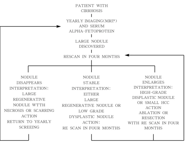
이형성결절을 어떻게 screening 하는 것이 바람직 한 지에 대해서는 아직 확실치 않으며, Fig 4는 최 근 제안된 한 예이다22).
이형성 결절을 조기 진단하는데 있어 문제가 되는 점은 통상의 검사방법이 대부분 비효과적이라는데 있으며, 아래 기술한 내용은 이형성 결절을 screen- ing시 고려하여야할 점이다.
1) 혈청 alpha-fetoprotein (AFP)을 검사하는 것이 도움이 될 것인가?
이형성 결절 및 이형성 결절에서 microscopic HCC가 발생된 early HCC 환자들의 혈청 AFP에 대
SCREENING PROTOCOL OF THE FUTURE ?
PATIENT WITH CIRRHOSIS YEARLY IMAGING(MRI?)
AND SERUM ALPHA-FETOPROTEIN
LARGE NODULE DISCOVERED RESCAN IN FOUR MONTHS
NODULE DISAPPEARS INTERPRETATION:
LARGE REGENERATIVE NODULE WTTH NECROSIS OR SCARRING
ACTION RETURN TO YEARLY
SCREEING
NODULE ENLARGES INTERPRETATION:
HIGH-GRADE DISPLASTIC NODULE
OR SMALL HCC ACTION ABLATION OR
RESECTION WITH RE SCAN IN FOUR
MONTHS NODULE
STABLE INTERPRETATION:
EITHER LARGE
REGENERATIVE NODULE OR LOW GRADE
DYSPLASTIC NODULE ACTION:
RE SCAN IN FOUR MONTHS
Fig. 4. With expected advances in imaging, detection of distinctive nodules in the setting of cirrhosis will likely be possible. Combined with new understandings of proliferative activity and malignant transformation in such nodules, these advances may make a more sensitiue screening protocol such as this one possible22
한 연구보고에 의하면 대부분의 환자 (82%)에서 혈 정 AFP 수치가 20ng/ml 이하로 정상이었으며, AFP 에 대한 면역조직화학염색을 시행하여 본 결과 이 형성 결절 부위에서는 AFP가 표지되지 않았다27, 40). 따라서 혈청 AFP 수치가 정상이어도 이형성 결절 또는 early HCC의 발생 가능성을 배제할 수 없다.
2) 이형성 결절의 혈액공급의 특성은?
이형성 결절의 형태구축학적 연구를 통해 이형성 결절의 혈관과 담관은 주위 간조직의 문맥역과 연 결되어 있음이 밝혀졌으며, 이형성 결절내에서는 주위 간경변과 비교하여 문맥(portal vein)의 총단 면적은 감소되어 있으나 동맥의 총단면적은 비슷하 거나 증가되어있는 반면, 간세포암종은 동맥혈만을 공급받는다23). 또한 신생혈관의 증식 즉, 담관을 동 반하지 않는 muscular artery의 증식이 간경변 결 절, 이형성 결절 저등도, 이형성 결절 고등도, 간세 포암종이 발생함에 따라 의의있게 증가됨이 보고되 었다38).
그러나 이형성 결절의 이중 혈액공급구조 (동맥 혈과 문맥혈의 공급)가 동맥혈의 공급으로만 바뀌 어도, 이것이 꼭 악성으로의 전환을 의미하지는 않 는다. 양성 종양인 간세포선종 (hepatocellular adenoma)은 동맥혈만 공급받는다41).
3) 영상진단학적 검사 방법은 얼마나 정확히 진단할 수 있을까?
초음파 검사, 전산화단층촬영 (computered
tomography, CT) 등의 통상의 영상 진단학적 검사 가 이형성 결절의 진단에 얼마나 도움이 될수있는 지는 아직 확실치 않으나, 영상진단 분야에서의 빠 른 발전은 매우 고무적이다. 최근의 연구보고에 의 하면 이형성결절 진단의 민감도(sensitivity)는 초음 파검사 23%, CT 4%, angiography 0%, CT arteriog- raphy 25%, CT arterial portography 40%, 수술중 초음파검사 54% 정도였으며, early HCC 진단의 민 감도는 초음파검사 64%, CT 58%, angiography 31%, CT arteriography 50%, CT arterial portogra- phy 71%, 수술중 초음파검사 88% 이다42). 각 병변 의 방사선학적 특징은 Table 3와 같으며42), 한가지 이상의 진단방법을 상호 보완적으로 적용시 더욱 진단율을 높일 수 있을 것으로 기대된다. 다음은 이 형성 결절의 영상진단에 대한 최근의 연구보고들이 다.
간의 결절에 대한 방사선학적 연구결과 간세포암 종은 portal venous flow가 없으므로 CT arterial portography상 clear defect로 나타나는데 반해, 이 형성 결절은 CT arterial portography상에서는 관찰 하기 어려우며 이는 이형성 결절이 주위의 간경변 결절처럼 portal venous blood flow를 받기때문인 것 같다. 이형성 결절의 4%에서만 동맥혈의 공급이 주위 간조직에서 보다 증가된 반면 간세포암종에서 는 94%에서 증가되어 있으며, 동맥내 lipiodol를 주 입후 CT를 시행하거나 동맥내 carbon dioxide microtubbles을 주입후 초음파검사를 시행하면 이 형성 결절은 어느 정도 저관류를 보인다43). Color Table 3. Imaging Characteristics Used to Differentiate Small Nodular Lesions Associated With Liver
Cirrhosis42
Imaging Technique Regenerating Dysplastic Nodule Early HCC Small HCC
Sonography Hypo - or isoechoic Hypo-or hyperechoic Hypo-, iso, or hyperechoic Hypo-, iso, or hyperechoic CT arterriography ND ND partial enhancement Enhancement
CT arterial portography ND ND Partial defect Defect Angiography ND ND partially hypervascular Hypernascular T1-weighted MR imaging Hypo - or isointense Hyperintense Iso - or hyperintense Hypo-, iso-, hyperintense T2-weighted MR imaging Hypointense Hypointense Isointense Hyperintense
HCC, hepatocellular carcinoma; ND, not detected.
Doppler flow imaging시 pulsating afferent arterial vessels이 관찰되면 HCC의 진단에 도움이 된다44). 이와같이 이형성 결절과 소간세포암종과의 감별에 혈류 공급 유형에 대한 조사가 도움이 될 것으로 생 각되나, 이러한 결절에서의 동맥혈 증가의 정확한 평가에 대해서는 좀더 연구가 필요하다.
MRI상에서 이형성 결절은 T1-weighted image 상 에서 hyperintensitiy를 보이는 반면, 소간세포암종 은 hyper-, iso- 또는 hypo-intensity를 보인다. T2- weighted image 상 에 서 는 이 형 성 결 절 은 hypointensity를 보이며, 소간세포암종은 hyperin- tensity를 보인다42, 45, 46)
. T1 weighted image상의 hyperintense pattern은 결절내의 지방변성 또는 출 혈의 소견과 연관이 있는 듯 하나, 조영신호강도에 영향을 미치는 병리학적 소견에 대해서는 앞으로 더 연구가 필요하다. 최근 thin section MR imaging 이 이형성결절을 발견하는데 효과적인 방법임이 보 고되었다47).
크기만을 기준으로 거대 증식결절, 저등도 이형성 결절, 고등도 이형성 결절 및 소간세포암종과의 감 별은 어려울 것으로 생각되나, 결절의 크기가 1.5cm 이상일 경우에는 악성 병변이 발생되었을 가능성이 높으며, 악성병변이 발생되지 않은 이형 성 결절은 대부분 장경이 1.2cm 이하이다18, 46, 48)
. 이 형성 결절내에 구리 또는 철의 축적, 지방변성 등의 특성이 관찰될 수 있으며, 이러한 소견이 방사선학 적 진단에 도움이 될 것으로 기대된다49).
4) 결절의 세침 생검의 유용성 및 진단의 정확성은 ?
최근 초음파검사하의 간의 세침생검은 점점 자주 쓰이는 진단방법의 하나로, 결절에서 정확히 생검 되었느냐 하는 것이 진단에 가장 중요하다. 생검된 검체 내에 이형성 결결 부위와 주변 간조직이 포함 되면 두 병변을 비교하여 검색할 수있으므로 진단 에 도움이 된다.세침 생검조직을 이용하여 이형성결절을 진단시 의 진단의 정확도 및 재현율에 대한 연구보고는 아 직 없으며, 악성에 대한 진단기준도 학자간에 다소 다르다. Nagato 등50)은 형태계측학적 연구를 통해
핵면적, 핵대 세포질의 비, 핵 밀도 등이 악성도의 진단에 중요하다고 하였으나, 실제로 생검조직의 진단시 언제나 도움이 되는 것은 아니다. 이러한 변 화가 재생결절을 구성하는 간세포에서도 반응성 변 화 (reactive change)로서 관찰될 수 있기 때문이 다.
이형성 결절의 치료 방침은 ?
이형성 결절, 특히 고등도의 이형성 결절일 경우, 그 생물학적 특성이 좀더 확실히 밝혀질 때까지는
"malignant" 또는 "potentially malignant"로 준하여 치료방침을 세우는 것이 권장된다. 이형성 결절의 치료방법은 수술적 절제 등 그 동안 소간세포암종 에 이용되었던 방법들이 실험적으로 실시되고 있으 며 특별히 권장되는 방법은 아직 없다.
이형성 결절은 문맥혈의 공급도 받기 때문에 대부 분의 경우 transarterial embolization (TAE)은 효과 적이지 못하다. 그러나 이형성 결절에서 간세포암 종이 발생된 경우, 특히 피막이 형성된 경우에는 TAE를 시도하고 있다51). 경피적 알코올 주입 (per- cutaneous ethanol injection)은 현재 일본에서 많 이 쓰이는 치료방법이다23).
고등도 이형성결절를 절제후에도 남아있는 간조 직에서 HCC가 발생될 가능성이 높으므로 간이식 도 하나의 치료방법이 될 수 있다. 소간세포암종으 로 간이식을 받은 환자에서 HCC의 재발율은 8- 15%인데 반하여52, 53, 54), 이형성결절로 간이식 수술 을 받은 환자에서의 HCC의 발생율은 아직 알려져 있지 않으며, 간이식의 시기도 중요하다. .
결 론
간암의 발생과정에는 일련의 동물실험에서 밝혀 졌듯이, 간암종의 형성 이전에 형태학적 변화를 동 반하거나 동반하지 않는 유전생물학적 변이가 선행 될 것으로 생각된다. 만성 간질환에서 발생되는 이 형성 결절, 특히 고등도의 이형성 결절은 전암병변 으로서 다단계 간암발생 과정의 조기 변화로 생각 된다. 이러한 병변에 대한 연구를 통해 인체의 간암 발생과정에 대한 이해를 높일 수 있을 것으로 생각
되며, 또한 전암 및 조기 간암종의 진단 및 치료의 새로운 도약이 기대된다.
감사의 글
간병리를 공부할 수 있도록 이끌어 주신 박찬일 교수님께 감사드리며, 많은 예의 이형성 결절을 검 색할 수 있도록 도와주신 Dr. Thung과 Dr. Theise 에게 고마운 마음을 전합니다.
참고 문헌
1. Arakawa M, Kage M, Sugihara S, Nakashima T, Suenaga M, Okuda K. Emergence of malignant lesions within an adenomatous hyperplastic nodule in a cirrhotic liver: observation in five cases. Gastroenterology 1986; 91: 198-208 2. Kaji K, Terada T, Naknuma Y. Frequent
occurrence of hepatocellular carcinoma in cirrhotic livers after surgical resection of atypical adenomatous hyperplasia (borderline hepatocellular leion): a follow-up study. Am J Gastroenterol 1994; 89:903-908
3. Takayama T, Makuuchi M, Hirohashi S, et al.
Malignant transformation of adnomatous hyperplasia to hepatocellular carcinoma.
Lancet 1990; 336: 1150-1153
4. Wada K, Kondo Y, Kondo F. Large regene- rative nodules and dysplastic nodules in cirrhotic livers : a histopathologic study.
Hepatology 1988; 8: 1684-1688
5. Sakamoto M, Hirohashi S, Shimosato Y. Early stages of multistep hepatocarcinogenesis:
adenomatous hyperplasia and early hepato- cellular carcinoma. Hum Pathol 1991;22:172- 178
6. Furuya K, Nakamura M, Yamamoto Y, Togei K, Otsuka H. Macroregenerative nodule of the liver. A clinicopathological study of 345 autopsy cases of chronic liver disease. Cancer 1988; 61: 99-105
7. Terada T, Terasaki S, Nakanuma Y. A clinicopathologic study of adenomatous hyperplasia of the liver in 209 consecutive cirrhotic livers examined by autopsy. Cancer 1993;72:1551-1556
8. Theise ND, Schwartz M, Miller C, Thung SN.
Macroregenerative nodules and hepatocellular carcinoma in fourty-four sequential adult liver explants with cirrhosis. Hepatology 1992; 16:
949-955
9. Ferrell L, Wright T, Lake J, Roberts J, Ascher N. Incidence and diagnostic features of macroregenerative nodules vs small hepato- cellular carcinoma in cirrhotic livers. Hepat- ology 1992;16:1372-1381
10. Hytiroglou P, Theise ND, Schwartz M, Mor E, Miller C, Thung SN. Macroregenerative nodules in a series of adult cirrhotic liver explants : issues of classification and nome- nclature. Hepatology 1995; 21: 703-708 11. Le Bail B, Belleannee G, Bernard P-H, Saric J,
Balabaud C, Bioulac-Sage P. Adenomatous hyperplasia in cirrhotic livers : histological evaluation, cellular density, and proliferative activity of 35 macronodular lesions in the cirrhotic explants of 10 adult French patients.
Hum Pathol 1995; 26: 897-906
12. Nagasue N, Akamizu H, Yukaya H, Yuuki I.
Hepatocellular pseudotumor in the cirrhotic liver. Report of three cases. Cancer 1984; 54:
2487-2494
13. Ferrell LD, Crawford JM, Dhillon AP, Scheuer PJ, Nakanuma Y. Proposal for standardized criteria for the diagnosis of benign, borderline and malignant hepatocellular lesions arising in chronic advanced liver disease. Am J Surg Pathol 1993;17:1113-1123
14. International Working Party. Terminology of nodular hepatocellular lesion. Hepatology
1995;22:983-993
15. Nakanuma Y, Terada T, Terasaki S, et al.
"Atypical adenomatous hyperplasia" in liver cirrhosis: low grade hepatocellular carcinoma or boderline lesion? Histopathology 1990;
17:27-35
16. Theise ND, Lapook JD, Thung SN. A macroregenerative nodule containing multi- ple foci of hepatocellular carcinoma in a non- cirrhotic liver Hepatology 1993;17: 993-996 17. Kanai T, Hirohashi S, Upton MP et al.
Pathology of small hepatocellular carcinoma.
A proposal for a new gross classification.
Cancer 1987; 60:810-819.
18. Okauda K. Hepatocellular carcimona:recent progress. Hepatology 1992;15:948-963 19. Winter TC, Takayasu K, Muramatsu Y, et al.
Early advanced hepatocellular carcinoma:
evaluation of CT and MR appearence with pathological correlation. Radilogy 1994;
192:379-387
20. Muramatsu Y, Nawano S, Takayasu K, et al.
Early hepatocellular carcinoma: MR imaging.
Radiology 1991;181:209-213
21. Kondo F, Ebara M, Suigiura N, et al.
Histologic features and clinical course of large regenerative nodules: evaluation of their precancerous potentiality. Hepatology 1990;12:592-598
22. Theise ND. Macroregenerative (dysplastic) nodules and hepatocarcinogenesis: theore- tical and clinical considerations. Seminars in Liver Dis 1995;15:360-371
23. Nakanuma Y, Terada T, Ueda K, Terasaki S, Nonomura A, Matsui O. Adenomatous hyper- plasia of the liver as a precancerous lesion.
Liver 1993;13:1-9
24. Tsuda H, Hirohashi S, Shimosato Y, Terada M, Hasegawa H. Clonal origin of atypical
adenomatous hyperplasia of the liver and clonal identify with hepatocellular carcinoma.
Gastroenterology 1988;95:1664-1666
25. Aihara T, Noguchi S, Sasaki Y, Nakano H, Monden M, Imaoka S. Clonal analysis of prec-ancerous lesion of hepatocellular carcinoma. Gastroenterology 1996;111:455- 461
26. Orsatti G, Theise ND, Thung SN, Paronetto F.
DNA image cytometric analysis of macroregenerative nodule (adenomatous hyperplasia) of the liver : evidence in support of their preneoplastic nature. Hepatology 1993; 17: 621-627
27. Terasaki S, Terada T, Nakanuma Y, Non- omura A, Unoura M, Kobayashi K. Argyro- philic nucleolar organizer regions and alpha- fetoprotein in adenomatous hyperplasia in human cirrhotic livers. Am J Clin Pathol 1991;
95: 850-857
28. Grigioni WF, D'Errico A, Bacci F, et al.
Primary liver neoplasms : evaulation of proliferative index using MoAb Ki67. J Pathol 1989; 158: 23-29
29. Terada T, Nakanuma Y. Cell proliferative activity of adenomatous hyperplasia in the liver and small hepatocellular carcinoma. An immunohistochemical study demonstrating proliferating cell nuclear antigen. Cancer 1992; 70: 591-598
30. Theise ND, Marcelin K, Goldfischer M, et al.
Low proliferative activity in macrorege- nerative nodules: evidence for an alternate hypothesis concerning human hepatocar- cinogenesis. Liver 1996;16:134-139
31. Nakanuma Y, Ohta G. Is Mallory body formation a preneoplastic change? A study of 181 cases of the liver bearing hepatocellular carcinoma and 82 cases of cirrhosis. Cancer
1984;55:2400-2404
32. Terada T, Hoso M, Nakanuma Y. Mallory body clustering in adenomatous hyperplasia in human cirrhotic livers: report of four cases.
Hum Pathol 1989;20:886-890
33. Terada T, Nakanuma Y. Survey of iron- accumulative macroregenerative nodules in cirrhotic livers. Hepatology 1989; 5:851-854 34. Terada T, Nakanuma Y. Iron-negative foci in
siderotic macroregenerative nodules in human cirrhotic livers: a marker of incipient pre-neoplastic or neoplastic lesions. Arch Pathol Lab Med 1989; 113: 916-920
35. Deugnier YM, Charalambous P, Le Quilleuc D, et al. Preneoplastic significance of hepatic iron -free foci in genetic hemochromatosis: a study of 185 patients. Hepatology 1993;18 :1363-1369
36. Terada T, Nakanuma Y. Expression of ABH blood group antigens, receptors of Ulex europaeus agglutinin I, and factor VIII-related antigen on sinusoidal endothelial cells in adenomatous hyperplasia in human cirrhotic livers. Hum Pathol 1991;22:486-493
37. Dhillon A, Colombari R, Savage K, Scheuer PJ. An immunohistochemical study of the blood vessels within primary hepatocellular tumors. Liver 1992;12:311-318
38. Park Y, Yang C-P, Thung SN, Theise ND.
Perisinusoidal cell activation, neoangiog- enesis, and sinusoidal "capillarization" in macroregenerative nodules: evidence for neoplastic progression. Hepatology 1995;22 :185A
39. Terada T, Nakanuma Y, Hoso M, Saito K, Sasaki M, Nonomura A. Fatty macroregenra- tive nodule in non-steatotic liver cirrhosis. A morphologic study. Virchows Arch A Pathol Anat 1989; 415:131-136
40. Theise ND, Fiel IM, Hytiroglou P, et al.
Macroregenerative nodules in cirrhosis are not associated with elevated serum or stainable tissue alpha-fetoprotein. Liver 1994;15:30-34
41. Goodman ZD, Mikel UV, Lubbers PR, et al:
Hepatocellular adenoma. Am J Surg Pathol 1987;11:191-6
42. Choi BI, Takayasu K, Han MC. Small hepatocellular carcinoma and associated nodular lesions of the liver: pathology, pathogenesis, and imaging findings. Am J Radiol 1993; 160:1177-1187
43. Mastsui O, Kadoya M, Kameyama T, et al.
Benign and malignant nodules in cirrhotic livers: distiction based on blood supply.
Radiology 1991;178:493-497
44. Tanaka S, Kitamra T, Fujita M , Kasugai H, Inoue A, Ishiguro S. Small hepatocellular carinoma : differentiation from adenomatous hyperplastic nodule with color Doppler flow imaging. Radiology 1992;182:161-165
45. Ebara M, Ohto M, Watanabe Y, et al. Diag- nosis of small hepatocellular carcinoma:
correlation of MR imaging and tumor histolo- gic studies. Radiology 1986;159:371-377 46. Matsui O, Kadoya M, Kameyama T, et al.
Adenomatous hyperplastic nodules in the cirrhotic liver: differentiation from hepatocell- ular carcinoma with MR imaging. Radiology 1989;173:123-126
47. Earls JP, Theise ND, Weinreb JC, et al.
Dysplastic nodules and hepatocellular carcinoma: thin section MR imaging of explanted cirrhotic livers with pathological correlation. Radiology 1996;210:207-214 48. Eguchi A, Nakamura O, Okudaira S, Sugihara
S, Kojiro M. Adenomatous hyperplasia in the vicinity of small hepatocellular carcinoma.
Hepatology 1992;15:843-848
49. Kitagawa K, Matsui O, Kadoya M, et al.
Hepatocellular carcinoma with excessive copper accumulation: CT and MR findings.
Radiology 1991;180:623-628
50. Nagato Y, Kondo F, Kondo Y, Ebara M, Ohto M. Histological and morphometrical indicat- ors for a biopsy diagnosis of well-differentia- ted hepatocellular carcinoma. Hepatology 1991;14:473-478
51. Ueda K, Saito K, Terada T, Nakamuma Y.
Selective necrosis of encapsulated malignant lesion within atypical adenomatous hyperp-
lasia of the liver following transarterial embolization. A report of two autopsy cases.
J Clin Gastroenterol 1991;13:709-714
52. Mazzaferro V, Regalia E, Doci R, et al. Liver transplantation for the treatment of small hepatocellular carcinoma in patients with cirrhosis. N Engl J Med 1996;334:693-699 53. Romani F, Belli LS, Rondinara GF, et al. The
role of transplantation in small hepatocellular carcinoma complicating cirrhosis of the liver.
J Am Coll Surg 1994; 178:379-384
54. Tan KC, Rela M, Ryder SD et al. Experience of orthotropic liver transplantation and