Comparative Analysis of the Result of Minimally Invasive Anterior Plating and Open Reduction and Internal Fixation in Humerus Shaft Simple Fracture
Sang-Hun Ko, Chang-Gyu Choe , Ju-Hyung Lee
Department of Orthopedic Surgery, Ulsan University Hospital, University of Ulsan College of Medicine, Ulsan, Korea
Background: This retrospective comparative study aims to evaluate the surgical outcomes and complications of two surgical methods for simple fractures of the humeral shaft; minimally invasive anterior plating and open reduction combined with internal fixation.
Methods: A total of 26 patients with humeral shaft simple fractures, who had surgery between June 2009 and September 2013 and were followed-up at least 12 months, were included in our analysis. They were divided into two groups; group 1 comprised of 12 pa- tients who underwent minimally invasive anterior plating and group 2 comprised of 14 patients who underwent an open reduction and internal fixation. The clinical outcomes, radiological results, and complications were compared and analyzed.
Results: We found that bone union was achieved in all patients, and the mean union periods were 20.7 ± 3.34 and 20.3 ± 3.91 weeks for groups 1 and 2, respectively. In most patients, we found that shoulder and elbow functions were recovered. At 12 months post-oper- ation, we found that the Korean Shoulder Scoring system, the University of California at Los Angeles score and Mayo elbow performance score were 91.4 ± 7.97, 33.4 ± 1.15, and 90.8 ± 2.23 for group 1, and 95.2 ± 1.53, 33.3 ± 1.43, and 90.17 ± 1.85 for group 2. In terms of complications, we found that 2 patients had radial nerve palsy after open reduction and internal fixation, but all cases spontane- ously resolved within 6 months. Complications such as infection and loss of fixation were not reported.
Conclusions: Both minimally invasive anterior plating and open reduction with internal fixation produced satisfactory outcomes in the treatment of simple fractures of the humeral shaft.
(Clin Shoulder Elbow 2015;18(2):75-79)
Key Words: Humeral fractures; Minimally invasive surgical procedure; Internal fracture fixation; Retrospective studies
Copyright © 2015 Korean Shoulder and Elbow Society. All Rights Reserved. pISSN 2383-8337
Clinics in Shoulder and Elbow Vol. 18, No. 2, June, 2015 http://dx.doi.org/10.5397/cise.2015.18.2.75
Received August 18, 2014. Revised December 14, 2014. Accepted December 19, 2014.
Correspondence to: Chang-Gyu Choe
Department of Orthopedic Surgery, Ulsan University Hospital, 877 Bangeojinsunhwan-doro, Dong-gu, Ulsan 682-714, Korea Tel: +82-52-250-7129, Fax: +82-52-235-2823, E-mail: [email protected]
Financial support: None. Conflict of interests: None.
Introduction
Humeral fractures compose a large proportion of around 5% to 8% of all fractures of the bone. Of these 30% to 50% are fractures of the humeral shaft meaning that around 1% to 3% of all fractures are fractures of the humeral shaft.1) Although in large most humeral shaft fractures are treated satisfactorily conserva- tively using functional braces,2) recent changes based on changes in prioritization of early joint exercises and rehabilitation are an increased treatment preference for surgical intervention and thereby an accelerated return to normal activity. In this study, we compared the clinical outcomes and complications of two
types of established surgical treatments for patients with simple fractures of the humeral shaft; minimally invasive anterior plating and open reduction with internal fixation (ORIF).
Methods
In this retrospective study, we enrolled 26 patients who un- derwent treatment for humeral shaft simple fractures between June 2009 and September 2013 and were able to participate in at least a 12-month follow-up. We divided the patients into two treatment groups; group 1 included 12 patients who underwent a minimally invasive plating approach and group 2 included
14 patients who underwent an ORIF. In our patient groups, the average patient age was 50.5 ± 18.1 years, and the ratio of sex was 12 males to 14 females (Table 1). We classified the simple fractures according to the AO classification; 6 patients had frac- tures of the proximal third half of the humeral shaft (AO class A);
16 patients had fractures of the middle third; and 4, the distal third. We also classified the fractures in terms of morphology using the AO classification; 12 patients had spiral fractures (AO class A1); 4, oblique fractures (AO class A2); and 10, transverse fractures (AO class A3). One patient from each treatment group showed preoperative radial nerve palsy. All treatment proce- dures were carried out by the authors.
In all cases, a 4.5-mm-wide limited contact locking compres- sion plate (LC-LCP; Synthes, Oberdorf, Swiss) and locking screws were used. An anterolateral portal was used for ORIF. First, a 10-cm-incision was created at the center of the fracture site. The underlying soft tissue was dissected to expose the deltoid and bicep muscles, after which the bicep muscles were temporarily pulled medially apart. Then, the brachial muscles were separat- ed to expose the site of fracture. Once the periosteum and the hematoma around the fracture site were removed, tools such as a bone reduction clamp were used to provisionally maintain the reduction, and subsequently a locking compression plate was used for the final fixation. The plate size used for the minimally invasive anterior plating was chosen so that at least 3 or 4 holes each could be placed on the distal and the proximal sides of the plate around the center of the fracture site. The spacing between the holes were made sufficient so that the stress enforced by the two screws closest to each other on either side of the main spic- ule, i.e., the distance between the two screws facing oppositely from the fracture, was within the effective range and could be distributed evenly across the entire plate (Fig. 1).
Irrespective of the treatment method, all patients were ad- ministered with post-operative long-arm splinting for 2 days from the operation date. We also periodically carried out a thorough physical examination with regards to damage of the medial nerve. After 2 or 3 days, the shoulder and elbow range of motion (ROM) exercises were cautiously begun along with ROM exercises of the metacarpopharangeal joints. For the as- sessment of the clinical outcome, we used the Korean Shoulder Scoring system (KSS), the American Shoulder and Elbow Society, and the the University of California at Los Angeles scores (UCLA) to measure shoulder function at the final postoperative follow- up. One specialist radiologist assessed all the radiological findings and determined the duration of bone union.
Statistical Analysis
For all statistical analyses, the SPSS for Windows release 17.0 (SPSS Inc., Chicago, IL, USA) was used. To compare the results of the two treatment groups, the independent t-test and the Mann-Whitney test were used.
Results
We found that all patients achieved bone union. The aver- age duration of bone union was 20.7 ± 3.34 weeks in group 1 and 20.3 ± 3.91 weeks in group 2. A comparative analysis did not show a statistically significant difference between these two values. The shoulder and elbow functions improved to a satis- factory level in most patients. At the 12th postoperative month, group 1 patients showed an average KSS of 91.4 ± 7.97, an average UCLA of 33.4 ± 1.15, and an average Mayo elbow performance score of 90.8 ± 2.23. The respective values for group 2 were 95.2 ± 1.53, 33.3 ± 1.43, and 90.17 ± 1.85, and showed a significant improvement over those of group 1 (p<0.05). All other parameters did not show a statistically signifi- cant difference. The postoperative radiography showed that in group 1 the anteroposterior angular deformity was 2.4o ± 2.42o, the lateral angular deformity was 3.0o ± 3.30o, whereas in group 2 these value were significantly greater at 4.5o ± 2.73o and at 6.1o ± 4.16o, respectively. Nevertheless, neither group showed any angular deformity that was deemed as a misalignment (Table 2). We found 2 cases of radial nerve palsy in the two patients that underwent ORIF, but these were found to be spontaneously resolved at postoperative follow-up. Further, the preoperative findings of radial nerve palsy in two other patients were seen to be spontaneously resolved by the 6th postoperative month. Pos- sible complications such as infection and fixation loss were not found in either group.
Discussion
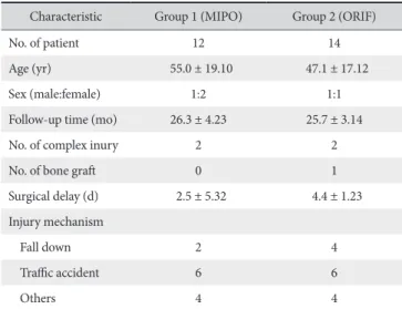
Table 1. Preoperative Demographics and Characteristic Data
Characteristic Group 1 (MIPO) Group 2 (ORIF)
No. of patient 12 14
Age (yr) 55.0 ± 19.10 47.1 ± 17.12
Sex (male:female) 1:2 1:1
Follow-up time (mo) 26.3 ± 4.23 25.7 ± 3.14
No. of complex inury 2 2
No. of bone graft 0 1
Surgical delay (d) 2.5 ± 5.32 4.4 ± 1.23 Injury mechanism
Fall down 2 4
Traffic accident 6 6
Others 4 4
C D B A
Fig. 1. Case treated by minimally invasive plate osteosynthesis technique. (A) Preoperative radiograph of a 15-year-old male shows transverse fracture on the distal 1/3 area of the humerus shaft. (B) Immediate postoperative radiograph shows satisfactory result. (C) Two month after operative radiograph shows callus formation. (D) Five month after operative radiograph shows radiologic union state.
Table 2. Period of Fusion and Radiological, Clinical Results
Characteristic Group 1 (MIPO) Group 2 (ORIF) p-value
Period of fusion (wk) 20.3 ± 3.91 20.7 ± 3.34 >0.05
Postoperative angulation (o)
Antero-posterior 4.5 ± 2.73 2.4 ± 2.42 0.01
Lateral 6.1 ± 4.16 3.0 ± 3.30 0.01
Korean Shoulder Scoring system 95.2 ± 1.53 91.4 ± 7.97 0.013
The University of California at Los Angeles score 33.3 ± 1.43 33.4 ± 1.15 >0.05
Mayo elbow performance score 90.17 ± 1.85 90.8 ± 2.23 >0.05
Values are presented as mean ± standard deviation.
MIPO: minimally invasive plate osteosynthesis, ORIF: open reduction with internal fixation.
can be broadly divided into either as conservative or as surgical, but as to which method is more effective is still under debate.
Sarmiento3) found that in terms of the occurrence of non and mal-union of fractures, the surgical method using the locking compression plate was better than the conservative treatment using functional braces. Interestingly, the likelihood of complica- tions such as infection or paralysis did not significantly change whether the patient received a conservative or a surgical treat- ment. However, when Gosler et al.4) carried out a multi-centre systematic analysis across 6 studies, they concluded that it was difficult to come down to a definitive conclusion as to which treatment method is superior. As such, we cannot come to a conclusive judgment as to the better treatment modality for simple fractures of the humeral shaft on the basis of the current literature. But recent emphasis on the importance of early joint exercises and rehabilitation, which is based on papers that have shown young and active patients with transverse or short oblique fractures have had a more delayed union after conservative treatment compared to the average union period for that re- spective age group,5,6) has naturally shifted the focus of research to surgical treatments.
Surgical procedures include external fixation, intramedullary fixation, open reduction, and compression plating, and each procedure has its own merits and pitfalls. Bae et al.7) have found that the mean union periods of fractured bones between these procedures do not show statistically significant differences. Of the above, the most common approach is the open reduction in combination with internal fixation. However, its associated complications such as non-union, deep tissue infection, and re-fracture after removal of the fixative material are serious dis- advantages.8,9) These complications are thought to be induced
by blockage of the periosteum blood flow when soft tissue is substantively dissected during massive surgical incision. In com- parison, although intramedullary fixation leads hardly to soft tis- sue damage at the fracture site, but can lead to shoulder pain or disabled ROM.10,11) Recently, the use of minimally invasive plat- ing for bone fixation has become widespread for fractures of the long bone, including the humerus. Its relative advantage over mechanical fixation is that it preserves the biological environ- ment better and through internal fixation achieve better bone union.12) Further, minimally invasive plating uses a small inci- sion site thus minimizes soft tissue dissection, and since this site is farther away from the fracture site, the likelihood of a deep tissue infection and bone grafting is lower.8) Lastly, esthetically, minimally invasive plating is preferable due to a small incision area (Fig. 2). In this comparative study, we did not find a signifi- cant difference in the clinical outcomes after minimally invasive plating and after open reduction and internal fixation in patients with humeral shaft simple fractures. Nevertheless, further study using an extended numbers of patient cases is required.
Conclusion
Using our parameters of clinical outcome, we found that both of the surgical treatment methodologies, the minimally invasive anterior plating approach and the open reduction and internal fixation approach, for humeral shaft simple fractures gave satisfactory results. We found that there were no significant differences in the clinical outcome in terms of duration of bone union, range of motion of the shoulder and elbow, and relief of pain between the two treatment groups.
References
1. Volgas DA, Stannard JP, Alonso JE. Nonunions of the humerus.
Clin Orthop Relat Res. 2004;(419):46-50.
2. Sarmiento A, Kinman PB, Galvin EG, Schmitt RH, Phillips JG.
Functional bracing of fractures of the shaft of the humerus. J Bone Joint Surg Am. 1977;59(5):596-601.
3. Sarmiento A. Outcome of nonoperative vs operative treat- ment of humeral shaft fractures: a retrospective study of 213 patients. Orthopedics. 2011;34(3):159; author reply 159.
4. Gosler MW, Testroote M, Morrenhof JW, Janzing HM.
Surgical versus non-surgical interventions for treating hu- meral shaft fractures in adults. Cochrane Database Syst Rev.
2012;1:CD008832.
5. Schemitsch EH, Bhandari M. Fractures of the diaphyseal hu- merus. In: Browner BD, Jupiter JB, Lebine AM, Trafton PG, eds. Skeletal trauma. 3rd ed. Toronto: WB saunders; 2003.
1481-511.
7. Bae SW, Kim WJ, Song BY, Choi NH, Lee JH. Postoperative functional assessments in adult humerus shaft fractures: com- parison among plates and screws, intramedullary nail and ex- ternal fixator. J Korean Soc Fract. 2001;14(2):228-35.
8. Farouk O, Krettek C, Miclau T, Schandelmaier P, Guy P, Tscherne H. Minimally invasive plate osteosynthesis and vascularity:
preliminary results of a cadaver injection study. Injury. 1997;28 Suppl 1:A7-12.
9. Perren SM. Evolution of the internal fixation of long bone frac- tures. The scientific basis of biological internal fixation: choos- ing a new balance between stability and biology. J Bone Joint
Surg Br. 2002;84(8):1093-110.
10. Flinkkilä T, Hyvönen P, Lakovaara M, Linden T, Ristiniemi J, Hämäläinen M. Intramedullary nailing of humeral shaft frac- tures. A retrospective study of 126 cases. Acta Orthop Scand.
1999;70(2):133-6.
11. Rommens PM, Blum J, Runkel M. Retrograde nailing of hu- meral shaft fractures. Clin Orthop Relat Res. 1998;(350):26- 39.
12. Byun YS. Minimally invasive plate osteosynthesis, MIPO. J Ko- rean Fract Soc. 2007;20(1):99-114.