Comparing the Use of Single and Double Interlocking Distal Screws on a Polarus Intramedullary Nail for Humeral Shaft Fractures
Hee Seok Yang, Jeong Woo Kim , Hong Je Kang, Jung Hyun Park, Yong Chan Lee, Kwang Mee Kim1
Department of Orthopaedic Surgery, Wonkwang University School of Medicine & Hospital, Iksan, 1Department of Nurshing, Chodang University, Muan, Korea
Background: Our aim was to make a comparative analysis of radiological and clinical outcomes of using either one or two interlocking distal screws on a Polarus intramedullary nail for the internal fixation of humeral shaft fractures.
Methods: From January 2008 to March 2014, we enrolled 26 patients with humeral shaft fractures who were operated on using intra- medullary nails. The patients were divided into 2 groups according to how many interlocking distal screws were used to lock the Polarus nail: in group 1, a single interlocking distal screw was used in 12 patients; and in group 2, double interlocking distal screws, in 14 pa- tients. We compared the degree of recovery of the displaced fracture fragments between the two groups. To compare the nonunion and shoulder function, we assessed each patient’s modified American Shoulder and Elbow Surgerns (ASES) score.
Results: We found that 10 of 12 fractures achieved union in group 1, and 13 of 14 fractures, in group 2. We did not find a meaningful difference in the time to bone union between the two groups. The percentage of recovery of displaced fracture fragments until union was 66.9% for group 1 and 59.41% for group 2. At the final follow-up, we found that the scores for shoulder joint modified ASES was 78.7 for group 1 and 80.7 for group 2.
Conclusions: Our results show that if locked appropriately, even a single screw on a Polarus nail can provide satisfactory radiological union and improved clinical outcome after intramedullary nailing of humeral shaft fractures.
(Clin Shoulder Elbow 2015;18(2):91-95)
Key Words: Humeral fractures; Intramedullary nailing; Bone screws Clinics in Shoulder and Elbow Vol. 18, No. 2, June, 2015
http://dx.doi.org/10.5397/cise.2015.18.2.91
Received October 28, 2014. Revised May 14, 2015. Accepted June 1, 2015.
Correspondence to: Jeong Woo Kim
Department of Orthopaedic Surgery, Wonkwang University School of Medicine & Hospital, 895 Muwang-ro, Iksan 570-711, Korea Tel: +82-63-859-1360 , Fax: +82-63-852-9329, E-mail: [email protected]
Financial support: This research was funded by grants from Wonkwang University. Conflict of interests: None.
Introduction
Humeral shaft fractures compose around 3% of fractures.
Mildly displaced humeral shaft fractures can be effectively treated conservatively.1,2) But more severe displacements, such as those seen in multiple, comminuted, and open fractures, require surgical treatments; the demand for which has been on the rise.3) Modalities of surgical treatment include locking plates, intramedullary nailing, external fixation, and etc. Although lock- ing plates provides swift functional recovery by providing strong fixation,4,5) intramedullary nailing which also provides sufficient fixation has the added advantages of minimal soft tissue dam- age and low infection rate, making it the preferred alternative treatment in recent yeras.6,7) However, intramedullary nailing is
associated with disadvantages such as technical difficulties and complications such as radial nerve injury. The technical difficulty demanded for the intramedullary nailing using distal interlocking screws can delay surgery time, exacerbate patient condition, and increase radiographic exposure to the surgeon.8) In general, most surgeons recommend the use of 2 distal interlocking screws to lock intramedullary nails; yet, in many reported incidents we find that surgeons have had to opt to using only one screw for surgical ease or to avoid injury to the radial nerve. To address the significance of the number of screws used in intramedullary nailing, we investigated the effect of the number of distal inter- locking screws used during intramedullary nailing on the clinical outcome of humeral shaft fractures.
Methods
In our retrospective study, we included 26 patients with type 12 humeral shaft fractures, according to the AO/OTA classifica- tion, who were operated on using intramedullary nails from January 2008 to March 2014. Those with pathologic fractures, with type III open fractures, with combined radial nerve injury, and who had had their treatment delayed by at least 2 weeks were excluded from the study. The patients were divided into 2 groups according to how many interlocking distal screws were used to lock the Polarus nail: in group 1, a single interlocking distal screw was used in 12 patients; and in group 2, double in- terlocking distal screws, in 14 patients.
Surgical Methods
All surgeries were performed by a single surgeon. Under general anesthesia, the patient was placed in a Fowler’s position on a beach chair; then, a 4–5 cm skin incision from the antero- lateral edge of the acromion to the posterior side was made. At the incision site, the deltoid muscle was temporarily separated to expose the greater tuberosity of the humeral bone. Another incision was made on the supraspinatus tendon at a position 1 cm medial of the posterior bicipital groove so that we could manually reconstruct the fracture with the aid of an image inten- sifier then insert a Polarus nail (Acumed Inc., Beaverton, OR, USA) into the intramedullary region using a guide pin. After we confirmed that the intramedullary nail was inserted correctly, we locked the nail in position by inserting either single or double 3.5-mm interlocking screws at the distal site without the use of target devices. With regard to the choice of screw numbers, we inserted, from the uppermost oval portal, one screw when it alone could prevent traction of the fractures, but if it could not, we inserted two screws. Postoperatively, an abduction brace was imposed for 4 weeks while elbow, wrist, and finger joint exer- cises were allowed.
Methods of Assessment
For each patient, we assessed the mechanism of injury using past medical records. Bone union was confirmed using radio- graphic findings and the time to bone union was noted. We deemed a successful union when the following criteria were ful- filled: 1) at least 3 sites of calluses on humeral cortical bone on either the anteroposterior or the lateral radiograms; and 2) no soreness at the site of fracture.
We measured the following radiological parameters: the mean postoperative distance between fracture fragments, the degree of recovery of the displaced fracture fragments, and the improvement in angulation of fracture fragments. To assess the improvement in angulation of fractures, we measured the im- mediate postoperative and post-union levels of angulation on
tion was measured as the angle that forms between the normal humeral cortical line and that of the most displaced fractured bone. The change in angulation was calculated as the percent- age difference between angle of postoperative angulation and the angle of post-union angulation over the angle of postopera- tive angulation. For the clinical and functional parameters, we assessed at every follow-up for soreness around the region of fracture, the range of motion of the shoulder such as maximum flexion, external rotation, and internal rotation, and lastly, the modified American Shoulder and Elbow Surgeons (ASES) score to assess patient shoulder function and the progress of treat- ment. Further, we kept a vigilant eye for any complications that arose during the follow-up period and executed the necessary secondary surgeries.
Our data did not show a normal distribution; thus, nonpara- metric tests were used for the statistical analyses. Comparative analysis of parameters gender and mechanism of injury were performed using the chi-square test, and parameters time to union and radiological markers, using the Mann-Whitney U test.
All statistical analyses were made using the SPSS program ver.
13.0 for Windows (SPSS Inc., Chicago, IL, USA).
Results
The mean age of the patients in group 1 was 65.1 years (range, 34–82 years) and in group 2, 59.8 years (range, 23–77 years).
The ratio of gender was 5 males to 7 females in group 1 and 9 males to 5 females in group 2. We divided the mechanism of in- jury into two broad categories: low-energy fractures, referring to injuries such as those from falls while standing; and high-energy
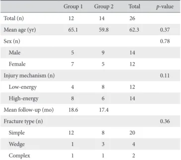
Table 1. Summary of Cases
Group 1 Group 2 Total p-value
Total (n) 12 14 26
Mean age (yr) 65.1 59.8 62.3 0.37
Sex (n) 0.78
Male 5 9 14
Female 7 5 12
Injury mechanism (n) 0.11
Low-energy 4 8 12
High-energy 8 6 14
Mean follow-up (mo) 18.6 17.4
Fracture type (n) 0.36
Simple 12 8 20
Wedge 1 3 4
Complex 1 1 2
Group 1: a single interlocking distal screw was used in 12 patients, Group 2:
fractures, referring to injuries such as those sustained from road accidents either as a driver or a pedestrian or from falls. How- ever, we found no significant difference in our parameters when they were compared according to mechanism of injury (p=0.11).
The mean follow-up period was 18.6 months (range, 12–27 months) for group 1 and 17.4 months (range, 10–29 months) for group 2 (Table 1, Fig. 1).
Through postoperative radiography, we found that 10 of 12 fractures (83.3%) achieved union in group 1, and 13 of 14 fractures (92.9%), in group 2. The mean postoperative distance between fracture fragments was 4.47 ± 3.88 mm in group 1 and 3.85 ± 3.32 mm in group 2. Two and 1 cases of nonunion were found in the respective groups, for which we had observed at least a 10-mm distance between the fracture fragments post- operatively. We carried out bone grafting to treat the nonunion.
Complications other than bone nonunion that occurred were also given secondary treatments accordingly: in group 1, we carried out arthroscopic capsular release for 2 cases of shoulder stiffness; and in group 2, we carried out arthroscopic capsular release for 1 case of shoulder stiffness and a screw exchange for a case of distal screw loosening (Fig. 2). We did not find a meaningful difference in the mean time to union between the two groups, which were 13.8 ± 2.9 weeks for group 1 and 14.2
± 4.2 weeks for group 2 (p=0.28). The percentage of recovery of displaced fracture fragments to union was 66.9% for group 1 and 59.41% for group 2. When we radiologically measured the change in mean angulations of fractures from postoperation to post-union, we found, from the anteroposterior view, a mean 62.90 ± 20.54% change in angulation in group 1 (postoperation, 7.98 ± 2.63o; and post-union, 3.44 ± 2.20o) and a mean 59.41
± 16.20% change, in group 2 (postoperation, 6.79 ± 3.22o; and post-union, 2.74 ± 1.92o). At the final follow-up, we found
A B C
Fig. 1. Closed humeral shaft fracture in a 63-year-old woman. (A) Preoperative radiographs demonstrating humeral shaft fracture. (B) Postoperative radiographs after closed reduction and internal fixation with the Polarus intramedullary nail with the single distal locking screw. (C) Obvious union with callus formation was found 14 weeks after the operation.
A B C
Fig. 2. Closed humeral shaft fracture in 78-year-old woman. (A) Preoperative radio- graphs demonstrating the fracture. (B) Post- operative radiographs after closed reduction and internal fixation with the Polarus intra- medullary nail with double distal screws. (C) The proximal distal targeting screw is failed.
Table 2. Summary of Results
Group 1 Group 2 p-value
Modified ASES score 78.7 80.7 0.58
Operation time (min) 151 116 0.02
Fracture site gap (mm) 4.47 ± 3.88 3.85 ± 3.32 0.42 Fracture site anlge (o) 6.78 ± 2.13 5.23 ± 2.42 0.56 Union period (wk) 13.8 ± 2.9 14.2 ± 4.2 0.28
Nonuion rate (%) 16.7 7.1 0.35
Group 1: a single interlocking distal screw was used in 12 patients, Group 2:
double interlocking distal screws, in 14 patients.
ASES: American Shoulder and Elbow Surgeons.
that the scores for shoulder joint modified ASES were 78.7 for group 1 and 80.7 for group 2 (Table 2).
Discussion
In this study we compared the outcomes of intramedullary nailing for humeral shaft fractures between those which had a single distal interlocking screws fixed to the Polarus nail and those which had double screws. We found that when appro- priately fixed the fixation of intramedullary nails with a single screw enables progression to satisfactory bone union and clini- cal improvement of the shoulder. Humeral shaft fractures, un- like fractures of shafts of other long bones, can be treated with good clinical outcomes using conservative measures.9,10) This is known to be because the axial force and muscle contraction help retain the correct alignment of the fracture and the large range of motion of the shoulders can acclimate to the changes after bone union.11) Sometimes, the condition of the shoulder can deteriorate during conservative treatment when the frac- ture displaces such as in a distraction. This delay in bone union may induce angular and rotational deformity and limited post- operative shoulder movement.12,13) For those unable to receive conservative treatment because of severely displaced fractures, comminuted fractures, co-sustained injuries that make early rehabilitation difficult, pathologic factures, and vascular dam- ages, surgical interventions should be considered.14,15) The main surgical interventions for humeral shaft fractures include plate fixation, intramedullary nailing, and external fixation; of these, the intramedullary nailing has received the most recognition for its efficacy in treating fractures of shaft of many long bones, such as the femoral and tibial bones.
Intramedullary nailing is known to be a relatively noninvasive method with a short surgery time, low infection rate, minimal soft tissue damage, and high resistance to flexion force.15,16) In this retrospective study, we show that a total of 23 out of 26 pa- tients who were treated using intramedullary nailing of humeral shaft fractures showed satisfactory bone union and favorable modified ASES scores, our clinical marker for shoulder function.
Despite these advantages, the technical difficulties of intramed- ullary nailing of the humeral bone such as the fixing of distal interlocking screws without the use of target devices exist. Other complications such as radial nerve injuries are incurred when soft tissue is not sufficiently protected. These technical difficulties and complications become more problematic when 2 screws, compared to 1, are used, causing delay of surgery time and sometimes even forcing surgeons to drop the use of one screw.
Although most authors recommend the use of 2 distal interlock- ing screws during intramedullary nailing,17) no studies, to the best of our knowledge, exist that actually present poor outcomes with a single screw.
that there is no significant difference in the results of the time to bone union, restoration of displaced fracture fragments, and shoulder function between patients who receives different num- bers of distal interlocking screws. In a study by Hajek et al.,18) they found that there is no difference in the resistance to twisting whether or not 1 or 2 distal interlocking screws were used in the intramedullary nailing of a femoral bone. But, because the femo- ral bone receives the burden of weight differently to the humeral bone this finding is not entirely applicable to humeral shaft frac- tures. Choy et al.19) used cadaveric humeral bones to compare the resistance to twisting after intramedullary nailing and report- ed that the number of distal interlocking screws did not make a difference to the resistance. From their study, we can conclude that there is no significant correlation between the number of distal interlocking screws and the stability of the rotational force.
Thus, we may consider distraction of fractures caused by strain from the upper body and not the rotational stability as the more important contributory factor to bone non-union after intra- medullary nailing of a humeral bone. In our sample of patients, we found a total of 3 cases of nonunion: 2 in group 1 and 1 in group 2. We noted that the postoperative distance between the fracture fragments was a least 10-mm in all 3 cases of nonunion, which was larger than the postoperative average. It is more likely that the cause of the nonunion is because of the distraction of the fracture site after the closed reduction of the fracture rather than the number of screws used. However, with a mild distrac- tion, when elbow exercises are commenced after surgery, micro- movements and axial force at the fracture site has been shown to promote callus formation–promoting bone healing.20)
Limitations of this study that should be addressed are the small sample size, the use of non-parametric tests, and the ab- sence of a quantitative or qualitative measure of the influence of the trauma-induced soft tissue. Nevertheless, literature on the clinical outcomes of using different numbers of distal interlock- ing screws during intramedullary nailing for humeral fractures is sparse, making our study which analyzed this is a meaningful addition to the current literature.
Conclusion
We conclude that if locked appropriately even a single screw on a Polarus nail can provide satisfactory radiological union and improved clinical outcome after intramedullary nailing of hu- meral shaft fractures.
References
1. Ekholm R, Adami J, Tidermark J, Hansson K, Törnkvist H, Ponzer S. Fractures of the shaft of the humerus. An epide- miological study of 401 fractures. J Bone Joint Surg Br. 2006;
2. Sarmiento A, Waddell JP, Latta LL. Diaphyseal humeral frac- tures: treatment options. Instr Course Lect. 2002;51:257-69.
3. Nam TS, Choi JW, Kim JH, Kim SY, Kim JJ, Chun JM. Nonunion of the humerus shaft. J Korean Fract Soc. 2005;18(3):294-8.
4. Lee HJ, Oh CW. Operative treatment of humerus shaft frac- ture: conventional open plating or minimally invasive plate osteosynthesis. J Korean Fract Soc. 2012;25(2):155-62.
5. Meekers FS, Broos PL. Operative treatment of humeral shaft fractures. The Leuven experience. Acta Orthop Belg. 2002;
68(5):462-70.
6. Chen F, Wang Z, Bhattacharyya T. Outcomes of nails versus plates for humeral shaft fractures: a Medicare cohort study. J Orthop Trauma. 2013;27(2):68-72.
7. Chen AL, Joseph TN, Wolinksy PR, et al. Fixation stability of comminuted humeral shaft fractures: locked intramedullary nailing versus plate fixation. J Trauma. 2002;53(4):733-7.
8. Levin PE, Schoen RW Jr, Browner BD. Radiation exposure to the surgeon during closed interlocking intramedullary nailing. J Bone Joint Surg Am. 1987;69(5):761-6.
9. Yang KH. Helical plate fixation for treatment of comminuted fractures of the proximal and middle one-third of the humer- us. Injury. 2005;36(1):75-80.
10. Riemer BL, Butterfield SL, D’Ambrosia R, Kellam J. Seidel intramedullary nailing of humeral diaphyseal fractures: a pre- liminary report. Orthopedics. 1991;14(3):239-46.
11. Spiguel AR, Steffner RJ. Humeral shaft fractures. Curr Rev Mus- culoskelet Med. 2012;5(3):177-83.
12. Garnavos C, Lasanianos N. Intramedullary nailing of combined/
extended fractures of the humeral head and shaft. J Orthop Trauma. 2010;24(4):199-206.
13. Lin J, Hou SM. Locked nailing of severely comminuted or seg- mental humeral fractures. Clin Orthop Relat Res. 2003;(406):
195-204.
14. Brumback RJ, Bosse MJ, Poka A, Burgess AR. Intramedullary stabilization of humeral shaft fractures in patients with multiple trauma. J Bone Joint Surg Am. 1986;68(7):960-70.
15. Lin J. Treatment of humeral shaft fractures with humeral locked nail and comparison with plate fixation. J Trauma. 1998;44(5):
859-64.
16. Hall RF Jr. Closed intramedullary fixation of humeral shaft frac- tures. Instr Course Lect. 1987;36:349-58.
17. Park JY, Oh JH, Kho DH, Jung JK. Intramedullary nail on the humeral fracture. J Korean Fract Soc. 2008;21(3):244-54.
18. Hajek PD, Bicknell HR Jr, Bronson WE, Albright JA, Saha S. The use of one compared with two distal screws in the treatment of femoral shaft fractures with interlocking intramedullary nailing.
A clinical and biomechanical analysis. J Bone Joint Surg Am.
1993;75(4):519-25.
19. Choy WS, Park YB, Park JH, Ann TG, Ahn JS, Choi SW. Tor- sional characteristics between single and double distal screws in the interlocking intramedullary nailing of humeral shaft frac- ture. J Korean Orthop Res Soc. 1999;2(2):111-6.
20. Carter DR, Beaupré GS, Giori NJ, Helms JA. Mechanobiology of skeletal regeneration. Clin Orthop Relat Res. 1998;(355 Suppl):S41-55.