Efficacy of Additive Trans-cuff Augmentation Sutures for Proximal Humeral Fractures Stabilized by Locking Plates in Elderly Patients
Nam Su Cho , Hee Seok Shim, Sang Hyeon Lee, Jong Wook Jeon, Yong Girl Rhee
Shoulder & Elbow Clinic, Department of Orthopaedic Surgery, Kyung Hee University College of Medicine, Seoul, Korea
Background: The purpose of our study was to evaluate the functional and radiologic outcomes of additive augmentation sutures through rotator cuff for proximal humeral fractures stabilized locking plate in elderly patients.
Methods: We enrolled 74 patients over the age of 60 years who received internal fixation using locking plates for proximal humeral fractures. Of these, 50 patients had additive augmentation sutures through rotator cuff. The mean age at the time of surgery was 72.1 years (range, 60–89 years), and the mean follow-up period was 17.5 months (range, 12–62 months). The humeral neck-shaft angle and humeral head height were used as radiological markers to assess the effect of additive augmentation sutures through rotator cuff. We al- located the patients who received additive augmentation sutures into group A and those who did not into group B.
Results: At the final follow-up, the mean Korean Showlder Society score and Constant scores were 88.96 ± 12.1 and 86.6 ± 11.9, respectively, in group A and 86.21 ± 11.8 and 85.3 ± 11.7, respectively, in group B (p=0.368, 0.271). At the final follow-up, the mean loss in humeral neck-shaft angle from the time of immediate postoperative measurement was 1.6o in group A and 4.8o in group B, whereas the mean loss in humeral head height was 0.82 mm in group A and 0.52 mm in group B (p=0.029, 0.178).
Conclusions: The surgical outcomes of internal fixation using locking plates for proximal humeral fractures were clinically and radiologi- cally good in elderly patients over the age of 60 years without any observable complications. Further, the loss of humeral head shaft angle at the final follow-up from its initial postoperative measurement was significantly smaller in patients who received an additive augmenta- tion suture than in those who did not. Thus, we conclude that augmentation sutures are a beneficial option for elderly patients that clini- cians can consider at the time of surgical decision making.
(Clin Shoulder Elbow 2015;18(2):68-74)
Key Words: Proximal humeral fracture; Locking plate osteosynthesis; Trans-cuff augmentation sutures; Clinical outcome; Radiologic out- come; Elderly patient
Clinics in Shoulder and Elbow Clinics in Shoulder and Elbow Vol. 18, No. 2, June, 2015
http://dx.doi.org/10.5397/cise.2015.18.2.68
Received October 2, 2014. Revised December 8, 2014. Accepted December 13, 2014.
Correspondence to: Nam Su Cho
Department of Orthopaedic Surgery, Kyung Hee University Hospital at Gangdong, 892 Dongnam-ro, Gangdong-gu, Seoul 134-727, Korea Tel: +82-2-440-6154, Fax: +82-2-440-7498, E-mail: [email protected]
Financial support: This research was supported by sports scientification of Convergent R&D Program through the National Research Foundation of Korea (NRF) funded by the Ministry of Science, ICT & Future Planning (NRF-2014M3C1B1033319). Conflict of interests: None.
Introduction
The incidence of proximal humeral fractures in the elderly is increasing due to an increase of sports activity and osteopo- rosis.1,2) Unstable and displaced proximal humeral fractures are generally accepted indications for operative treatment.3) Current treatments include osteosynthesis using proximal humeral nails and plates, tension band wiring, percutaneous or minimally invasive techniques such as pinning, intramedullary flexible nails, and screw osteosynthesis, and hemiarthroplasty.4-7) Surgical
treatment requires anatomical reduction and a stable fixation, which proves to be especially difficult in an osteoporotic bone.
Unreduced or poorly reduced fractures with varus angulation of the neck-shaft angle can be a cause of avascular necrosis of the humeral head.8,9)
With the advent of locking plates and screw fixation, a greater number of displaced proximal humeral fractures are being treat- ed with osteosynthesis.10-14) Locking plates may have a mechani- cal advantage over standard implants in osteoporotic bones.13,15) Nevertheless, complication rates after surgical stabilization re-
main high.11,16,17) Several studies report complications, of which the two most commonly reported are varus inclination and screw penetration. Gardner et al.13) reported that a varus col- lapse is typically caused by rotator-cuff forces, thereby suggesting that reduction maintenance necessitates some medial support.
Other than these, loss of fixation is another frequently observed complication and in locking plates this requires removal of the screws to avoid impending joint destruction.18)
Fixation loss is a problem often met after surgical treatment of displaced unstable proximal humeral fractures in elderly patients with osteoporosis. Despite many approaches and attempts to overcome this problem no definitive consensus operation exists.
One reason for the loss of fixation is an insufficient anchorage of screws in the humeral head. Liew et al.19) also found screw purchase to be significantly greater when screws were placed into the medial subchondral bone and cautioned about relying on fixation in the superior humeral head. When necessary, the fixative construction should be augmented with heavy sutures, which passes adjacent to bony fragments, goes through rotator cuff tissue, and winds back to the fixation implant to provide maximal implant-fragment stability.20) In other words, the displac- ing force of the rotator cuff must be reduced through additive fiber cerclages. In patients who received plate fixations without additive fiber-cerclages, subacromial impingement and tuberosi- ty displacement are observed more frequently than in those who received plate fixations with additive fiber-cerclages.21) However, to date, the effect of additive augmentation sutures in proximal humeral fractures stabilized by locking plates is clinically unclear.
The purpose of our study was to evaluate the functional and radiologic outcomes following a trans-cuff additive augmenta- tion sutures in proximal humeral fractures stabilized by locking plates in elderly patients. In addition, we sought to investigate the clinical benefits of implementing additive augmentation sutures in these patients. We hypothesize that the mechanical support provided by additive augmentation sutures through the rotator cuff is important for establishing a stable construct.
Methods
Patient Selection
Of the 125 patients who underwent surgical treatment for dislocated unstable proximal humeral fractures between May 2007 and June 2013, 74 patients over the age of 60 years who were able to participate in at least a year follow-up study were enrolled into our study. All 74 patients received an internal fixa- tion using locking plates, and in 50 patients we carried out a further additive augmentation suture through the rotator cuff.
The mean age of the patients at the time of operation was 72.1
± 14.8 years (range, 60 to 89 years) and the mean follow- up period was 17.5 months (range, 12 to 62 months). Sixteen patients (21.6%) were males and 58 patients (78.4%) were fe-
males. All selected patients were operated on within a week of injury and the mean time interval from injury to operation was 4.5 ± 1.2 days. We divided the patients according to whether they received or did not receive an additive augmentation su- ture through the rotator cuff; 50 patients who received additive augmentation sutures were allocated to group A and 24 patients who did not were allocated to group B (Table 1).
Preoperative and Postoperative Evaluations
Postoperative clinical evaluations were performed regu- larly on an outpatient basis (at 2 weeks, 6 weeks, 3 months, 6 months, 9 months, and 12 months postoperatively and at the last follow-up), and the results of the last follow-up were ana- lyzed. Postoperative subjective pain score was measured using the visual analog scale (VAS). For postoperative shoulder range of motion (ROM), forward flexion, external rotation at the side, and internal rotation to the back were assessed. The Korean Shoulder Society score (KSS) and Constant score22) were used for clinical assessment.
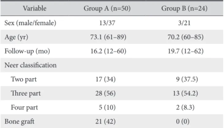
Preoperatively, routine roentgenograms with antero-posterior, lateral, and axillary views were taken followed using computed tomography. All patients were evaluated with plain x-rays on a regular basis postoperatively until bone union was observed. We measured the neck-shaft angle and humeral head height as our radiological parameters and compared these values at the im- mediate postoperation and at the final follow-up (Fig. 1).
Operative Techniques
All operations were performed by a single surgeon (C.N.S).
During surgery, the patient was placed in a beach chair position with the bed inclined at about 30o. The sterilized ipsilateral arm was not fixed and was rested on a removable, height-adjustable tray table. Before draping, the portable C-arm (image intensifier) was optimally positioned to take radiographic images.
To begin with, we made an incision starting just inferior to the
Table 1. Patients Demographics
Variable Group A (n=50) Group B (n=24)
Sex (male/female) 13/37 3/21
Age (yr) 73.1 (61–89) 70.2 (60–85)
Follow-up (mo) 16.2 (12–60) 19.7 (12–62)
Neer classification
Two part 17 (34) 9 (37.5)
Three part 28 (56) 13 (54.2)
Four part 5 (10) 2 (8.3)
Bone graft 21 (42) 0 (0)
Values are presented as number only, median (range), or number (%).
Group A: patients who received additive augmentation sutures, Group B: pa- tients who did not receive additive augmentation sutures.
coracoid process and extending towards the proximal area of the insertion of the deltoid using the deltopectoral approach. During the procedure, we gained sufficient exposure through a partial release of the anterior deltoid insertion and the abduction of the arm. First, the head of the humerus was reconstructed using a few sutures or cerclage wires or both if required as described by Hertel,23) which in their words is akin to reconstructing a broken eggshell. We held the larger fragments like tuberosities into their anatomical positions using pointed reduction clamps and fixed them using Kirschner wires (K-wires). If the medial calcar was fractured, we made sure that they were held reduced as much as possible as this could cause postoperative varus displacement during healing.
Next, the fracture was reduced by ligamentotaxis, which is the manipulation of the upper arm and the buttressing effect of the plate (Periarticular Proximal Humeral Locking Plate; Zimmer Inc., Warsaw, IN, USA). Taking into consideration of the extent of the fracture, the desired length of the plate was chosen; usu- ally plate length that can accommodate approximately 3 to 4 holes distal to the fracture is sufficient. We created space for the plate close to the bone via blunt insertion of the plate along the distal fragment of the fracture and along the humeral shaft. The proximal humerus was usually already exposed and the frac- ture clearly visualized from this approach. Through traction and manipulation, we restored length of the plate and aligned it to its desired position, the ideal position being the upper tip of the plate around 5 mm inferior to the greater tuberosity. In case of a residual valgus deformity of the humeral head, the plate was placed slightly higher than the tip of the greater tuberosity.
To fix the correctly positioned locking plates with screws, we temporarily stabilized the plate by holding it down with a thumb on the proximal fragment and drilled a non-locking hole just distal to the fracture and perpendicular to the bone fragment.
As the screw is tightened, the reduction of the valgus deformity
gradually occurs as the shaft that is usually displaced medially also lateralizes to a reduced state. Thus, this was done carefully and the alignment was constantly checked with a C-arm. Once the screw is tightened, the angulation is usually reduced to an acceptable position. If required, a few K-wires were temporally inserted into the proximal part of the plate to maintain a hold on the proximal humerus. These loosely inserted wires help main- tain plate position to the head while allowing correction.
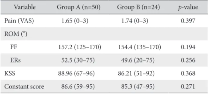
After the first screw is screwed in, the plate should already buttress the bone with its anatomically contoured curves, and the fracture is reduced. We then carefully placed the calcar screw at the surgical neck to catch the medial calcar of the fracture that helps resist postoperative varus angulation. Once this was done, the rest of the screws were inserted while checking with C-arm imaging. Care was taken to ensure none of the screws were in the joint, which was thoroughly checked with the C-arm in fluo- roscopic mode. Lastly, in selected patients we made additive augmentation sutures through the rotator cuff by anchoring the cuffs to holes on the plate using sutures (Fig. 2), which is antici- pated to maximize function of the shoulder after healing.
Comminuted fractures were held reduced with the help of bone clamps, K-wires, and sutures. All cases were checked post- reduction with the C-arm before closure to confirm a successful reduction, correct plate placement, and no protruding screws in the joint. In cases of poor bone quality, we performed a fixation using allografting during the insertion of screws.
Postoperative Rehabilitation
All patients were provided with a shoulder sling from the operation room. Passive motion was started from postoperative day 1 with pendulum exercises and then assisted-arm flexion by the aid of the contralateral hand. Active motion was begun after 6 weeks postoperation.
Fig. 1. Radiological assessments. (A) Neck shaft angle. (B) Humeral head height.
B A
Statistical Analysis
An independent t-test was used to compare the VAS score, ROM, KSS, Constant score, and the radiological results between the two groups. Statistical significance was set to an α-level of 0.05 with 95% confidence intervals. For all statistical analyses, the Statistical Package for the Social Sciences (SPSS) software package ver. 17.0 (SPSS Inc., Chicago, IL, USA) was used.
Results
Pain
We found that the mean subjective pain score, VAS, im- proved from 4.01 ± 2.6 at 2 weeks postoperation to 1.65 ± 1.7 at the last follow-up in group A (p=0.037) and from 3.96 ± 2.81 to 1.74 ± 1.9 in group B (p=0.042) (Table 2).
Range of Motion
In group A, we found that the mean active ROM for forward flexion changed from 76.3o (range, 50o–110o) at 2 weeks post- operation to 157.2o (range, 125o–170o) at the last follow-up; ex-
ternal rotation at the side, from 31.7o (range, 15o–45o) to 52.5o (range, 30o–75o); and internal rotation to the back, from L3 (range, L1–L5) to T11 (range, T8–L3) (p=0.021, 0.015, 0.027).
At the same time-points, in group B, the mean ROM for forward flexion changed from 77.4o (range, 50o–120o) to 154.4o (range, 135o–170o), external rotation at the side, from 30.9o (range, 10o– 50o) to 49.6o (range, 20o–75o), and internal rotation to the back, from L2 (range, L1–S1) to T11 (range, T9–L3) (p=0.014, 0.007, 0.036). In terms of the ROM at the last follow-up, we found no statistically significant differences between the 2 groups (p=0.194, 0.256, 0.672, respectively) (Table 2).
Clinical Assessments
At the last follow-up, we found that the mean KSS and Con- stant scores were 88.96 ± 12.1 and 86.6 ± 11.9, respectively, in group A and 86.21 ± 11.8 and 85.3 ± 11.7, respectively, in group B. There were no statistically significant differences be- tween the 2 groups (p=0.368, 0.271) (Table 2).
Radiologic Results
We found that the mean immediate postoperative humeral neck-shaft angle was 132.9o ± 12.2o for group A and 133.7o ± 13.9o for group B. The respective values at the final follow-up were 131.3o ± 13.9o and 129.9o ± 12.5o, respectively (p=0.352, 0.134). The mean loss of humeral neck-shaft angle at the final follow-up compared to the immediate postoperative fixation was 1.6o ± 0.7o in group A and 4.8o ± 1.2o in group B (p=0.029) (Table 3).
We found that the mean immediate postoperative height of the humeral head was 9.04 ± 3.2 mm in group A and 10.16
± 2.8 mm in group B, and later, the respective values for the mean final follow-up was 8.22 ± 3.2 mm and 9.64 ± 4.2 mm, respectively (p=0.271, 0.263). Across these two time-points, the mean loss of humeral head height was 0.82 ± 0.5 mm for group A and 0.52 ± 1.1 mm for group B (p=0.178) (Table 3).
Fig. 2. (A) Additive augmentation sutures through holes on the locking plate. (B) Ad- ditive augmentation sutures through the rotator cuff are done by anchoring the cuffs to holes on the plate with the help of sutures.
A B
Table 2. Comparison of Postoperative Clinical Outcomes between Group A and Group B
Variable Group A (n=50) Group B (n=24) p-value
Pain (VAS) 1.65 (0–3) 1.74 (0–3) 0.397
ROM (o)
FF 157.2 (125–170) 154.4 (135–170) 0.194
ERs 52.5 (30–75) 49.6 (20–75) 0.256
KSS 88.96 (67–96) 86.21 (51–92) 0.368
Constant score 86.6 (59–95) 85.3 (47–95) 0.271 Values are presented as median (range).
Group A: patients who received additive augmentation sutures, Group B:
patients who did not receive additive augmentation sutures, VAS: visual ana- logue scale, ROM: range of motion, FF: forward flexion, ERs: external rotation at the side, KSS: Korean Shoulder Society score.
Complications
In our series, we did not observe any complications such as varus failure, screw penetration, delayed union, nonunion, in- fection, avascular necrosis, and implant failure in neither group at the last follow-up.
Discussion
Proximal humeral fractures are closely associated with os- teoporosis and commonly occur in over elderly patients over the age of 60 years. Stabilizing and promoting fracture healing of displaced unstable fractures in an osteoporotic bone requires optimal treatment, which there is no consensus of as of yet.
Various techniques have been used to stabilize fractures of the proximal part of the humerus, including intramedullary nails, plate-and-screw osteosynthesis, tension band wiring, percutane- ous pin fixation, and hemiarthroplasty.6,11,15,17,18) Of these, the locking proximal humerus plate, or osteosynthesis, was designed to maintain a stable fracture reduction even in an osteoporotic bone. Advantages of the locking proximal humerus plate include gentle fracture reduction using indirect maneuvers, a high resis- tance to avulsion even in patients with poor bone stock because of the combination of fixed-angle screw-plate locking and three- dimensional placement of screws in the humeral head, pos- sibility of early exercise, and a short period of immobilization because of the high initial stability achieved.24) The advent of locking plates has helped enhance the healing rate of many frac- tures that come combined with osteoporosis.
However, a retrospective study by Egol et al.25) on 51 con- secutive patients showed that proximal humerus fracture fixation using locking plates was associated with early complication and with their risk factors. In their study, a total of 12 patients (24%) had complications; screws penetration in 8 patients (16%), os- teonecrosis in 2 (4%), early fixation failure in 2 (4%), and hetero- topic ossification in 1 (2%). Similarly, when Clavert et al.26) per-
formed locking plate fixation in 73 proximal humeral fractures 8.2% of patients exhibited surgery-related secondary dislocation and 5.5% exhibited nonunion, which led to a significantly poor postoperative Constant score. They concluded that although us- ing locking plates in weak bones enables more fixation security, the increase in complication rate may outweigh the benefits especially patients with severe osteoporosis, comminuted frac- tures, or with head split, in which case an arthroplasty may be a more appropriate treatment option.
Keeping a locking plate fixed for as long as it is needed for the union of a fracture is not possible in all patients. Secondary dis- placement or secondary head deformities have been reported in more than 20% of elderly patients.11,16,17,23) Failure in fixation are induced by rotator-cuff forces.13) Consequently, it was postulated that fixation constructs should be augmented, for example, with sutures passing adjacent to bony fragments, through rotator cuff tissue, and back to the fixation implant, to provide maximum implant-fragment stability.20) Additive fiber-cerclages should reduce displacing forces of the rotator cuff and secure tuberosi- ties after their anatomical reduction by acting as a stable circular platform on which head fragments can rest.27) As such, in a study on the effects of additive fiber cerclages on stable plate fixations, the authors found that subacromial impingement and tuberos- ity displacement were observed more frequently in individuals without fiber cerclages than in those with.21) However, even with additive augmentation sutures, we cannot eliminate the displac- ing forces of the rotator cuff completely. Voigt et al.28) found no significant difference when additive fiber-cerclages were per- formed or not performed for a 24 unstable 3-part fracture model with an intact rotator cuff in terms of interfragmentary motion.
Until now, we could not find literature regarding the clinical effects of additive augmentation sutures besides a few biome- chanical studies. To the best of our knowledge, our study is the first to test whether an additive augmentation suture through the rotator cuff for proximal humeral fractures stabilized by locking plates has a clinically significant advantage in elderly patients.
In this study, we found that proximal humeral fractures in the elderly patients that were internally fixed using locking plates were clinically and radiologically successful according to our chosen parameters. Regardless of whether additive augmenta- tion sutures were made, we observed union in both groups and a satisfactory functional outcome. Further, postoperative ROM of the shoulders was improved almost as well as its contralateral counterpart. There were no signs of postoperative varus failure or screw penetration. However, at the final follow-up, we found that the loss of neck-shaft angle was greater in the group B than in group A. Thus, in case of severe dislocation or comminution of the fracture especially in elderly patients with concomitant osteoporosis, we believe that making additive trans-cuff sutures after internal fixation will provide a sturdier fixation and should be considered as a strong surgical candidate.
Table 3. Comparison of Radiologic Outcomes according to Subgroups Variable Group A (n=50) Group B (n=24) p-value NSA (o)
Immediate postoperative 132.9 133.7 0.352
At the last follow-up 131.3 129.9 0.134
Angle loss 1.6 4.8 0.029
HH height (mm)
Immediate postoperative 9.04 10.16 0.271
At the last follow-up 8.22 9.64 0.263
Height loss 0.82 0.52 0.178
Group A: patients who received additive augmentation sutures, Group B:
patients who did not receive additive augmentation sutures, NSA: neck-shaft angle, HH: humeral head.
Our study has a few limitations. First, being retrospective in nature, our study has limitations related retrospective studies but we tried to minimize these limitations by conducting a retrospec- tive analysis of the prospectively collected patients’ data. Sec- ond, since the technique was chosen depending on operative findings a presence of selection bias cannot be eliminated. How- ever, the additive augmentation sutures technique, though per- formed in osteoporotic bones or in more unstable and severely displaced fracture patterns, presented similar clinical results and fracture healing to the suture negative technique. Nevertheless, this confirmed its efficacy for the treatment of osteoporotic com- plex fractures. In addition, the definition of elderly person was somewhat subjective because the value was an arbitrary cutoff.
Finally, the neck-shaft angle and the humeral head height were measured in the antero-posterior view of proximal humerus, the radiologic findings for which may be affected by many factors.
Conclusion
We found good clinical and radiological outcomes without complications after internal fixation using locking plates for the treatment of proximal humeral fractures in elderly patients over the age of 60 years. However, at the final follow-up, we found that in patients who did not received additive augmentation su- tures on the rotator cuff a loss in humeral neck-shaft angle of the humeral bone that was greater in those who received additional sutures. Thus, we believe additional strengthening sutures are vi- able options for elderly patients receiving surgical treatment.
References
1. Palvanen M, Kannus P, Niemi S, Parkkari J. Update in the epi- demiology of proximal humeral fractures. Clin Orthop Relat Res. 2006;442:87-92.
2. Court-Brown CM, Caesar B. Epidemiology of adult fractures: a review. Injury. 2006;37(8):691-7.
3. Neer CS 2nd. Displaced proximal humeral fractures. I. Classifi- cation and evaluation. J Bone Joint Surg Am. 1970;52(6):1077- 89.
4. Cornell CN. Tension-band wiring supplemented by lag-screw fixation of proximal humerus fractures: a modified technique.
Orthop Rev. 1994;Suppl:19-23.
5. Hintermann B, Trouillier HH, Schäfer D. Rigid internal fixation of fractures of the proximal humerus in older patients. J Bone Joint Surg Br. 2000;82(8):1107-12.
6. Jaberg H, Warner JJ, Jakob RP. Percutaneous stabilization of unstable fractures of the humerus. J Bone Joint Surg Am.
1992;74(4):508-15.
7. Schippinger G, Szyszkowitz R, Seibert FJ. Current concepts in the treatment of proximal humeral fractures. Curr Orthop.
1997;11(3):203-14.
8. Robinson CM, Amin AK, Godley KC, Murray IR, White TO. Modern perspectives of open reduction and plate fixation of proximal humerus fractures. J Orthop Trauma.
2011;25(10):618-29.
9. Aggarwal S, Bali K, Dhillon MS, Kumar V, Mootha AK. Dis- placed proximal humeral fractures: an Indian experience with locking plates. J Orthop Surg Res. 2010;5:60.
10. Plecko M, Kraus A. Internal fixation of proximal humerus frac- tures using the locking proximal humerus plate. Oper Orthop Traumatol. 2005;17(1):25-50.
11. Agudelo J, Schürmann M, Stahel P, et al. Analysis of efficacy and failure in proximal humerus fractures treated with locking plates. J Orthop Trauma. 2007;21(10):676-81.
12. Gardner MJ, Lorich DG, Werner CM, Helfet DL. Second- generation concepts for locked plating of proximal humerus fractures. Am J Orthop (Belle Mead NJ). 2007;36(9):460-5.
13. Gardner MJ, Weil Y, Barker JU, Kelly BT, Helfet DL, Lorich DG.
The importance of medial support in locked plating of proxi- mal humerus fractures. J Orthop Trauma. 2007;21(3):185-91.
14. Rose PS, Adams CR, Torchia ME, Jacofsky DJ, Haidukewych GG, Steinmann SP. Locking plate fixation for proximal humeral fractures: initial results with a new implant. J Shoulder Elbow Surg. 2007;16(2):202-7.
15. Edwards SL, Wilson NA, Zhang LQ, Flores S, Merk BR. Two- part surgical neck fractures of the proximal part of the hu- merus. A biomechanical evaluation of two fixation techniques.
J Bone Joint Surg Am. 2006;88(10):2258-64.
16. Südkamp N, Bayer J, Hepp P, et al. Open reduction and internal fixation of proximal humeral fractures with use of the locking proximal humerus plate. Results of a prospec- tive, multicenter, observational study. J Bone Joint Surg Am.
2009;91(6):1320-8.
17. Schliemann B, Siemoneit J, Theisen Ch, Kösters C, Weimann A, Raschke MJ. Complex fractures of the proximal humerus in the elderly: outcome and complications after locking plate fixation. Musculoskelet Surg. 2012;96 Suppl 1:S3-11.
18. Jost B, Spross C, Grehn H, Gerber C. Locking plate fixation of fractures of the proximal humerus: analysis of complica- tions, revision strategies and outcome. J Shoulder Elbow Surg.
2013;22(4):542-9.
19. Liew AS, Johnson JA, Patterson SD, King GJ, Chess DG. Effect of screw placement on fixation in the humeral head. J Shoul- der Elbow Surg. 2000;9(5):423-6.
20. Smith AM, Mardones RM, Sperling JW, Cofield RH. Early com- plications of operatively treated proximal humeral fractures. J Shoulder Elbow Surg. 2007;16(1):14-24.
21. Hente R, Kampshoff J, Kinner B, Füchtmeier B, Nerlich M.
Treatment of dislocated 3- and 4-part fractures of the proximal humerus with an angle-stabilizing fixation plate. Unfallchirurg.
2004;107(9):769-82.
22. Constant CR, Murley AH. A clinical method of functional as-
sessment of the shoulder. Clin Orthop Relat Res. 1987;(214):
160-4.
23. Hertel R. Fractures of the proximal humerus in osteoporotic bone. Osteoporos Int. 2005;16 Suppl 2:S65-72.
24. Kettler M, Biberthaler P, Braunstein V, Zeiler C, Kroetz M, Mutschler W. Treatment of proximal humeral fractures with the PHILOS angular stable plate. Presentation of 225 cases of dislocated fractures. Unfallchirurg. 2006;109(12):1032-40.
25. Egol KA, Ong CC, Walsh M, Jazrawi LM, Tejwani NC, Zucker- man JD. Early complications in proximal humerus fractures (OTA Types 11) treated with locked plates. J Orthop Trauma.
2008;22(3):159-64.
26. Clavert P, Adam P, Bevort A, Bonnomet F, Kempf JF. Pitfalls and complications with locking plate for proximal humerus frac- ture. J Shoulder Elbow Surg. 2010;19(4):489-94.
27. Nho SJ, Brophy RH, Barker JU, Cornell CN, MacGillivray JD.
Management of proximal humeral fractures based on current literature. J Bone Joint Surg Am. 2007;89 Suppl 3:44-58.
28. Voigt C, Hurschler C, Rechi L, Vosshenrich R, Lill H. Additive fiber-cerclages in proximal humeral fractures stabilized by locking plates: no effect on fracture stabilization and rotator cuff function in human shoulder specimens. Acta Orthop.
2009;80(4):465-71.