Clinical and Radiological Results of Reverse Total Shoulder Arthroplasty Using a 25-mm Glenoid Baseplate
Ji Min Lee, In Bo Kim , Dong Wook Jung
Department of Orthopedic Surgery, Bumin Busan Hospital, Busan, Korea
Background: The size of the baseplate used in reverse total shoulder arthroplasty (RTSA) tends to be larger than the average size of the glenoid in the Korean population. The mismatch between the sizes of the baseplate and the patient’s glenoid may result in improper fixation of the glenoid baseplate. This in turn may lead to the premature loosening of the glenoid component. Thus, we evaluated the short-term results of using a 25-mm baseplate in RTSA.
Methods: Seventeen patients with cuff tear arthropathy underwent RTSA with a 25-mm baseplate. The mean age of the patients was 70.1 years, and the mean follow-up period was 14.0 months. We evaluated clinical outcomes preoperatively and postoperatively: the range of shoulder motion, the American Shoulder and Elbow Surgeons (ASES) score, and the Korean Shoulder Society (KSS) score.
Results: We found that the mean ASES score and KSS improved from 35.0 to 74.4 (p<0.001) and from 46.9 to 71.8 (p<0.001) with RTSA. The mean forward elevation and abduction, external rotation also improved from 78.6° to 134.3° (p<0.05) and from 66.6° to 125.0° (p<0.05), from 20.2° to 28.4° (p=0.43). Postoperative complications were seen in 12% of patients, but neither the loosening of the glenoid baseplate nor inferior scapular notching were observed.
Conclusions: In sum, the results of using a 25-mm baseplate in RTSA were similar to those of previous reports. Even though the out- comes are those of a short-term follow-up, neither the loosening of the glenoid baseplate nor the scapular notching were observed.
(Clin Shoulder Elbow 2015;18(4):242-247)
Key Words: 25-mm baseplate; Reverse total shoulder arthroplasty; Glenoid size of the Korean population
Copyright © 2015 Korean Shoulder and Elbow Society. All Rights Reserved. pISSN 2383-8337
Clinics in Shoulder and Elbow Vol. 18, No. 4, December, 2015 http://dx.doi.org/10.5397/cise.2015.18.4.242
Received October 4, 2015. Revised December 8, 2015. Accepted December 12, 2015.
Correspondence to: In Bo Kim
Department of Orthopedic Surgery and Joint Center, Bumin Busan Hospital, 59 Mandeok-daero, Buk-gu, Busan 46555, Korea Tel: +82-51-330-3082, Fax: +82-51-330-5041, E-mail: [email protected]
Financial support: None. Conflict of interests: None.
Introduction
Rotator cuff arthropathies and massive irreparable tears of the rotator cuff are two of the most challenging works encountered by orthopedic surgeons. Arthroscopic debridement, subacromial decompression, partial repair, tendon transfer, and total shoulder arthroplasty have been used for the surgical treatment of these lesions.1-6) For total shoulder arthroplasty, the traditional total shoulder arthroplasty provides relief of pain, but to anticipate a satisfactory clinical outcome is difficult, for the dysfunctional rotator cuff places an eccentric glenoid wear that may lead to premature loosening of the glenoid component.7-9)
In 1985, Grammont proposed reverse total shoulder ar- throplasty (RTSA) as a treatment for rotator cuff deficiency. The
principle of RTSA is that it displaces the center of rotation medi- ally and distally to increase the leverage of the deltoid muscle and to enhance the stability of the prosthesis. Past studies have proven that patients present with favorable clinical outcomes at the early and at the intermediate follow-ups following RTSA.10-13) However, RTSA have been associated with complications such as hematoma, infection, scapular notching, instability, acromial fractures, and component loosening at a high rate.14-16) Especially, scapular notching and loosening of the glenoid baseplate are the most common complications associated with RTSA; the preva- lence of scapular notching in RTSA patients has been reported to be 0% to 96%, and that of glenoid baseplate loosening to be 0.4% to 11.2%.17)
The glenoid baseplate that is currently used for RTSA tends
to be large for the average glenoid in Asian patients. The result- ing mismatch in size leads to insufficient fixation of the glenoid baseplate by the employment of current screws and, thereby, to premature loosening of the glenoid baseplate.18) To address this issue, the authors investigated the effect of RTSA using a smaller than the orthodox glenoid baseplate on the short-term clinical and radiological outcomes in patients with rotator cuff arthropa- thy or with massive irreparable rotator cuff tears. In addition, we report the mean glenoid size of the Korean patients.
Methods
Between February 2011 and March 2014, we treated a total of 41 patients with either rotator cuff arthropathy (Fig. 1) or a massive irreparable rotator cuff tear (Fig. 2) using RTSA and a 25-mm baseplate (Aequalis Reverse Shoulder; Tornier, Edina,
MN, USA). Of the 41 patients, the 20 patients who were able to participate in at least a 1-year follow-up of clinical and radio- logical examinations were enrolled. We excluded the 2 patients with severe glenoid deficit, in whom we performed a parallel Latarjet procedure, and 1 patient who had a glenoid nonunion due to a previous trauma, giving a total of 17 patients who were enrolled in our study. The mean age of the patients at the time of surgery was 70.1 ± 5.1 years (range, 61–78 years), and the mean duration of follow-up was 14.0 ± 5.81 months (range, 12–28 months). The study comprised 2 men and 15 women, and the arthropathy involved the left arm in 4 patients and the right arm in 13 patients. Three patients complained of persistent pain, and we diagnosed their symptoms as a retear of a past cuff tear, for which they had all received an arthroscopic repair;
these patients were still included in this study.
In all the patients, we used the deltopectoral approach for
A B
C D E
Fig. 1. An anteroposterior radiograph (A) and computed tomography images (B) of a 69-year-old female with glenohumeral joint osteoarthritis. (C) Magnetic resonance imaging of this patient showed cuff tear arthropathy. (D) We used a 25-mm glenoid baseplate during reverse total shoulder arthroplasty to treat cuff tear arthropathy. (E) An X-ray image at the final follow-up shows no evidence of scapular notching and of loosening of the component.
A B C
Fig. 2. (A) A preoperative X-ray of a 66-year-old patient with pseudoparalysis shows no osteoarthritic change. (B) Magnetic resonance imaging images of this patient shows a massive rotator cuff tear. (C) An X-ray taken at the final follow-up does not indicate scapular notching.
RTSA. We detached the subscapularis muscle and the anterior capsule from their site of attachment, after which we inserted the prosthesis and sutured the incision ensuring that they were re-attached anatomically. Following humeral preparation, we made a partial incision of the capsule and removed the remnant labrum so that the glenoid could be exposed entirely. Then, we positioned the baseplate in a way that allows maximal posterior tilt of the glenoid and so that it can be positioned inferiorally. In all the patients, we used a 25-mm glenoid baseplate and a 36- mm glenosphere, and for the stabilization of the glenoid base- plate we secured it with 4 screws. After drainage and wound re- pair, we immobilized the patient’s shoulder using an abduction brace. We restricted active shoulder motion and only allowed motion of the elbow, wrist, and hand. We allowed passive shoul- der motion for 6 weeks after which we ceased brace application and initiated muscle strengthening of the deltoid and external rotator muscles.
To assess the preoperative and the final follow-up clinical out- comes we measured the following shoulder ROMs, forward el- evation, abduction, and external rotation, and shoulder function using the American Shoulder and Elbow Surgeons (ASES) score and the Korean Shoulder Society (KSS) score. To assess radio- logical parameters, we analyzed preoperatively and at the final follow-up the results of magnetic resonance imaging (MRI) and computed tomography (CT) using the anteroposterior (AP) view and the axial view of the shoulder. Through the radiographs, we sought to see whether the two complications commonly associ- ated with RTSA, scapular notching and loosening of the glenoid baseplate, had occurred.
To analyze the glenoid size in the Korean patients, we exam- ined the CT scan images of those who had received a CT scan
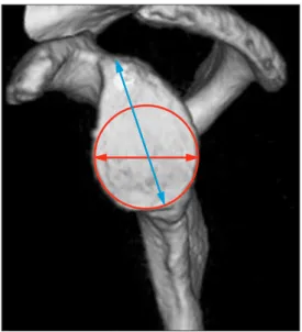
for rotator cuff arthropathies. A total of 56 patients, 12 men and 44 women, with a mean age of 71.4 ± 4.9 years (range, 61–80 years) were assessed. Taking the bare spot as the center of a hy- pothetical circle, we measured the maximal superoinferior (SI) diameter of the glenoid possible within the given space at the en face view of the 3-dimension CT images. Further, we measured the AP diameter of this hypothetical glenoid circle when its maxi- mal AP alignment was parallel to the sub-glenoid line (Fig. 3). The SI and the AP diameters were measured twice in all the patients by the same observer, and the mean and the standard deviation of the two measurements were compared to those of previous studies. The pre- and post-operative results were analyzed statis- tically using the Wilcoxon signed rank test, and statistical signifi- cance was set to p<0.05.
All statistical tests were run on IBM SPSS Statistics ver. 20.0 (IBM Co., Armonk, NY, USA).
Results
We found that the glenoids of the female patients had a mean SI diameter of 33.1 ± 2.0 mm and an AP diameter of 24.7 ± 1.4 mm and that the glenoids of the male patients had a mean SI diameter of 34.9 ± 1.3 mm and an AP diameter of 26.8 ± 1.0 mm. Altogether, the mean SI diameter of the gle- noid in all the patients was 33.4 ± 2.0 mm and the mean AP diameter, 25.0 ± 1.5 mm (Table 1). The anterior cortical screw showed a mean length of 25.7 mm, the posterior cortical screw, 19.8 mm, the superior locking screw, 33.5 mm, and the inferior locking screw, 33.3 mm. We found that the mean ASES score increased significantly from a preoperative score of 35.0 ± 12.1 to a final follow-up score of 74.4 ± 10.7 (p<0.001). The mean KSS score also improved significantly from a preoperative score of 46.9 ± 15.7 to a final follow-up score of 71.8 ± 12.4 (p<0.001). For the shoulder ROM, we found that the forward elevation of a preoperative value of 78.6o ± 32.2o increased sig- nificantly to a final follow-up value of 134.3o ± 18.6o (p<0.001) and that the abduction of a preoperative value 66.6o ± 9.5o increased significantly to a final follow-up value of 125.0o ± 14.2o (p<0.001). Only the change in the preoperative external rotation of 20.2o ± 10.4o to a final follow-up value of 28.4o ± 9.2o did not show a significant improvement (p=0.43) (Table 2).
At the final follow-up X-ray, we did not find evidence for loos- ening of the glenoid baseplate or scapular notching in any of the
Fig. 3. A computed tomography image of the glenoid was taken in the en face view. The bare spot was used as the center of the hypothetical circle with which the glenoid size was measured.
Table 1. Measurement of the Glenoid Size (mm)
Variable Superoinferior diameter Anteroposterior diameter
Male 34.9 ± 1.3 26.8 ± 1.0
Female 33.1 ± 2.0 24.7 ± 1.4
Total 33.4 ± 2.0 25.0 ± 1.5
Values are presented as mean ± standard deviation.
patients. Intraoperatively, we found an indentation of the meta- diaphysis of the proximal humerus in 2 patients (12%) whilst loosening of the humeral component was found in none. Other complications such as hematoma, infection, instability, acromial fracture, and humeral component loosening were not observed.
Discussion
In our study, we treated patients with rotator cuff arthropa- thies or with massive irreparable rotator cuff tears using a 25- mm baseplate rather than using the more established 29-mm baseplate to perform RTSA. We found that the clinical outcomes of RTSA in our study were similar to those of previous studies. In any of the patients did we find complications commonly associ- ated with RTSA such as scapular notching and loosening of the baseplate component, but because our study design comprised a relatively short follow-up period the possibility that with a lon- ger follow-up patients may present with complications cannot be overruled.
Scapular notching is the most common complication associ- ated with RTSA occurring in 0% to 96% of patients. Studies have shown that the longer the follow-up the greater the prevalence of scapular notching. Scapular notching, erosion of the lower scapular neck, relates to a mechanical collision that occurs between the scapula and the humeral component during ad- duction and during rotation motions of the arm.11) Polyethylene components of RTSA have been shown to induce osteolysis, thereby, accelerating scapular notching.19) A massive scapular notching can not only influence clinical outcome but also in- duce loosening of the glenoid component.20) In their results of a 44-month follow-up study, Sirveaux et al.11) found that 65%
of 77 patients presented with postoperative scapular notching.
Lévigne et al.21) found that 62% of 337 patients had scapular notching and that the longer follow-up the greater the chances were to detect the prevalence of scapular notching.
The factors to do with the glenoid component that are caus- ative of scapular notching include the extent of glenosphere
offset, the tilt of the humeral component, and the position of the glenoid component. Studies have investigated the optimal design of the glenoid component, of the component position, and of the screw fixation in the aim to increase stability of the impacted glenoid component and to decrease the incidence of scapular notching, and thereby glenoid component loosen- ing.22-25) Frankle et al.26) found that by offsetting the center of ro- tation externally they did not observe scapular notching even at the 33rd month of follow-up. However, when the rotator center is externally displaced a greater than usual torque is placed on the glenoid component which makes patients with osteoporosis more susceptible to glenoid component loosening. Rather, it has been shown that the perpendicularity of the humeral com- ponent is more efficient at decreasing the collision between the humeral prosthesis and the scapula than the tilting of the humeral component, but the former can cause instability during abduction of the shoulder. Nyffeler et al.27) suggested that plac- ing the glenoid baseplate more inferiorally can reduce the risk of scapular notching. To increase stability of the glenoid com- ponent after fixation, the size and shape of the glenoid must be taken into consideration as well as other factors influencing the cementation of the component. For instance, glenoid bone loss or glenoid erosion can be observed in most patients with rotator cuff arthropathies or with massive irreparable rotator cuff tears;
this decreases the surface area of contact between the glenoid and the glenoid baseplate and the bony support. Such factors contribute to the loss in initial fixation of the glenoid component.
The glenoid size in women is significantly smaller than that in men, which is especially true in the Asian population. The com- monly used 29-mm baseplate in RTSA, which may be appro- priate for men, tends to be too large for the average glenoid in Asian women.18,28,29) The success of initial fixation of the glenoid baseplate is influenced largely by screw position and the integrity of the glenoid bony structure. The consequence of a relatively small glenoid in comparison to the glenoid baseplate is loosening of the glenoid component resulting from an insufficient surface area for contact between the bony structure and the component and from insufficient fixation of the AP screws. DiStefano et al.25) proposed the following ideal screw lengths for the fixation of the glenoid baseplate: a 35 mm superior screw, a 34 mm inferior screw, a 29 mm anterior screw, and a 19 mm posterior screw. Through biomechanical studies on freshly frozen cadav- ers, Chae et al.18) found that a 25-mm glenoid baseplate can be used with longer fixation screws than a 29-mm glenoid base- plate. By using longer fixation screws, the use of 25-mm glenoid baseplate essentially increases the primary stability of the glenoid component and decreases the chances of collision between the scapular bone and the component, which is a problem espe- cially incurred by Asians with small glenoids. Ji et al.28) reported that in 42 patients who received RTSA using a 29-mm glenoid baseplate, 35% of patients exhibited scapular notching. In their Table 2. Functional Outcome
Variable Preoperative Postoperative p-value Clinical scores
ASES 35.0 ± 12.1 74.4 ± 10.7 <0.001
KSS 46.9 ± 15.7 71.8 ± 12.4 <0.001
Range of motion (o)
Forward elevation 78.6 ± 32.2 134.3 ± 18.6 <0.001 Abduction 66.6 ± 9.5 125.0 ± 14.2 <0.001 External rotation 20.2 ± 10.4 28.4 ± 9.2 0.43 Values are presented as mean ± standard deviation.
ASES: American Shoulder and Elbow Surgeons, KSS: Korean Shoulder Soci- ety.
study the use of a large glenoid baseplate compared to the small glenoid meant that the following screw lengths were used: a 35 mm superior screw, a 30 mm inferior screw, a 26 mm anterior screw, and a 21 mm posterior screw. They found that only 60%
of patients ended up with stable screw fixation. These findings highlight the need for a more appropriate glenoid component during RTSA for Asian patients.
In this study, we used a 25-mm glenoid baseplate and posi- tioned it as inferiorally as possible in the glenoid and then imple- mented the RTSA. In terms of the clinical outcome, we observed a similar extent of improvement of symptoms as those seen in previous studies. We found that scapular notching did not appear in all 17 patients; this low incidence may be because, like in the study by Chae et al.,18) of the use of a small glenoid baseplate that may have increased the chances of the collision between the scapular bone and the humeral component dur- ing adduction and rotation. We also found that loosening of the glenoid component were not seen in any of the 17 patients. But the postoperative findings were assessed only until 14 months, which is a relatively short period to assess occurrence of post- operative outcomes and complications. Yet we also believe that the low rate of complications and early glenoid baseplate stability may be attributed to the use of the Korean glenoid size matched-glenoid baseplate, which provided an average screw length of 33.5 mm at the superior plate, 33.3 mm, at the infe- rior plate, 25.7 mm, at the anterior plate, and 19.8 mm, at the posterior plate. In any of the patients did we find complications commonly associated with RTSA such as scapular notching and loosening of the baseplate component, but because our study design comprised a relatively short follow-up period the possibil- ity that with a longer follow-up patients may present with com- plications cannot be overruled.
Moon et al.30) reported that the glenoids in the Korean women have a mean SI diameter of 30.0 ± 2.3 mm and a mean AP diameter of 24.5 ± 0.8 mm. Ji et al.28) reported that glenoids in Korean women of age greater than 60 years have a mean radius of 13.5 ± 1.7 mm. In accordance to these studies, we found that the glenoids in female patients with rotator cuff arthropa- thies have a mean SI diameter of 33.1 ± 2.0 mm and a mean AP diameter of 24.7 ± 1.4 mm. Altogether, the findings of other authors and those of ours show that the average glenoid size in Koreans, especially that of Korean women, is small and that the use of the 29-mm glenoid baseplate thus far would have resulted in a large mismatch in the glenoid and baseplate size.
In comparison, the 25-mm glenoid baseplate, which is of better match to the glenoids of the Korean population, can be antici- pated to provide a better glenoid baseplate fixation.
There are several limitations to this study. For instance, the study encompassed a small sample number, the putative ad- vantages of the 25-mm glenoid baseplate relative to that of the 29-mm glenoid baseplate were not investigated, and the mean
follow-up period was only 14.0 months. Considering that the prevalence of scapular notching and glenoid baseplate loosen- ing may increase with time, this follow-up period was probably a less-than-sufficient period with which to conclude an accurate prevalence of complications. Thus, prospective studies with a greater study population number and a longer follow-up are needed. Another limitation is that although our study addressed patients with rotator cuff arthropathies we did not consider the potential error introduced by varying extents of glenoid bone loss or of glenoid curvature on baseplate fixation and on the clinical outcomes. Thus, further studies are required to delineate such influences in a systematic approach. In addition, because the number of patients who were followed-up for more than 1 year was only 20 (48.8%) in our study, the low N number means that a possibility of selection bias due to an over-representation of patients with favorable outcomes cannot be ruled out. This can be overcome by carrying out a subsequent study that ana- lyzes the postoperative results of the rest of the patients who were not initially enrolled in the study because they had not participated in the 1-year follow-up.
Conclusion
In sum, we found that RTSA using a 25-mm glenoid base- plate for patients with rotator cuff arthropathy or with massive irreparable rotator cuff tears is associated with favorable clinical outcomes. Complications such as scapular notching and loosen- ing of the baseplate were not found, but given that this was a short-term follow-up study a longer observance of the patients may be needed. Considering that the glenoid of the Korean pa- tients, which has a mean AP diameter of 25.0 ±1.5 mm, exhib- its a comparable match in size to the 25-mm glenoid baseplated used in this study, we believe that the 25-mm glenoid baseplate with its greater congruence to the average Korean glenoid size may provide the basis for enhanced clinical outcomes after RTSA.
References
1. Burkhart SS. Arthroscopic treatment of massive rotator cuff tears. Clinical results and biomechanical rationale. Clin Orthop Relat Res. 1991;(267):45-56.
2. Gartsman GM. Massive, irreparable tears of the rotator cuff.
Results of operative debridement and subacromial decom- pression. J Bone Joint Surg Am. 1997;79(5):715-21.
3. Gerber C, Vinh TS, Hertel R, Hess CW. Latissimus dorsi trans- fer for the treatment of massive tears of the rotator cuff. A pre- liminary report. Clin Orthop Relat Res. 1988;(232):51-61.
4. Kempf JF, Gleyze P, Bonnomet F, et al. A multicenter study of 210 rotator cuff tears treated by arthroscopic acromioplasty.
Arthroscopy. 1999;15(1):56-66.
5. Rockwood CA Jr, Williams GR Jr, Burkhead WZ Jr. Débride- ment of degenerative, irreparable lesions of the rotator cuff. J Bone Joint Surg Am. 1995;77(6):857-66.
6. Neer CS 2nd, Watson KC, Stanton FJ. Recent experi- ence in total shoulder replacement. J Bone Joint Surg Am.
1982;64(3):319-37.
7. Zuckerman JD, Scott AJ, Gallagher MA. Hemiarthroplasty for cuff tear arthropathy. J Shoulder Elbow Surg. 2000;9(3):169- 72.
8. Sarris IK, Papadimitriou NG, Sotereanos DG. Bipolar hemiar- throplasty for chronic rotator cuff tear arthropathy. J Arthro- plasty. 2003;18(2):169-73.
9. Matsen FA 3rd, Boileau P, Walch G, Gerber C, Bicknell RT.
The reverse total shoulder arthroplasty. J Bone Joint Surg Am.
2007;89(3):660-7.
10. Grammont PM, Baulot E. Delta shoulder prosthesis for rotator cuff rupture. Orthopedics. 1993;16(1):65-8.
11. Sirveaux F, Favard L, Oudet D, Huquet D, Walch G, Molé D.
Grammont inverted total shoulder arthroplasty in the treat- ment of glenohumeral osteoarthritis with massive rupture of the cuff. Results of a multicentre study of 80 shoulders. J Bone Joint Surg Br. 2004;86(3):388-95.
12. Frankle M, Siegal S, Pupello D, Saleem A, Mighell M, Vasey M.
The reverse shoulder prosthesis for glenohumeral arthritis as- sociated with severe rotator cuff deficiency. A minimum two- year follow-up study of sixty patients. J Bone Joint Surg Am.
2005;87(8):1697-705.
13. Werner CM, Steinmann PA, Gilbart M, Gerber C. Treatment of painful pseudoparesis due to irreparable rotator cuff dysfunc- tion with the Delta III reverse-ball-and-socket total shoulder prosthesis. J Bone Joint Surg Am. 2005;87(7):1476-86.
14. Frankle MA, Teramoto A, Luo ZP, Levy JC, Pupello D. Glenoid morphology in reverse shoulder arthroplasty: classification and surgical implications. J Shoulder Elbow Surg. 2009;18(6):874- 85.
15. Wierks C, Skolasky RL, Ji JH, McFarland EG. Reverse total shoulder replacement: intraoperative and early postoperative complications. Clin Orthop Relat Res. 2009;467(1):225-34.
16. Boileau P, Watkinson D, Hatzidakis AM, Hovorka I. Neer Award 2005: The Grammont reverse shoulder prosthesis:
results in cuff tear arthritis, fracture sequelae, and revision ar- throplasty. J Shoulder Elbow Surg. 2006;15(5):527-40.
17. Macaulay AA, Greiwe RM, Bigliani LU. Rotator cuff deficient arthritis of the glenohumeral joint. Clin Orthop Surg. 2010;
2(4):196-202.
18. Chae SW, Kim SY, Lee H, Yon JR, Lee J, Han SH. Effect of
baseplate size on primary glenoid stability and impingement- free range of motion in reverse shoulder arthroplasty. BMC Musculoskelet Disord. 2014;15:417.
19. Nyffeler RW, Werner CM, Simmen BR, Gerber C. Analysis of a retrieved delta III total shoulder prosthesis. J Bone Joint Surg Br. 2004;86(8):1187-91.
20. Boileau P, Watkinson DJ, Hatzidakis AM, Balg F. Grammont re- verse prosthesis: design, rationale, and biomechanics. J Shoul- der Elbow Surg. 2005;14(1 Suppl S):147S-61S.
21. Lévigne C, Boileau P, Favard L, et al. Scapular notching in reverse shoulder arthroplasty. J Shoulder Elbow Surg. 2008;
17(6):925-35.
22. Harman M, Frankle M, Vasey M, Banks S. Initial glenoid com- ponent fixation in “reverse” total shoulder arthroplasty: a bio- mechanical evaluation. J Shoulder Elbow Surg. 2005;14(1 Suppl S):
162S-7S.
23. Virani NA, Harman M, Li K, Levy J, Pupello DR, Frankle MA.
In vitro and finite element analysis of glenoid bone/baseplate interaction in the reverse shoulder design. J Shoulder Elbow Surg. 2008;17(3):509-21.
24. Gutiérrez S, Greiwe RM, Frankle MA, Siegal S, Lee WE 3rd.
Biomechanical comparison of component position and hard- ware failure in the reverse shoulder prosthesis. J Shoulder El- bow Surg. 2007;16(3 Suppl):S9-S12.
25. DiStefano JG, Park AY, Nguyen TQ, Diederichs G, Buckley JM, Montgomery WH 3rd. Optimal screw placement for base plate fixation in reverse total shoulder arthroplasty. J Shoulder Elbow Surg. 2011;20(3):467-76.
26. Frankle M, Levy JC, Pupello D, et al. The reverse shoulder prosthesis for glenohumeral arthritis associated with severe rotator cuff deficiency. a minimum two-year follow-up study of sixty patients surgical technique. J Bone Joint Surg Am.
2006;88 Suppl 1 Pt 2:178-90.
27. Nyffeler RW, Werner CM, Gerber C. Biomechanical relevance of glenoid component positioning in the reverse Delta III total shoulder prosthesis. J Shoulder Elbow Surg. 2005;14(5):524-8.
28. Ji JH, Jeong JY, Song HS, et al. Early clinical results of reverse total shoulder arthroplasty in the Korean population. J Shoul- der Elbow Surg. 2013;22(8):1102-7.
29. Phonphok P, Kulkamthorn N. Assessment of approximate gle- noid size in Thai people. J Med Assoc Thai. 2014;97 Suppl 2:
S14-8.
30. Moon YL, Ha SH, Noh KH. Normal glenoid size of the Korean in 7th and 8th decades. J Korean Shoulder Elbow Soc. 2008;
11(1):37-40.