Clinical and Radiological Outcome after Surgical Treatment in Displaced Clavicular Midshaft Fracture
Heui-Chul Gwak, Jung-Han Kim
Department of Orthopaedic Surgery, Inje University Busan Paik Hospital, Inje University College of Medicine, Busan, Korea
Background: The first purpose of this study is to compare the clinical and radiological outcomes of surgical treatment for displaced mid- shaft clavicle fracture (Robinson type 2B1 vs. 2B2) with 3.5-mm low profile clavicular locking compression plate. The second purpose is to evaluate the difference of the results depending on the presence of accompanying injuries.
Methods: Forty-nine patients who underwent an operation for the fractures were reviewed retrospectively. Fracture patterns were classi- fied according to group 2B1 and 2B2 using Robinson’s classification. For radiological outcome, time to union after operation was evalu- ated and for clinical outcome, American Shoulder and Elbow Society (ASES) score, University of California in Los Angeles (UCLA) score, visual analogue scale (VAS), and range of motion (ROM) were evaluated from preoperative period to last follow-up period.
Results: The mean time for union was not significantly different in the 2B1 group and 2B2 group (p=0.062). No statistically significant difference in ASES score, UCLA score, and VAS was observed between 2B1 and 2B2 (p=0.619, p=0.896, p=0.856, respectively). In ROM, significant higher mean forward flexion and abduction was observed in 2B2 (p=0.025, p=0.017, respectively) and there was no difference in external rotation and external rotation at shoulder 90o abduction position (p=0.130, p=0.180, respectively). There was no significant difference in clinical outcomes according to the accompanying injuries.
Conclusions: There was no difference in clinical and radiological outcome between Robinson 2B1 and 2B2 type fracture after the op- eration. Accompanying injuries may not affect the clinical result of displaced midshaft clavicle fractures.
(Clin Shoulder Elbow 2016;19(2):73-77)
Key Words: Clavicle; Fracture, closed; Displacement; Treatment outcome
Clinics in Shoulder and Elbow Clinics in Shoulder and Elbow Vol. 19, No. 2, June, 2016
http://dx.doi.org/10.5397/cise.2016.19.2.73
Received July 10, 2015. Revised December 19, 2015. Accepted January 6, 2016.
Correspondence to: Jung-Han Kim
Department of Orthopaedic Surgery, Inje University Pusan Paik Hospital, 75 Bokji-ro, Busanjin-gu, Busan 47392, Korea Tel: +82-51-890-6996, Fax: +82-51-891-1906, E-mail: [email protected]
Financial support: None. Conflict of interests: None.
Introduction
The clavicle is one of the most frequently fractured bones in the body, accounting for 5% of all fractures and 44% of all shoulder fractures and midshaft clavicular fracture is the most common fracture site.1-3) The clavicle acts as a mechanical strut connecting axial skeleton and upper extremities by maintain- ing appropriate length and position of the scapula. Therefore, clavicle fracture affects the length and the position. The fracture causes functional problem. In particular, delayed union and nonunion was more related to displaced fractures than nondis- placed fractures,2) and he comminuted fracture showed worse prognosis.
With the development of plates such as the low-profile lock- ing compression plate (LCP), good results have been reported after surgical treatment in displaced fractures.4,5) However, there are few reports on results after surgical treatment comparing simple displaced fracture patterns with displaced and commi- nuted patterns using low-profile LCP, and there are few reports on the treatment results of clavicular fractures with accompany- ing injuries.
The purpose of this study is to compare the clinical and ra- diological outcome after surgical treatment in displaced clavicle midshaft fracture using low-profile LCP. In addition, we wanted to examine the difference in results depending on the presence of accompanying injuries.
Methods
Institutional review board approval was obtained for this re- port (IRB #15-0004). Patients diagnosed as displaced clavicular midshaft fracture who underwent open reduction and internal fixation using low-profile LCP between 2010 and 2013 in Busan Paik Hospital were reviewed retrospectively. Patients with history of previous ipsilateral shoulder surgery, degenerative change in shoulder joint, neurovascular injury, or open fracture, and those with a follow-up period of less than 12 months were excluded.
Forty nine patients were finally included as subjects. Subjects were divided into the 2B1 group and 2B2 group. ‘2B1’ group (Robinson type 2B1 clavicular fracture6)) included patients a with simple or wedge comminuted fracture pattern and the ‘2B2’
group (Robinson type 2B2 clavicular fracture6)) included those with an isolated or comminuted segmental fracture pattern.
Thirty-four patients were male, and 15 patients female. The mean age was 45.1 ± 13.2 years (17–74 years) (Table 1). The 2B1 included 31 cases of clavicular midshaft fracture, and 2B2 included 18 cases. Causes of fracture were traffic accident in 24 cases (20 in 2B1, 4 in 2B2), fall from height in 6 cases (1 in 2B1, 5 in 2B2), simple fall in 15 cases (8 in 2B1, 7 in 2B2), and direct blow by an object in 4 cases (2 in 2B1, 2 in 2B2), respectively.
In 31 cases of Robinson 2B1 type fractures, 14 cases were initially classified as 2A2 fractures but changed to 2B1 fractures during conservative treatment (Fig. 1). For radiological assess- ment, the bone union period was compared using radiographic evidence, such as callus formation and trabecular bridging across the fracture site by analyzing both clavicle anteriorposterior and clavicle cephalic tilting views during the follow-up period. For the comparison of clinical outcome, American Shoulder and El- bow Society (ASES) score, University of California in Los Angeles (UCLA) score, visual analogue scale (VAS), and range of motion (ROM) were evaluated from the preoperative period to the last follow-up period. Clinical outcome according to accompanying injuries was also investigated.
Surgical Technique
We used superior plating fixation for clavicular midshaft fracture. The patient underwent the operation in beach chair position. A padding bump was placed between the scapulae, allowing the injured shoulder girdle to fall posteriorly. A linear in- cision was made centered over the fracture site, followed by an incision on the deltotrapezial fascia along its attachment to the anterior clavicle. For segmented fractures or comminuted frac- tures with large bony fragments, Kirchner wires were used to fix the segments temporarily or lag screws were used to fix the seg- ments. For small bony fragments we used MERSILENE® Polyester Fiber Suture (Ethicon, West Somerville, NJ, USA) to fasten them.
Then the fracture was reduced and held with bone clamps. A 3.5-mm low profile clavicular LCP (Acumed, Hillsboro, OR, USA) was fit to the superior edge of the clavicle. Screws were in- serted from superior to inferior, taking care to avoid injury to the neurovascular structures. After surgery, the operated extremity was placed in an arm sling. Pendulum exercise was started the day after the operation, and gentle passive ROM exercise within 1 week postoperative. Return to full activities was allowed at 3 months postoperative.
Statistical Analysis
Mann-Whitney test was used to assess the difference of time of bony union, ASES score, UCLA score, VAS and ROM (forward flexion, abduction, internal rotation, internal rotation at shoul- der 90o abduction) between 2B1 group and 2B2 group. Chi- square test was used to evaluate the incidence of refracture after implant removal between the 2B1 group and 2B2 group. Data entry and analysis were performed using IBM SPSS Statistics soft- ware ver. 22.0 (IBM CO., Armonk, NY, USA) and p-value <0.05 was considered significant.
Table 1. Demographic Characteristics of 2B1 Group and 2B2 Group Variable Robinson’s classification
Total Group 2B1 Group 2B2
Case of fracture 31 (57.1) 18 (42.9) 49 (100) Age (yr) 43.2 ± 12.1 (18–74) 49.4 ± 14.2 (17–71) 45.1 ± 13.2 (17–74) Sex
Male 23 11 34
Female 8 7 15
Values are presented as number (%), mean ± standard deviation (range), or number only.
Fig. 1. (A) Initial both clavicle anteriorposterior view. Robinson 2A1 type fracture was found. Conservative treatment using figure of eight brace was done. (B) Two weeks after conservative treatment. Fracture was displaced and now it is classified as Robinson 2B1 type.
A
B
Results
Radiological Result
The mean period from injury to operation was 11.3 days (1–92 days) and the mean follow-up period was 12.3 months (12–34 months). Among 49 patients, 44 patients (27 in 2B1, 17 in 2B2) completed radiologic follow-up until bony union.
The mean time for union was 2.9 ± 1.1 months. The mean time for union was 2.6 ± 1.3 months in 2B1 type fractures and 3.3 ± 0.8 months in Robinson 2B2 type fractures. There was no statistically significant difference (Mann-Whitney test, p=0.062).
Clinical Result
Among 49 patients, all patients completed the follow-up questionnaire, VAS and ROM measurement. The mean UCLA scores in 2B1 group and 2B2 group were 31.6 ± 5.4 and 32.1
± 4.3, respectively. The mean ASES scores in the 2B1 group and 2B2 group were 84.5 ± 17.5 and 88.9 ± 16.1, respectively.
The mean VAS in the 2B1 and 2B2 group was 1.2 ± 1.3 and 1.0 ± 1.1, respectively. No statistically significant difference in ASES score, UCLA score and VAS was observed between 2B1 and 2B2 (Mann-Whitney test, p=0.619, p=0.896, p=0.856, respectively).
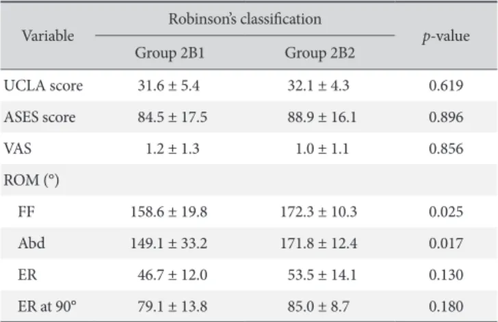
In ROM, significant higher mean forward flexion and ab- duction was observed in 2B2 (Mann-Whitney test, p=0.025, p=0.017, respectively) and there was no difference in external rotation and external rotation at shoulder 90o abduction posi- tion (Mann-Whitney test, p=0.130, p=0.180, respectively) (Table 2).
In cases without accompanying injuries, the mean time of surgical treatment after injury was 4.9 days (1–13 days). In cases with accompanying injuries, the mean time of surgical treatment after injury was 14.6 days (1–92 days).
Thirty-three cases (20 in 2B1, 13 in 2B2) presented with ac- companying injuries except fracture. Regarding combined frac- ture, 12 cases (11 in 2B1, 1 in 2B2) had accompanying fracture (Table 3). The clinical outcomes according to the accompanying injuries are listed in Table 4–6.
Hardware removal was done in 27 cases (16 in 2B1, 11 in 2B2). The mean time to hardware removal was 14.2 months (12–23 months). Regarding complication, there were 6 cases
Table 2. Clinical Results between 2B1 Group and 2B2 Group Variable Robinson’s classification
p-value
Group 2B1 Group 2B2
UCLA score 31.6 ± 5.4 32.1 ± 4.3 0.619
ASES score 84.5 ± 17.5 88.9 ± 16.1 0.896
VAS 1.2 ± 1.3 1.0 ± 1.1 0.856
ROM (°)
FF 158.6 ± 19.8 172.3 ± 10.3 0.025
Abd 149.1 ± 33.2 171.8 ± 12.4 0.017
ER 46.7 ± 12.0 53.5 ± 14.1 0.130
ER at 90° 79.1 ± 13.8 85.0 ± 8.7 0.180
Values are presented as mean ± standard deviation.
UCLA: University of California in Los Angeles, ASES: American Shoulder and Elbow Society, VAS: visual analogue scale, ROM: range of motion, FF:
forward flexion, Abd: abduction, ER: external rotation.
Table 3. Accompanying Injuries in 2B1 Group and 2B2 Group Accompanying injury Robinson’s classification
Total Group 2B1 Group 2B2
Except fracture (n) 20 13 33
Fracture (n) 11 1 12
Table 4. UCLA Score and Results Depending on the Presence of Accompany- ing Injury
Robinson’s classification AI No AI p-value
Group 2B1 30.9 ± 6.1 33.2 ± 3.6 0.632*
Group 2B2 31.8 ± 4.6 33.7 ± 2.3 0.413*
p-value 0.642* 0.715*
Values are presented as mean ± standard deviation.
UCLA: University of California in Los Angeles, AI: accompanying injury.
*Mann-Whitney test.
Table 5. ASES Score and Results Depending on the Presence of Accompany- ing Injury
Robinson’s classification AI No AI p-value
Group 2B1 82.0 ± 16.1 90.0 ± 20.7 0.245*
Group 2B2 87.8 ± 17.2 93.9 ± 10.6 0.634*
p-value 0.643* 0.547*
Values are presented as mean ± standard deviation.
ASES: American Shoulder and Elbow Society, AI: accompanying injury.
*Mann-Whitney test.
Table 6. VAS and Results Depending on the Presence of Accompanying Injury
Robinson’s classification AI No AI p-value
Group 2B1 1.4 ± 1.4 1.1 ± 1.1 0.456*
Group 2B2 0.7 ± 1.2 0.7 ± 1.2 0.852*
p-value 0.642* 0.476*
Values are presented as mean ± standard deviation.
VAS: visual analogue scale, AI : accompanying injury.
*Mann-Whitney test.
of complications (1 in 2B1, 5 in 2B2), including 1 case of metal breakage in Robinson 2B2 fractures and 5 cases of refracture (1 in 2B1, 4 in 2B2) after hardware removal (Fig. 2). Higher inci- dence of refracture after implant removal was observed for 2B2 clavicular fracture than 2B1 clavicular fracture (chi-square test, p=0.036).
Discussion
In our study, all cases of Robinson 2B1 and 2B2 clavicular fracture showed bony union. In addition, there was no differ- ence in clinical outcomes including ASES, UCLA scores, and VAS. There was no difference in the mean time for union be- tween Robinson 2B1 and 2B2 clavicular fracture. However, the mean time for union in Robinson 2B2 clavicular midshaft frac- ture was seemingly longer than that of Robinson 2B1 clavicular fracture. In addition, higher incidence of refracture after implant removal was observed for 2B2 clavicular fracture than 2B1 cla- vicular fracture. In general, the initial morphology of the fracture provide the best indication of the risk of delayed or nonunion, irrespective of the mechanism of injury.7) Displaced fracture of the clavicular midshaft shows a higher rate of delayed and non-
union than nondisplaced fracture patterns.8-14) However, with the development of plates such as low-profile LCP, more stable fixation is possible and good results have been reported after surgical treatment in clavicle midshaft fracture.4,5) In our study, there was no significant difference in nonunion rate between 2B1 and 2B2. However, there was a tendency of longer union term in Robinson 2B2 fracture than in Robinson 2B1 fracture.
Poigenfürst et al.15) reported on 122 patients who underwent cla- vicular plating and there were 4 cases of refracture. Schwarz and Höcker16) reported that 1 of 19 patients who underwent plate removal had a refracture. For prevention of refracture, Jupiter and Ring17) and Poigenfürst et al.15) suggested leaving a plate in place for 12 to 18 months and placement of restrictions against contact sports for 2 to 3 months after plate removal. Although the cause of the refracture is beyond this study, this finding sug- gests that if removal of the plate is planned, especially in Robin- son 2B2 fractures, radiographs and even computed tomography images should be thoroughly evaluated for union progression and removal can be delayed until radiological evidence of defi- nite bony union appears.
Midshaft clavicular fractures are caused by different trau- ma mechanisms. A high energy injury is common reported mechanism of injur that produces a midshaft fracture of the clavicle.18-21) In addition, high energy injury like that resulting from a traffic accident is increasing and survivorship of patients from high energy trauma is also improving.6) As a result, cases of clavicular fracture with accompanying injuries rather than an isolated clavicular fracture are increasing. In our study, of a total of 49 patients, there were 24 cases of traffic accident which is al- most 50% of the cases and 33 cases had accompanying injuries.
In cases of patients with accompanying injuries like intracranial hemorrhage, hemopneumothorax or visceral injury, these prob- lems were treated with priority and as a result, timing of surgical treatment was delayed. However, there was no significant differ- ence in clinical result depending on the presence of accompany- ing injury. These findings suggest that surgical treatment may be an effective treatment option even in case of delayed operation due to accompanying injuries. In addition, in 14 cases, the initial Robinson classification was changed from 2A2 to 2B1 during conservative treatment. This implies that, in case of conservative treatment, the maintenance state of the treatment should be monitored serially.
There are several limitations in our study. First, this study had a retrospective design. Second, during the patient selection, the number of patients who met all the requiring factors including the completion of questionnaires and over 12 months of follow- up was relatively small and could cause selection bias. Third, in determining the timing of fracture union, accurate radiological interpretation was difficult due to the presence of the plate.
Fourth, the number of patients included in the study was rela- tively small. Fourth, the accompanying injuries being treated at Fig. 2. (A) Initial X-ray image. Initial fracture pattern was Robinson 2B2 type.
(B) Six months after plating for Robinson 2B2 type fracture. Bony union was notified. (C) Hardware removal was done and there was no evidence of re- fracture. (D) Refracture was occurred at the site of initial fracture after hard- ware removal.
A
B
C
D
the time of the last follow-up period can affect the functional outcome of clavicular fracture, for example, patients with neuro- surgical problem.
Conclusion
The presence of comminution in displaced clavicular mid- shaft fracture has no effect on the clinical and radiological out- come. In addition, accompanying injuries may not affect the clinical result of clavicular midshaft fractures.
References
1. Johnson EW Jr, Collins HR. Nonunion of the clavicle. Arch Surg. 1963;87(6):963-6.
2. Paffen PJ, Jansen EW. Surgical treatment of clavicular fractures with Kirschner wires: a comparative study. Arch Chir Neerl.
1978;30(1):43-53.
3. Khan LA, Bradnock TJ, Scott C, Robinson CM. Fractures of the clavicle. J Bone Joint Surg Am. 2009;91(2):447-60.
4. Haidukewych GJ. Innovations in locking plate technology. J Am Acad Orthop Surg. 2004;12(4):205-12.
5. Perren SM. Evolution and rationale of locked internal fixator technology. Introductory remarks. Injury. 2001;32 Suppl 2:B3-9.
6. McKee MD, Schemitsch EH, Stephen DJG, Kreder HJ, Yoo D, Harrington J. Functional outcome following clavicle fractures in trauma patients. J Trauma Inj Infect Crit Care. 1999;47(3):616.
7. Hill JM, McGuire MH, Crosby LA. Closed treatment of dis- placed middle-third fractures of the clavicle gives poor results.
J Bone Joint Surg Br. 1997;79:537-9.
8. Neer CS 2nd. Nonunion of the clavicle. J Am Med Assoc.
1960;172:1006-11.
9. Kulshrestha V, Roy T, Audige L. Operative versus nonoperative management of displaced midshaft clavicle fractures: a pro- spective cohort study. J Orthop Trauma. 2011;25(1):31-8.
10. Eskola A, Vainionpää S, Myllynen P, Pätiälä H, Rokkanen P.
Outcome of clavicular fracture in 89 patients. Arch Orthop Trauma Surg. 1986;105(6):337-8.
11. Wilkins RM, Johnston RM. Ununited fractures of the clavicle. J Bone Joint Surg Am. 1983;65(6):773-8.
12. Wick M, Müller EJ, Kollig E, Muhr G. Midshaft fractures of the clavicle with a shortening of more than 2 cm predispose to nonunion. Arch Orthop Trauma Surg. 2001;121(4):207-11.
13. Davids PH, Luitse JS, Strating RP, van der Hart CP. Operative treatment for delayed union and nonunion of midshaft cla- vicular fractures: AO reconstruction plate fixation and early mobilization. J Trauma. 1996;40(6):985-6.
14. Robinson CM, Court-Brown CM, McQueen MM, Wakefield AE. Estimating the risk of nonunion following nonoperative treatment of a clavicular fracture. J Bone Joint Surg Am. 2004;
86(7):1359-65.
15. Poigenfürst J, Rappold G, Fischer W. Plating of fresh clavicular fractures: results of 122 operations. Injury. 1992;23(4):237-41.
16. Schwarz N, Höcker K. Osteosynthesis of irreducible fractures of the clavicle with 2.7-MM ASIF plates. J Trauma. 1992;33(2):
179-83.
17. Jupiter JB, Ring D. Fractures of the clavicle. In: Iannotti JP, Wil- liams GR, eds. Disorders of the shoulder: diagnosis and man- agement. Philadelphia: Lippincott Williams & Wilkins; 1999.
1427-70.
18. Canadian Orthopaedic Trauma Society. Nonoperative treat- ment compared with plate fixation of displaced midshaft clavicular fractures. A multicenter, randomized clinical trial. J Bone Joint Surg Am. 2007;89(1):1-10.
19. Moseley HF. The clavicle: its anatomy and function. Clin Or- thop Relat Res. 1968;58:17-27.
20. Robinson CM. Fractures of the clavicle in the adult. Epidemiol- ogy and classification. J Bone Joint Surg Br. 1998;80(3):476-84.
21. Stanley D, Trowbridge EA, Norris SH. The mechanism of cla- vicular fracture. A clinical and biomechanical analysis. J Bone Joint Surg Br. 1988;70(3):461-4.