Arthroscopic-assisted Reduction and Percutaneous Screw Fixation for Glenoid Fracture with Scapular Extension
Se Jin Kim, Sung Hyun Lee1, Dae Woong Jung, Jeong Woo Kim1
Department of Orthopedic Surgery, St. Carollo Hospital, Suncheon, 1Institute of Wonkwang Medical Science and Department of Orthopedic Surgery, Wonkwang University Hospital, Iksan, Korea
Background: To evaluate the clinical and functional outcomes of arthroscopic-assisted reduction and percutaneous screw fixation for glenoid fractures with scapular extension, and investigate the radiologic and clinical benefits from the results.
Methods: We evaluated patients treated with arthroscopic-assisted reduction and percutaneous screw fixation for glenoid fractures with scapular extension from November 2008 to September 2015. Fractures with displacement exceeding one-fourth of the anterior-articular surface or more than one-third of the posterior-articular surface in radiographic images were treated by surgery. Clinical assessment was conducted based on range of motion, Rowe score, and Constant score of injured arm and uninjured arm at last follow-up.
Results: Fifteen patients with Ideberg classification grade III, IV, and V glenoid fracture who underwent arthroscopic-assisted reduc- tion using percutaneous screw fixation were retrospectively enrolled. There were no differences in clinical outcomes at final follow-up compared to uninjured arm. Bone union was seen in all cases within five months, and the average time to bone union was 15.2 weeks.
Ankylosis in one case was observed as a postoperative complication, but the symptoms improved in response to physical therapy for six months. There was no failure of fixation and neurovascular complication.
Conclusions: We identified acceptable results upon radiological and clinical assessment for the arthroscopic-assisted reduction and per- cutaneous fixation. For this reason, we believe the method is favorable for the treatment of Ideberg type III, IV, and V glenoid fractures.
Restoration of the articular surface is considered to be more important than reduction of fractures reduction of the scapula body.
(Clin Shoulder Elbow 2017;20(3):147-152)
Key Words: Arthroscopy; Fracture fixation; Scapula; Glenoid cavity
Copyright © 2017 Korean Shoulder and Elbow Society. All Rights Reserved. pISSN 2383-8337
Clinics in Shoulder and Elbow Vol. 20, No. 3, September, 2017 https://doi.org/10.5397/cise.2017.20.3.147
Received June 5, 2017. Revised July 24, 2017. Accepted July 30, 2017.
Correspondence to: Jeong Woo Kim
Department of Orthopedic Surgery, Wonkwang University Hospital, 895 Muwang-ro, Iksan 54538, Korea Tel: +82-63-859-1360, Fax: +82-63-852-9329, E-mail: [email protected]
IRB approval (No. WKUH 201608-HR-092).
Financial support: None. Conflict of interests: None.
Introduction
Glenoid fractures with scapular extension are uncommon in- juries, but may result in chronic shoulder instability and degener- ative arthritis as an intra-articular fracture.1) Anatomical reduction and stable fixation of the articular surface are needed to prevent these complications and recover an ample range of motion in the shoulder joint.2) Although open reduction is conducted con- ventionally, an open surgery is more likely to increase morbidity related to shoulder joint and postoperative complications.
Favorable results have been reported after arthroscopic- assisted reduction and internal fixation for glenoid fractures with
scapular extension in recent studies. However, most of these studies were case reports that dealt with a type of fracture, while there have been very few outcome studies. Therefore, this study was conducted to evaluate the clinical and functional outcomes of arthroscopic-assisted reduction and percutaneous screw fixa- tion for glenoid fractures with scapular extension, as well as to investigate the radiologic and clinical benefits of the results.
Methods
Institutional Review Board at Wonkwang University Hospital approval was obtained before starting the study. We conducted
distribution by age and sex, causes of injury, fracture site, con- comitant injuries and surgical methods. The inclusion criteria were as follows: (1) glenoid fracture with scapular extension, (2) those who have undergone arthroscopic-assisted reduction and percutaneous screw fixation, and (3) minimum follow-up of 12 months. Exclusion criteria were as follows: (1) patients with a neurologic deficit due to major nerve injury, (2) patients with preexisting disease of injured arm, and (3) patients who have undergone open surgery. We indicated a surgical treatment of the glenoid fracture based on a displacement >5 mm, and pa- tients with displacement exceeding one-fourth of the anterior- articular surface or more than one-third of the posterior-articular
Operative Procedures
Surgery was performed under general anesthesia. For this op- eration, patients were placed in a beach chair position in order to facilitate the traditional open approach. The arthroscope was then inserted into the standard posterior portal. Upon observ- ing the anterior glenoid through the standard anterosuperior portal, a working cannula was inserted into the rotator interval portal. The joint was then sufficiently irrigated with saline solu- tion to remove the intra-articular hematoma before further ex- amination. Hematoma and bone fragments were subsequently debrided with an arthroscopic shaver, after which the stability
A B
C D
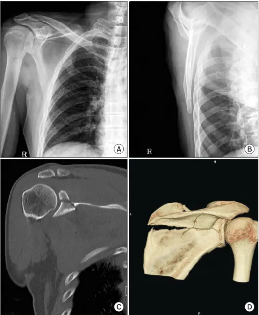
Fig. 1. (A, B) Simple radiologic evaluation showed an Ideberg classification type V glenoid fracture with scapular extension in 68-year-old man. (C, D) The three- dimensional computed tomography scan confirmed the configuration of displaced fracture.
of the fracture site was identified with a probe (Fig. 2A). A guide wire was then placed in the middle of fracture line (based on the fracture patterns) through the skin in the lateral portal of the lateral edge acromion, the subclavian portal or the Neviaser por- tal while maintaining fracture reduction using 1.6 mm K-wires, raspatories and probes. Next, a 4.0 mm cannulated screw was installed over the guide pin to complete the fixation. At the time of cannulated screws fixation, an additional 1.6 mm K-wire was fixed to prevent loss of reduction at the fracture site. Reduction was performed using additional cannulated screws and 1.6 mm K-wires according to the fracture patterns unless good reduc- tion was achieved with a cannulated screw. The gap between fracture fragments was identified based on arthroscopic findings, and the fixation was once again thoroughly checked under ar- throscopy to confirm that the screw did not violate the articular surface (Fig. 2B). Concomitant lesions of the rotator cuff or long head of biceps tendon were also treated at the same time. The incisions were closed in standard fashion, and a shoulder abduc- tion orthosis was placed for postoperative use. From the day after surgery, patients were instructed to conduct pendulum ex- ercises and passive range of motion exercises (including forward elevation to 60° and abduction to 60°) while wearing a shoulder abduction orthosis. At 4 weeks after surgery, the range of mo- tion had gradually increased to 90° of forward elevation, 90° of abduction and 10° of external rotation, and patients were in- structed to conduct active range of motion exercises as often as possible. After 6 weeks, the patients were permitted to perform active range of motion exercises in all directions and strength exercises without wearing the shoulder abduction orthosis.
Evaluation Methods
Outpatient follow-up was conducted at 3 weeks, 6 weeks, 3 months, 6 months, and 12 months after surgery. Patients were observed once a year after 12 months. If callus formation was observed on anterior-posterior and lateral radiographs they were regarded as bony unions. In such cases, we examined the time to bony union. Clinical assessment was conducted using range of motion, Rowe score, and the Constant score of the injured
arm and uninjured arm at last follow-up. Statistical analysis was performed with IBM SPSS Statistics version 23.0 (IBM Co., Ar- monk, NY, USA), with a confidence interval of 95%. Data were presented as the averages and standard deviations. An indepen- dent-samples t-test was used to compare clinical scores, with a p<0.05 considered to indicate statistical significance.
Results
Out of a total of 32 cases with glenoid fractures, 15 cases that underwent arthroscopic procedure were identified (12 men and 3 women). The mean age of the patients was 53.2 years (range, 28–68 years) and the causes of injury were traffic crashes in six cases and falls in nine cases. All cases involved closed fractures.
In this study, glenoid fractures were divided into six major types according to the modified Ideberg classification,3) with type I and II without scapular extension being excluded. There were four cases of type III, 5 cases of type IV, and 6 cases of type V fractures. In 10 cases, surgery was performed within 3 weeks;
however, in another 5 cases, surgical treatment was postponed because of multiple, major and even life-threatening injuries.
The mean delay from injury to surgery was 18.87 days (range, 7–27 days). In this study, a delay in treatment may not influence clinical outcomes, but these were not deemed statistically signifi- cant. There were concomitant injuries such as clavicle fractures (two cases), acromioclavicular joint injury (one case), tear of the long head of biceps tendon (one case), partial thickness tear of the supraspinatus (one case), and other fractures (e.g., multiple rib fractures, maxillary fractures, spine fractures). Clavicle frac- tures or acromioclavicular joint injuries were treated by open reduction and internal fixation with plate at the same time, and accompanying SSSC injuries did not influence clinical outcomes.
Based on the arthroscopic findings at the time of surgery, the labrum and capsulolabral complex were found to be intact in all cases. Biceps tenotomy was performed for a case with a tear of more than 50% of the tendon thickness, whereas debridement was performed for another case with a partial tear of less than 50% of the supraspinatus thickness (Table 1). The average ob-
A B
Fig. 2. Arthroscopic image of the right shoul- der during arthroscopic-assisted reduction.
The viewing portal was the posterior portal, the working portal was the anterior portal, and a cannulated screw was fixed through the Neviaser portal. (A) After debridement was performe on the fracture site. (B) Reduc- tion state of fracture by use of probe.
servation period was 19.8 months (range, 12–29 months). Bone union was seen in all cases within 5 months, and the average time to bone union was 15.2 ± 2.2 weeks (range, 12–20 weeks) (Fig. 3). At the last follow-up assessment, the active range of mo- tion consisted of forward elevation to 161.3° ± 7.9° (contralateral side, 162.7° ± 8.0°; p=0.649), external rotation to 66.3° ± 7.9°
(contralateral side, 68.3° ± 7.2°; p=0.478), and internal rota- tion to T9 ± 2 (contralateral side, T8 ± 1; p=0.531). The aver- age Rowe score and Constant score upon clinical assessment were 89.0 ± 7.4 points (contralateral side, 90.7 ± 5.9 points;
p=0.501) and 93.5 ± 2.4 points (contralateral side, 93.7 ± 2.1 points; p=0.872), respectively. Ankylosis in 1 case was observed as a postoperative complication, but the symptoms improved in response to physical therapy for 6 months. There was no failure of fixation or neurovascular complications.
Discussion
Glenoid fractures are more likely to have complications such as post-traumatic arthritis and chronic shoulder instabil- ity because they are intra-articular fractures of the shoulder.4) Although accurate anatomical reduction is necessary for their treatment, the rate of complications is high when conservative treatment or open surgery is used before the development of arthroscopic surgery.3,5) Recently, minimal incision surgery rather than traditional open reduction has been used. Accordingly,
since accurate evaluation of intra-articular fractures and perfect reduction became possible by using supplemental arthroscopy, minimal incision surgery can be a favorable treatment. However, better techniques for performing even in the limited range of vi- sion are needed for arthroscopic surgery. In certain cases, direct manipulation of bone fragments may be difficult. There is a dis- advantage in that arthroscopic surgery may produce injuries of the suprascapular nerve, which is located in the inner and upper portal of the articular surface of the glenohumeral joint.
In theory, arthroscopic surgery is useful for the diagnosis and treatment of any intra-articular fractures. Indeed, it is useful for distal radial fractures,6) tibial plateau fractures,7) ankle fractures,8) and distal femoral condyle fracture of the knee9) in real practice.
According to a previous report, percutaneous external fixation using K-wire was performed by Carro et al.10) and Gigante et al.11) as an auxiliary arthroscopic surgery for treatment of glenoid fractures of the shoulder, while screw fixation for intra-articular fractures of the anterior glenoid was performed by Cameron.12)
Arthroscopic-assisted fragments fixation for glenoid fractures with scapular extension is mainly conducted using screws for internal fixation. During insertion of screws, there is the potential for injuries of the suprascapular nerve and blood vessels passing through the lateral border of the scapular spine in the posterior portion of the scapula, whereas the risk for injuries of the ce- phalic vein and musculocutaneous nerve in the anterior portion of the scapula should be considered. Marsland and Ahmed13) re-
1 M/28 Fall from height III 175/80/T6 100 98 - 12 12
2 M/50 Traffic accident III 160/75/T9 95 94 Clavicle fracture 20 18
3 M/68 Fall from height V 165/70/T8 90 95 - 16 24
4 M/46 Fall from height IV 160/70/T9 85 93 - 14 24
5 M/52 Fall from height V 155/60/T10 80 91 Biceps tendon tear 15 29
6 F/56 Traffic accident V 150/55/T11 80 88 - 16 25
7 M/58 Traffic accident IV 160/60/T9 90 93 - 14 18
8 M/60 Traffic accident III 165/65/T8 95 94 - 14 16
9 M/41 Fall from height IV 170/75/T7 100 96 Clavicle fracture 18 22
10 F/42 Fall from height V 145/50/T12 75 92 - 12 18
11 F/53 Traffic accident V 170/70/T8 95 96 Rotator cuff tear 15 17
12 M/49 Fall from height III 165/65/T9 90 94 AC joint injury 18 24
13 M/51 Traffic accident IV 160/65/T10 85 92 - 15 12
14 M/38 Fall from height V 165/70/T9 90 95 - 14 16
15 M/56 Fall from height IV 155/65/T11 85 92 - 15 22
M: male, F: female, ROM: range of motion, FE: forward elevation, ER: external rotation, IR: internal rotation, AC: acromioclavicular.
*Glenoid fractures were divided into six major types according to the modified Ideberg classification.3)
ported that superior and posterior percutaneous approaches are safe between the clock times 7:40 to 2:50, except for the areas of the coracoid (1:50–2:00) and acromion (9:35–10:55).
We obtained favorable results in terms of radiological and clinical assessments. Specifically, lesions of other intra-articular structures were identified using an arthroscope for glenoid frac- tures with scapular extension, while fixation with cannulated screws through the skin was performed in the subclavian portal or lateral portal of the lateral edge f the acromion used for ar- throscopic shoulder surgery. This study was conducted to inves- tigate glenoid fractures with scapular extension. Although severe displacement of the scapular body is accompanied at the time of arthroscopic treatment for this type of fracture, it is important to focus on the repair of articular surfaces. Even in the case of frac- ture of the scapular body with severe displacement, favorable results can be obtained by means of non-operative treatments;
thus, we believe that the first goal in this arthroscopic surgery is the reduction of articular surfaces. This approach is useful to avoid larger incisions and comorbidity and to reduce the articular surfaces by direct observation, even though treatment for accompanying fracture of scapular bodies is not performed.
Furthermore, arthroscopic treatment is useful for reducing post- operative pain and achieving early return to activities for the patient, as well as for diagnosing and treating associated intra- articular lesions.
It should be noted that there were several limitations to this study. First, the study included only a few cases because glenoid fractures with scapular extension are rare. Second, the fractures in all cases corresponded to those located in the superior por- tion of the glenoid labrum. In general, the fractures located in the underside of the glenoid labrum are difficult to access with an arthroscope. However, arthroscopic fixation in the scapular
A B
C D
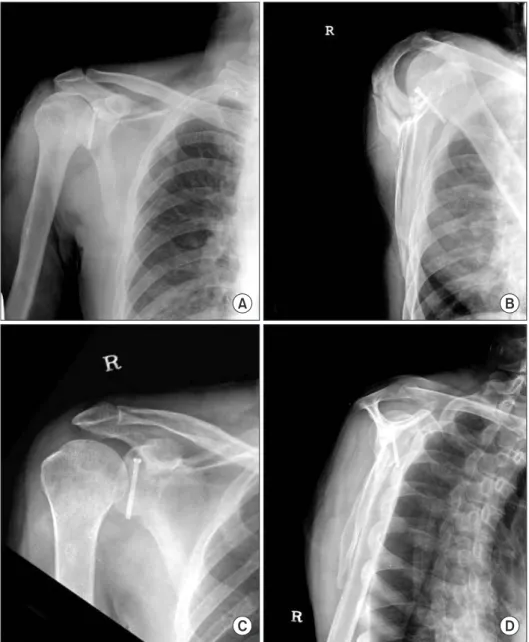
Fig. 3. (A, B) Postoperative radiologic image of the right shoulder. (C, D) After 16 weeks later, bone union was confirmed.
fractures, we believe that more indications would be secured from further studies.
In conclusion, we found that arthroscopic-assisted surgery is one of the favorable operative procedures for glenoid fractures with scapular extension. The treatment provides accurate fixa- tion strength because it enables observation of the site of glenoid fractures through the arthroscope. Additionally, during the op- eration, lavage for articular surfaces is conducted with treatment for concomitant injuries of intra-articular and other anatomical regions at the same time.
Conclusion
We identified acceptable results in the radiological and clinical assessment for arthroscopic-assisted reduction and per- cutaneous fixation. For this reason, we believe the method is favorable for the treatment of Ideberg type III, IV, and V glenoid fractures. Restoration of the articular surface is considered to be more important than reduction of the fractures of the scapula body.
References
1. Hardegger FH, Simpson LA, Weber BG. The operative treat- ment of scapular fractures. J Bone Joint Surg Br. 1984;66(5):
725-31.
2. Ko SH, Jeon HM, Shin SM. Arthroscopy assisted percutane- ous reduction and screw fixation of a displaced intra-articular glenoid fracture: a case report. J Korean Shoulder Elbow Soc.
2010;13(1):127-31.
1992;74(2):299-305.
5. Imatani RJ. Fractures of the scapula: a review of 53 fractures. J Trauma. 1975;15(6):473-8.
6. Geissler WB, Freeland AE. Arthroscopically assisted reduction of intraarticular distal radial fractures. Clin Orthop Relat Res.
1996;(327):125-34.
7. Jennings JE. Arthroscopic management of tibial plateau frac- tures. Arthroscopy. 1985;1(3):160-8.
8. Miller MD. Arthroscopically assisted reduction and fixation of an adult Tillaux fracture of the ankle. Arthroscopy. 1997;13(1):
117-9.
9. McCarthy JJ, Parker RD. Arthroscopic reduction and internal fixation of a displaced intraarticular lateral femoral condyle fracture of the knee. Arthroscopy. 1996;12(2):224-7.
10. Carro LP, Nuñez MP, Llata JI. Arthroscopic-assisted reduction and percutaneous external fixation of a displaced intra-articu- lar glenoid fracture. Arthroscopy. 1999;15(2):211-4.
11. Gigante A, Marinelli M, Verdenelli A, Lupetti E, Greco F.
Arthroscopy-assisted reduction and percutaneous fixation of a multiple glenoid fracture. Knee Surg Sports Traumatol Ar- throsc. 2003;11(2):112-5.
12. Cameron SE. Arthroscopic reduction and internal fixation of an anterior glenoid fracture. Arthroscopy. 1998;14(7):743-6.
13. Marsland D, Ahmed HA. Arthroscopically assisted fixation of glenoid fractures: a cadaver study to show potential applica- tions of percutaneous screw insertion and anatomic risks. J Shoulder Elbow Surg. 2011;20(3):481-90.
14. Tuman JM, Bishop JA, Abrams GD. Arthroscopic reduction and internal fixation of an inferior glenoid fracture with scapular extension (Ideberg V). Arthrosc Tech. 2015;4(6):e869-72.