Comparison between Accurate Anatomical Reduction and
Unsuccessful Reduction with a Remaining Gap after Open Reduction and Plate Fixation of Midshaft Clavicle Fracture
Joon Yub Kim, Jung Soo Choe, Seok Won Chung1
Department of Orthopedic Surgery, Myongji Hospital, Seonam Univeristy School of Medicine, Goyang, 1Department of Orthopedic Surgery, Konkuk University Medical Center, Seoul, Korea
Background: The purpose of this study is to compare the radiological and clinical outcomes after open reduction and plate fixation of midshaft clavicle fractures between patients who achieved successful anatomical reduction and those who had a remaining fracture gap even after open reduction and plate fixation, and were thus treated with additional demineralized bone matrix (DBM).
Methods: This retrospective analysis was conducted on 56 consecutive patients who underwent open reduction and internal fixation using a locking compression plate for acute displaced midshaft clavicle fractures, and who underwent radiographic and clinical outcome evaluations at least 6 months postoperatively. The outcomes between those who achieved perfect anatomical reduction without rem- nant gap (n=32) and those who had a remaining fracture gap even after open reduction and plate fixation treated with additional DBM (n=24) were evaluated.
Results: There were no differences in the use of lag screws or wiring and operation time (all p>0.05) between those with and without remnant gap. No difference in the average radiological union time and clinical outcomes (satisfaction and Constant score) was observed between the two groups (all p>0.05). However, significantly faster union time was observed for AO type A fracture compared with other types (p=0.012), and traffic accident showed association with worse clinical outcomes compared with other causes of injury.
Conclusions: Surgical outcome of midshaft clavicle fracture was more affected by initial fracture type and event, and re-reduction and re-fixation of the fracture to obtain a perfect anatomical reduction spending time appears to be unnecessary if rigid fixation is achieved.
(Clin Shoulder Elbow 2016;19(1):2-7)
Key Words: Clavicle; Fracture fixation; Fracture healing; Demineralized bone matrix gel
Copyright © 2016 Korean Shoulder and Elbow Society. All Rights Reserved. pISSN 2383-8337
Clinics in Shoulder and Elbow Vol. 19, No. 1, March, 2016 http://dx.doi.org/10.5397/cise.2016.19.1.2
Received December 19, 2014. Revised April 2, 2015. Accepted April 4, 2015.
Correspondence to: Seok Won Chung
Department of Orthopedic Surgery, Konkuk University Medical Center, Konkuk University School of Medicine, 120-1 Neungdong-ro, Gwangjin- gu, Seoul 05030, Korea
Tel: +82-2-2030-7604, Fax: +82-2-2030-7748, E-mail: [email protected] Financial support: None. Conflict of interests: None.
Introduction
Clavicle fracture is one of the most common bone injuries with an incidence of up to 10% of all fractures.1) Most clavicle fractures involve the midshaft, which is the thinnest part with the least soft tissue, and more than half of midshaft fractures are displaced.2,3) Due to the reported unsatisfactory results after non- operative treatment, operative fixation has been the recent treat- ment trend for the displaced midshaft clavicle fracture.4,5) Plate fixation is the standard operative method for midshaft clavicle
fracture, and many authors have demonstrated the effectiveness of plate fixation with excellent results.6,7)
Nevertheless, plate fixation for midshaft clavicle fractures, par- ticularly the comminuted fractures, could still be a challenging issue to the nonexpert. Not infrequently, we observe the unsuc- cessful reduction with notable gap formation and poor fixation strength, causing malunion or nonunion, after plate fixation of the midshaft clavicle fracture. Demineralized bone matrix (DBM) is a gel-type allogeneic bone and a commercially available alter- native to autogenous bone graft. DBM was suggested as a mate-
rial which enhances revascularization, endochondral ossification, and osteoinduction by various bioactive molecules in DBM,8) and its efficacy in treatment of acute bone defects from fractures as well as bone cavities or nonunion was well demonstrated.9,10)
Thus, the purpose of this study was to compare the radiologi- cal and clinical outcomes after open reduction and plate fixation of midshaft clavicle fractures between patients who achieved successful anatomical reduction and those who had a remain- ing fracture gap even after open reduction and plate fixation, and were thus treated with an additional DBM. We postulated that even though a fracture gap remains after open reduction and plate fixation, successful results could still be achieved with supplementation of DBM.
Methods
Demographics
This retrospective analysis was conducted on 56 consecutive patients who underwent open reduction and internal fixation using a compression plate with additional interfragmentary lag screws for acute midshaft clavicle fractures between March 2013 and May 2014. Indications for surgery were complete displacement, marked shortening of the clavicle (>2 cm), or comminuted fractures. Exclusion criteria were set as pathologic and open facture, fracture in the proximal or distal third of the clavicle, multiple injuries of the shoulder girdle, previous mal- union or nonunion on the same clavicle, and postoperative follow-up of less than 6 months. The cause of injury was a slip
Fig. 1. (A) Th e radiograph shows successful anatomical reduction without remnant fracture gap. (B) Th e radiograph shows reduction with a notable fracture gap.
In patients with a notable fracture gap even aft er reduction and plate fi xation, 1 ml of demineralized bone matrix was injected into the gap site.
A B
Table 1. Demographic and Clinical Data
Variable Remnant gap (–) group (n=32) Remnant gap (+) group (n=24) p-value
Age (yr) 39.9 ± 18.3 43.6 ± 15.6 0.423
Gender (male:female) 26:6 19:5 >0.999
Follow-up period (mo) 8.3 ± 4.3 8.6 ± 4.1 0.765
Cause of injury (n) 0.461
Slip down 21 11
Fall down 2 1
Traffi c accident 8 10
Impact during fi ght 0 1
Fall of an object 1 1
Type of fractures (A:B:C)* 14:3:15 9:0:15 0.223
No. of fracture fragment 2.9 ± 2.1 3.5 ± 1.7 0.257
Values are presented as mean ± standard deviation or number only. All patients in the remnant gap (+) group were treated with an additional demineralized bone matrix during the operation. Th e mean values were compared using Student’s t-test for the continuous variables and χ2 test or Fisher’s exact test for the categorical variables.
*Th e fracture pattern of a midshaft clavicle fracture was classifi ed according to the AO and OTA classifi cation (type A, simple pattern; B, wedge shape pattern; C, segmental or comminuted pattern fracture).
down in 32 cases, fall down in 3 cases, traffic accident in 18 cases, impact during a fight in 1 case, and a falling object on the clavicle in 2 cases. The type of midshaft clavicle fracture was classified according to the AO and OTA classification (type A, simple pattern; B, wedge shape pattern; C, segmental or com- minuted pattern fracture).11) Twenty-three cases fell under type A, 3 under type B, and 30 under type C. All surgeries were per- formed within 2 days after trauma. In 32 cases, perfect anatomi- cal reduction was achieved without remnant gap (remnant gap [–] group). However, in 24 cases, the reduction was not perfect and some notable gap remained (mean, 1.63 ± 1.33 mm) even after reduction and plate fixation (remnant gap [+] group). The remnant gap was measured as the mean of the shortest and lon- gest perpendicular gap distances. In these cases, 1 ml of DBM (BongenerTM; Daewoong Bio Inc., Hwaseong, Korea) was ad- ditionally applied into the fracture gap site (Fig. 1). As suggested in the previous study, to increase bone healing, even though the remnant gap was not large in several cases, the bone substitute was still injected if there was any remnant gap.12)
There were 26 males and 6 females (mean age, 39.9 ± 18.3 years) in the remnant gap (–) group, and 19 males and 5 females (mean age, 43.6 ± 15.6 years) in the remnant gap (+) group.
The mean follow-up period of the remnant gap (–) group was 8.3
± 4.3 months and that of the remnant gap (+) group was 8.6 ± 4.1 months. The demographic and clinical data of each group are shown in Table 1.
Operative Technique
All surgeries were performed by a single surgeon under gen- eral anesthesia in a beach chair position. The surgeries were performed according to the AO principles. The fracture site was exposed through the superior transverse incision. After iden- tification of fracture fragments, the fractures were reduced as anatomically as possible. Comminuted fragments were secured
with lag screws if possible (Fig. 2A). In addition, in the case of an unreducible comminuted fragment, wiring with 0.9 mm (stainless steel) wire was performed for more secure reduction (Fig. 2B). Three kinds of locking compression plates (Acumed, Hillsboro, OR, USA; Synthes, Oberdorf, Netherlands; Tradimed- ics, Gwangju, Korea) were used according to the shape and curve of the clavicle. In all cases, the locking compression plate was positioned on the anterosuperior surface of the clavicle. To obtain maximum fixation strength, three screws were used in the proximal and distal fragments, respectively, regardless of the plate used. Even if any notable gap remained after reduction and plate fixation, re-reduction and re-fixation of the fracture to make a perfect anatomical reduction were not performed if there was no problem with the stability. Instead, 1 ml of DBM (BongenerTM; Daewoong Bio Inc.) was additionally injected into the gap site (Fig. 1). After filling the gap with DBM, no further ir- rigation was performed in order not to lose the DBM. Bone graft was not performed in any cases.
For postoperative protection, an arm sling for postoperative 4 weeks was recommended; however, passive stretching exercise was initiated one day after the operation and light daily activities in a tolerable range were allowed. The patients were followed up at 2 weeks, 6 weeks, 3 months, and 6 months postopera- tively, with regular X-ray checkups.
Outcome Evaluation
For radiological evaluation, the bone union period was evaluated by clavicular X-ray series (anteroposterior and 45˚
cephalic and caudal tilting view). Bone union was determined by adequate callus formation bridging across the fracture site on all three plain radiographs.13) Even in anatomically reduced frac- tures, we could check a hairline like fracture line, and in these cases, bone union was determined by observance of bone con- tinuity in all three plain radiographs.
Fig. 2. Postoperative anteroposterior radiographs aft er open reduction and plate fi xation. Comminuted fragments were secured with lag screws if possible (A), and if an unreducible comminuted fragment existed, wiring with 0.9 mm (stainless steel) wire was performed for more secure reduction (B).
A B
For clinical evaluation, the satisfaction score and Constant shoulder score were evaluated at least postoperative 6 months.
The satisfaction score was evaluated by asking the question,
“How satisfied are you with the results of the operation? Zero means you are completely dissatisfied with the result, 10 means you are completely satisfied with the results, please tell us the score from 0 to 10.” The Constant shoulder score is a 100-point scale for assessment of functional outcome after treatment of a shoulder injury, composed of pain, activities of daily living, strength, and range of motion. Dumbbell (Ban Suk Sports, Na- myangju, Korea) weights from 1 kg to 12 kg with 1 kg intervals were used to evaluate the strength of the Constant score.
Statistical Analysis
The mean values were compared using Student’s t-test for the continuous variables and 2 test or Fisher’s exact test for the cat- egorical variables to determine the differences between patients who used DBM and those who did not. All statistical analyses were performed using PASW for Windows ver. 18.0 (IBM Co., Armonk, NY, USA). Statistical significance was set at p≤0.05.
Results
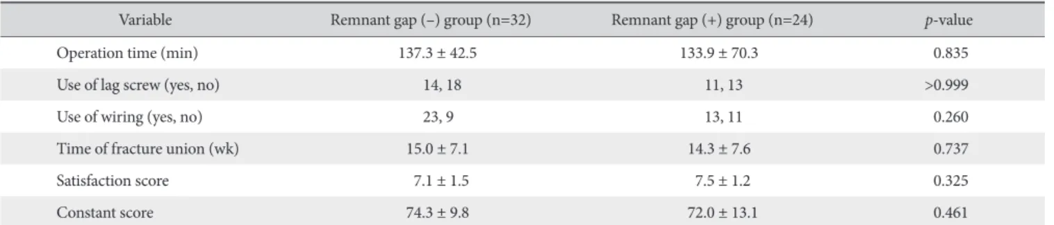
With resepct to the surgery overall, there were no differ- ences in the use of lag screws or wiring and operation time (all p>0.05) were observed between those with and without remnant fracture gap (137.3 ± 42.5 minutes and 133.9 ± 70.3 minutes in remnant gap (–) group and remnant gap (+) group, respectively; p=0.835) (Table 2).
The average radiologic union time was 15.0 ± 7.1 weeks in the remnant gap (–) group and 14.3 ± 7.6 weeks in the remnant gap (+) group, and was not statistically different between groups (p=0.737) (Table 2). However, the average time to achieve union was significantly faster in the AO type A fracture (11.3 ± 4.9 weeks) compared with the type B (18.0 ± 9.5 weeks) or type C fracture (16.9 ± 7.7 weeks) (p=0.012). The cause of in- jury did not affect the average radiologic union time (p=0.177).
There was no difference in the outcomes, regardless of the presence of the remnant gap. The satisfaction score was 7.12 ± 1.51 in the remnant gap (–) group and 7.50 ± 1.21 in the rem- nant gap (+) group, showing no significant difference between groups (p=0.325), and the Constant score was 74.3 ± 9.8 in the remnant gap (–) group and 72.0 ± 13.1 in the remnant gap (+) group, indicating no significant difference between groups (p=0.461) (Table 2). The type of fracture also did not affect the patient satisfaction or Constant score (all p>0.05). However, the fracture caused by traffic accident showed association with worse clinical outcomes compared with other cause of injury (patient satisfaction score: 6.77 ± 1.89 and 7.52 ± 1.03, re- spectively, p=0.061; Constant score: 69.1 ± 14.9 and 75.4 ± 8.5, respectively, p=0.049).
Five patients in the remnant gap (–) group, and 2 in the remnant gap (+) group (p=0.686) complained of paresthesia around the incision site. No other complications such as non- union, malunion, implant fracture, or infection occurred.
Discussion
In this study, we showed satisfactory outcomes regardless of the remnant fracture gap by the use of DBM. That is, there was no difference in outcomes between patients who achieved suc- cessful anatomical reduction and those who had remnant frac- ture gap and were treated with additional DBM.
In addition, even though the operation time was relatively long with average 135.4 ± 59.5 minutes as the surgeon was not highly experienced, the operation time does not appear to affect outcomes with regard to union time. Altamimi and McKee14) reported that the average radiographic union time after plate fixation of displaced midclavicle fracture was 16.4 weeks, and Khorami et al.15) reported that the fracture was united after 19.2 weeks from surgical plate fixation of displaced midshaft clavicle fracture. In this study, the average radiographic union time was 14.7 weeks, which was compatible with those previous stud- ies.14,15) In addition, the union time was not affected by the pres-
Table 2. Radiological and Clinical Outcomes
Variable Remnant gap (–) group (n=32) Remnant gap (+) group (n=24) p-value
Operation time (min) 137.3 ± 42.5 133.9 ± 70.3 0.835
Use of lag screw (yes, no) 14, 18 11, 13 >0.999
Use of wiring (yes, no) 23, 9 13, 11 0.260
Time of fracture union (wk) 15.0 ± 7.1 14.3 ± 7.6 0.737
Satisfaction score 7.1 ± 1.5 7.5 ± 1.2 0.325
Constant score 74.3 ± 9.8 72.0 ± 13.1 0.461
Values are presented as mean ± standard deviation or number only. All patients in the remnant gap (+) group were treated with an additional demineralized bone matrix during the operation. Th e mean values were compared using Student’s t-test for the continuous variables and 2 test or Fisher’s exact test for the categorical variables.
ence of the remnant fracture gap, which was additionally treated with DBM (15.0 weeks vs. 14.3 weeks, p=0.737). The very few complication events of this study except several paresthesias also support the successful outcomes of this case series.
We do not know the exact reason why this case series showed successful outcomes regardless of the remnant fracture gap. However, we think strong fixation by the use of a locking compression plate and additional treatment of DBM may be the reason. Locking compression plate has the advantage of strong fixation due to locking between the screw and plate as well as blood supply preservation due to minimal contact between plate and cortical bone.16,17) In this study, stable fixation was achieved in all cases by use of a locking compression plate, in spite of the remnant notable fracture gap in several cases. On the other hand, if a stable fixation is not achieved and there is too much motion, delayed union or even non-union could occur.18) Thus, we believe that the rigid fixation may lead to successful outcome without delay in bone union time, regardless of remnant fracture gap.
In addition, the effect of DBM applied in a fracture gap may affect the comparably successful outcome in patients with rem- nant fracture gap. The mobilization of various growth factors of DBM can cause differentiation of mesenchymal stem cells to the osteoblastic pathway, chemotaxis of osteoblastic precursor cells, and induction of osteoblast function in the grafted area.19,20) We think these actions may offset the possibility of nonunion or de- layed union coming from the remaining fracture gap.
Rather than the remaining fracture gap, the initial injury mechanism seems to be associated more with treatment out- comes. In this study, type A fracture by AO classification showed faster bone union time compared to other types, and the frac- tures that occurred by traffic accident showed worse clinical scores. Type A fracture is a simple non-comminuted fracture.
Thus, the operation for type A fracture is less problematic, and rigid internal fixation and bone union from direct cortical remod- elling via the provision of stable compression across the fracture may be more probable in that fracture type, which could lead to successful bone union.
Therefore, as application of DBM to the remaining fracture gap after open reduction and plate fixation in displaced mid- clavicle fracture does not affect surgical outcomes, we think that re-reduction and re-fixation of the fracture to make a perfect an- atomical reduction spending time is unnecessary if rigid fixation is acquired. However, we were not able to independently dem- onstrate whether that comparably successful result regardless of remaining fracture gap is due to the effect of DBM. Further study with a well-controlled prospective design, such as the use of DBM in groups with similar fracture gap size, may be needed to confirm the effect of DBM on gap healing that remains even after open reduction and plate fixation.
There are several limitations. First, three types of locking com-
pression plate were used. Even though the plate was selected according to the shape and curve of the clavicle and no differ- ence was found in outcomes according to the type of plates, this may limit the general application of the results of this study. Sec- ond, other factors such as range of shoulder motion, shoulder strength, or pain score, were not evaluated, which might influ- ence the result of treatment. Third, the number of patients was relatively small, and the minimum 6-month follow-up period may not be sufficient for complete evaluation of the outcomes and any chronic complications. Finally, due to the retrospective nature of this study, potential bias cannot be excluded.
Conclusion
Both the patients who achieved accurate anatomical reduc- tion and those who had a remaining fracture gap even after open reduction and internal fixation of a displaced midclavicle fracture treated with DBM showed successful bone union and favourable clinical outcomes. Re-reduction and re-fixation of the fracture for removal of a remaining gap and for achievement of a perfect anatomical reduction spending time appears to be un- necessary if rigid fixation is achieved.
References
1. O’Neill BJ, Hirpara KM, O’Briain D, McGarr C, Kaar TK. Clav- icle fractures: a comparison of five classification systems and their relationship to treatment outcomes. Int Orthop. 2011;
35(6):909-14.
2. Postacchini F, Gumina S, De Santis P, Albo F. Epidemiology of clavicle fractures. J Shoulder Elbow Surg. 2002;11(5):452-6.
3. Nordqvist A, Petersson C. The incidence of fractures of the clavicle. Clin Orthop Relat Res. 1994;(300):127-32.
4. Postacchini R, Gumina S, Farsetti P, Postacchini F. Long-term results of conservative management of midshaft clavicle frac- ture. Int Orthop. 2010;34(5):731-6.
5. McKee MD, Pedersen EM, Jones C, et al. Deficits following nonoperative treatment of displaced midshaft clavicular frac- tures. J Bone Joint Surg Am. 2006;88(1):35-40.
6. Smekal V, Oberladstaetter J, Struve P, Krappinger D. Shaft frac- tures of the clavicle: current concepts. Arch Orthop Trauma Surg. 2009;129(6):807-15.
7. Zlowodzki M, Zelle BA, Cole PA, Jeray K, McKee MD;
Evidence-Based Orthopaedic Trauma Working Group. Treat- ment of acute midshaft clavicle fractures: systematic review of 2144 fractures: on behalf of the Evidence-Based Orthopaedic Trauma Working Group. J Orthop Trauma. 2005;19(7):504-7.
8. Martin GJ Jr, Boden SD, Titus L, Scarborough NL. New for- mulations of demineralized bone matrix as a more effective graft alternative in experimental posterolateral lumbar spine arthrodesis. Spine (Phila Pa 1976). 1999;24(7):637-45.
9. Ziran BH, Smith WR, Morgan SJ. Use of calcium-based de- mineralized bone matrix/allograft for nonunions and posttrau- matic reconstruction of the appendicular skeleton: preliminary results and complications. J Trauma. 2007;63(6):1324-8.
10. Oakes DA, Lee CC, Lieberman JR. An evaluation of human demineralized bone matrices in a rat femoral defect model.
Clin Orthop Relat Res. 2003;(413):281-90.
11. Marsh JL, Slongo TF, Agel J, et al. Fracture and dislocation classification compendium - 2007: Orthopaedic Trauma As- sociation classification, database and outcomes committee. J Orthop Trauma. 2007;21(10 Suppl):S1-133.
12. Caneva M, Botticelli D, Stellini E, Souza SL, Salata LA, Lang NP. Magnesium-enriched hydroxyapatite at immediate im- plants: a histomorphometric study in dogs. Clin Oral Implants Res. 2011;22(5):512-7.
13. Jiang H, Qu W. Operative treatment of clavicle midshaft frac- tures using a locking compression plate: comparison between mini-invasive plate osteosynthesis (MIPPO) technique and conventional open reduction. Orthop Traumatol Surg Res.
2012;98(6):666-71.
14. Altamimi SA, McKee MD; Canadian Orthopaedic Trauma
Society. Nonoperative treatment compared with plate fixation of displaced midshaft clavicular fractures. Surgical technique. J Bone Joint Surg Am. 2008;90 Suppl 2 Pt 1:1-8.
15. Khorami M, Fakour M, Mokarrami H, Arti HR, Nasab AM, Shahrivar F. The comparison of results of treatment of midshaft clavicle fracture between operative treatment with plate and non-operative treatment. Arch Bone Jt Surg. 2014;2(3):210-4.
16. Haidukewych GJ. Innovations in locking plate technology. J Am Acad Orthop Surg. 2004;12(4):205-12.
17. Perren SM. Evolution and rationale of locked internal fixator technology. Introductory remarks. Injury. 2001;32 Suppl 2:B3-9.
18. Phillips AM. Overview of the fracture healing cascade. Injury.
2005;36 Suppl 3:S5-7.
19. Edwards JT, Diegmann MH, Scarborough NL. Osteoinduc- tion of human demineralized bone: characterization in a rat model. Clin Orthop Relat Res. 1998;(357):219-28.
20. Rabie AB, Wong RW, Hägg U. Composite autogenous bone and demineralized bone matrices used to repair defects in the parietal bone of rabbits. Br J Oral Maxillofac Surg. 2000;38(5):
565-70.