Clinical Results of the Arthroscopic “Multiple Pulled Suture”
Technique for Large or Comminuted Bony Bankart Lesion
Byung-Ill Lee, Byoung-Min Kim1 , Duk-Hwan Kho1, Hyeung-June Kim1
Department of Orthopedic Surgery, Soonchunhyang University Seoul Hospital, Seoul, 1Department of Orthopedic Surgery, Konkuk University Chungju Hospital, Chungju, Korea
Background: Arthroscopic fixations for large and comminuted bony Bankart lesions are technically difficult. We developed an ar- throscopic multiple pulled suture (MPS) technique to restore large and comminuted bony Bankart lesions.
Methods: Ten patients (mean age, 49.8 years; range, 31–79 years) underwent bony Bankart repair using the illustrated MPS technique and were then followed for a mean of 27.3 months. A plain radiograph series and three-dimensional computed tomography scans were taken at the initial clinical evaluation and 3 months postoperatively. Outcome measurements included the American Shoulder and El- bow Surgeons (ASES) score, Rowe score, University of California at Los Angeles (UCLA) score, and subjective patient satisfaction, along with surgical complications.
Results: Union of an osseous fragment with the glenoid rim was confirmed in all patients on a computed tomography scan 3 months af- ter operation. The osseous fragment was restored to proper articular congruence and reduction. The affected shoulder was stable in nine of the 10 patients. One patient presented with a redislocation after a sports injury 3 years postoperatively. The ASES, Rowe, and UCLA scores improved at the final evaluation, and median patient satisfaction at the final follow-up was 9 of 10 points (range, 6–10 points).
Conclusions: The arthroscopic MPS technique for bony Bankart lesions with large or comminuted osseous fragments was a relatively easy and safe method for stable fixation of the osseous fragment. Therefore, the arthroscopic MPS technique resulted in good restoration of stability with high patient satisfaction and low complication rates.
(Clin Shoulder Elbow 2017;20(3):138-146)
Key Words: Shoulder; Bony Bankart lesion; Instability; Arthroscopic Clinics in Shoulder and Elbow Vol. 20, No. 3, September, 2017 https://doi.org/10.5397/cise.2017.20.3.138
Received July 5, 2017. Revised July 20, 2017. Accepted July 30, 2017.
Correspondence to: Byoung-Min Kim
Department of Orthopedic Surgery, Konkuk University Chungju Hospital, 82 Gugwon-daero, Chungju 27376, Korea Tel: +82-43-840-8250, Fax: +82-43-844-7300, E-mail: [email protected]
IRB approval (No. SCHUH-2014-10-018-005).
Financial support: None. Conflict of interests: None.
Introduction
Bony Bankart lesions are avulsions of the labroligamentous complex associated with an anterior glenoid rim fracture.1) These lesions have the potential to result in persistent glenohumeral joint instability if the fragment is displaced. An anterior glenoid rim fracture is present in 16% of primary shoulder dislocations and 23% of recurrent dislocations.2) However, a study using three-dimensionally reconstructed computed tomography (3D- CT) revealed that the prevalence of osseous lesions was as high as 50% in shoulders with recurrent anterior glenohumeral insta- bility.3)
The osseous contour of the glenoid is more important than soft tissue status in the midrange of motion. In a cadaver study, peak translation force of the shoulder and humeral head lat- eral translation decreased as the size of the osseous defect increased.4) In a histological study, all bony fragments in bony Bankart lesions were viable and were used to treat fractured glenoid defects.5) Thus, primary fixation should be attempted to decrease the glenoid defect, regardless of the bony fragment size.
Various surgical techniques6-14) to treat bony Bankart lesions have been described. Arthroscopic or open reduction is per- formed to fix the bony Bankart lesion to the glenoid depend-
ing on the size of the bony fragment. Open surgical treatment often requires extensive exposure and muscular dissection with the risk of postoperative joint stiffness, muscular weakness, and pain. Therefore, many surgeons believe that these lesions should be reconstructed arthroscopically by reducing the fragment to the labroligamentous complex. An arthroscopic technique has many advantages, such as anatomic repair with a direct view, decreased invasiveness and tissue trauma, and a concomitant pathological diagnosis. Many arthroscopic methods have been used;8,10,13,15-18) however, they all have major limitations, includ- ing technical challenge.
Our intention was to develop a minimally invasive ar- throscopic technique that provides less technical challenge and a more stable anatomic glenoid reduction. In our previous report, we described the “multiple pulled suture (MPS) technique” with a knotless suture anchor.19) In the present study, we investigated the clinical outcomes and complications after arthroscopic fixa- tion of bony Bankart lesions using the MPS technique. We hy- pothesized that this technique would provide good restoration of stability with high patient satisfaction and low complication rates.
Methods
We retrospectively collected and reviewed data for this study.
Ten patients (mean age, 49.8 years; range, 31–79 years; three women and seven men) with anterior shoulder instability due to a bony Bankart lesion were treated from January 2009 to June 2014 using the arthroscopic MPS technique.19) The indications for the MPS technique were: (1) anterior glenoid loss <25%
except to the osseous fragment or (2) the bony fragment was
>20% of the diameter of the inferior circle on the glenoid or comminuted in an acute or a chronic bony unstable Bankart lesion. The mean follow-up was 27.3 months (range, 6–50 months). None of the patients reported pain, apprehension, or other symptoms suggestive of a subluxation during sports or work activities before the first trauma. All patients had sustained trauma-related dislocations, where the dominant shoulder was affected in seven (70.0%) of 10 patients. The arthroscopic repair was performed within 3 months (acute; range, 6–60 days) of the initial injury in eight shoulders (80.0%), and at a later time (chronic; range, 95–545 days) in two shoulders (20.0%). Among the patients in the acute group, seven (87.5%) of eight patients had a single dislocation, and one (12.5%) of eight patients had two or more frank dislocations. One of two patients in the chronic group had six dislocations, and one of two had 15 dislo- cations before surgery.
Radiographic Evaluation
A plain radiograph series and 3D-CT scans were taken at the initial clinical evaluation and 3 months postoperatively. Glenoid
bone loss was evaluated with an en face view of the glenoid on a 3D-CT scan preoperatively. The size of the osseous defect as a percentage of the glenoid rim was calculated as a ratio of the width of the osseous defect to the diameter of an assumed outer fitted circle based on the inferior portion of the glenoid.20,21) An osseous fragment at the anteroinferior quadrant of the glenoid rim was confirmed in all patients by CT scan. The mean glenoid bone defect was 25.68% (range, 21%–31%) of the diameter of the inferior glenoid circle. Two (20.0%) of 10 patients had partial absorption of the osseous fragment or erosion of the glenoid rim. Nine patients (90.0%) had Hill-Sachs lesions. Five patients (50.0%) had Hill-Sachs lesions at a depth > than 10% (range, 11%–20%) of the humeral head diameter. The length-width ra- tio of the osseous fragment was measured as a ratio of the length between the upper and lower endpoints of the glenoid fracture margin to the diameter of an assumed outer fitted circle based on the inferior portion of the glenoid.22) The length-diameter ra- tio was 0.81 (range, 0.65–1.00). Union of the osseous fragment was evaluated with a two-dimensional axial image of a 3D-CT scan 3 months postoperatively.
Outcome Measurement
Data describing the history of shoulder subluxations, num- ber of dislocations, more subtle feelings of subluxations, and the activities in which instability occurred both preoperatively and postoperatively were collected. Instability was tested with the anterior apprehension and anterior relocation tests with the patient in the supine position. Preoperative and postoperative outcome measurements included the American Shoulder and Elbow Surgeons (ASES) score, the Rowe score, the University of California at Los Angeles (UCLA) score, and subjective patient satisfaction (by visual analogue scale), along with surgical compli- cations.
Arthroscopic Findings
In all patients, diagnostic arthroscopy confirmed a bony lesion of the anteroinferior glenoid rim and a humeral head impression fracture. Patients were classified by Bigliani classification.23) There were seven Bigliani type I lesions, one Bigliani type II lesion, and two Bigliani type IIIa lesions. In seven patients with Bigliani type I lesions, the mean size of the osseous fragment was 26.4% (range, 21%–31%) of the diameter of the inferior glenoid circle, and two of seven patients with Bigliani type I lesions had comminuted osseous fragments. In two patients with Bigliani type IIIa lesions, the mean glenoid defect was 25%, and small osseous fragments were observed. A superior labrum anterior and posterior lesion was found in four (40.0%) of the 10 patients and was treated with debridement. Three patients (30.0%) had full-thickness tears of the supraspinatus. One of the three patients was treated by repair, and two patients were treated with debridement be- cause of an unrepairable condition and old age.
Surgical Technique
All patients underwent induction of general anesthesia and an interscalene brachial plexus block. The patient was then placed in the lateral decubitus position and the involved arm was abducted at 70° with 25° forward elevation and 5 kg of distal traction. A 4 mm arthroscope was introduced through a standard posterior portal. The glenohumeral joint was inspected, the pathologic lesions were identified, and the anterosuperior and anteroinferior portals were prepared. Once the diagnostic arthroscopy and assessment of the bony Bankart lesion was completed, the anterosuperior portal was used to provide the arthroscopic view.
1) Fragment management
After identifying the bony fragment, the surrounding labrolig- amentous complex was mobilized and prepared using elevators, rasps, shavers, and an abrader through the anteroinferior portal.
The fragment was then confirmed to be reduced to the articular margin.
2) Loading of suture material
We inserted a suture hook (Linvatec., Largo, FL, USA) via the anteroinferior portal using a shuttle relay system, then secured the labroligamentous complex, starting from the anteroinferior edge (Fig. 1A). As the shuttle relay was unthreaded from the suture hook, a grasper was used to retrieve the shuttle relay through the posterior portal. The initial #2 suture material (Et- hibond; Ethicon, Somerville, NJ, USA) was hooked to the shuttle relay hole and then redirected to pass through the attached labroligamentous complex. Both ends of the suture were re-
trieved through the posterior portal (Fig. 1B). Four to six sutures were placed in the labroligamentous complex using the same method (Fig. 1C). When tension was applied to loaded sutures, the operator checked the amount of capsular placation and mo- bility of the fragment for reduction under arthroscopic control (Fig. 1D). The sutures were then brought out through the antero- inferior portal.
3) Insertion of the knotless anchor
The arthroscope was inserted through the anterosuperior portal. The fixation point was 2 to 3 mm further down the ar- ticular margin at the 2 or 10 o’clock position on the glenoid surface. One pilot hole was punched as the fixation point via the anteroinferior portal, after which all suture strands were threaded through the eyelet of a Pushlock (Arthrex, Naples, FL, USA) anchor. After applying appropriate tension for reduction and applying constant tension, a knotless anchor was advanced completely into the pilot hole (Fig. 1E). The suture limb was trimmed after the insertion. As described above, we achieved arthroscopic reduction and fixation by compressing the bony fragment back into the fracture bed.
Postoperative Regimen
The shoulder joint was immobilized postoperatively for 3 weeks using a shoulder abduction brace, after which passive mobilization was conducted. After 6 weeks, patients were al- lowed to start an active exercise and passive external rotation.
Non-contact sports were permitted after 12 weeks. Finally, a full return to throwing or contact sports was allowed after 6 months,
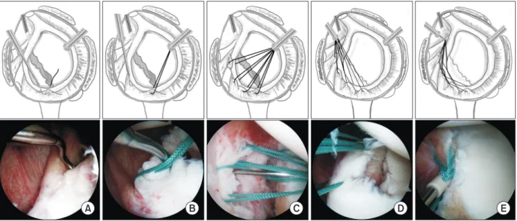
A B C D E
Fig. 1. Description of the multiple pulled suture technique. (A) The suture hook load with a shuttle-relay penetrates the torn labroligamentous complex associ- ated with an anterior glenoid rim fracture. The first suture is placed in the detached anteroinferior labroligamentous complex. (B) Both ends of the suture are retrieved through the posterior portal. (C) The passed suture remains, and three to six sutures are placed in the detached labroligamentous complex at regular intervals. (D) After confirming appropriate tension for reduction and applying constant tension, (E) a knotless anchor is completely advanced into the pilot hole.
The fixation point is below the articular margin at the 2 or 10 o’clock position on the glenoid surface.
depending on individual functional recovery status.
Statistical Analysis
Statistical analyses were conducted using the IBM SPSS ver. 20.0 statistical package (IBM Co., Armonk, NY, USA). The Wilcoxon signed-rank test was used to compare differences be- tween preoperative and postoperative outcome measures. The level of significance was set at p<0.05.
Results
The mean number of sutures used per repair to secure the glenoid fragment and labrum back into anatomic position was 3.4 (range, 3–5). Only one knotless anchor (Pushlock Anchor;
Arthrex) was used to fix the osseous fragment. Complications were observed in two patients. One patient complained of irrita- tion of his shoulder and clicking during exercise. A secondary ar- throscopic exploration revealed a disrupted suture on the labral side, but it had not slipped from the knotless anchor. The labro- ligamentous complex had healed well, so the disrupted suture was removed. The other patient presented with a redislocation after a sports injury 3 years postoperatively. This patient had Big- liani type IIIa lesions with a small bony fragment and a Hill-Sachs lesion. We recommended the Latarjet operation to reconstruct the glenoid defect.
Radiographic Results
Union of the osseous fragment with the glenoid rim was con- firmed in all of 10 patients (100%) by CT scan 3 months post- operatively. The osseous fragment had restored proper articular congruence and reduction.
Clinical Outcomes
The affected shoulders in nine (90%) of 10 patients were stable, and the anterior apprehension and anterior relocation tests were negative. The mean ASES score at the final evalua- tion improved from 52.1 (range, 27–89) preoperatively to 94.8 (range, 89–100) postoperatively (p=0.008). The mean Rowe score improved from 27.5 (range, 0–65) preoperatively to 89.0 (range, 35–100) postoperatively (p=0.008). The mean UCLA score improved from 18.0 (range, 7–24) preoperatively to 31.9 (range, 24–35) postoperatively (p=0.007). Overall, median pa- tient satisfaction at the final follow-up was 9 of 10 points (range, 6–10). One patient who redislocated his shoulder in a sports injury reported a lower satisfaction of 6 out of 10 (Table 1).
Cases 1) Case 1
A 38-year-old man fell eight days prior to the visit, which had caused a dislocated shoulder for the first time. The dislocation was reduced at another institution. An examination revealed no
sign of hyperlaxity; however, range of motion was limited due to pain.
Plain anteroposterior radiographs showed a bony fragment at the inferior part of the glenoid. A 3D-CT scan revealed a bony Bankart lesion at the anteroinferior part of the glenoid rim with- out a Hill-Sachs lesion. The bony fragment measured 9×19 mm with glenoid bone loss of 28% (quantification of glenoid bone loss based on glenoid rim distance) (Fig. 2A). The Rowe score was 65 preoperatively (graded from 0–100, with a high score indicating good function). Additionally, the patient had a preop- erative score of 56 on the ASES and 16 on the UCLA shoulder scale.
Although the size of the glenoid defect was >25%, the bony fragment was not absorbed and a Bigliani type I lesion was pres- ent. The bone fragment was prepared for mobilization by ar- throscopy (Fig. 2B). The large bone fragment was reduced with- out difficulty when the three sutures on the labroligamentous complex were pulled. The bony fragment was fixed successfully using only a knotless anchor (Fig. 2D).
A CT scan at the 3 months follow-up revealed articular con- gruence and union of the fragment (Fig. 2C). The patient did not complain of any pain at the 41 months follow-up, and had full range of motion without instability. At this time, the Rowe score had improved to 100, the ASES score had improved to 100 and the UCLA score had improved to 35. Patient satisfaction at the final follow-up was 10 points.
2) Case 2
A 58-year-old man presented with right shoulder pain and a tingling sensation with hypothesis on the right upper extrem- ity after accident. Although his shoulder was dislocated 5 years ago, he has not felt any instability symptom until this accident.
Electromyography revealed an incomplete brachial plexus injury.
The tingling sensation and hypoesthesia improved progressively during 8 weeks before the surgery.
Magnetic resonance imaging (MRI) showed a massive full thickness tear, atrophy of the rotator cuff, degenerative changes in the glenoid bone, and a displaced glenoid rim fragment. A 3D-CT evaluation revealed that the bony fragment was 4.8×16.8 mm in size, and that glenoid bone loss was 21% (Fig. 3A).
Range of motion was limited preoperatively, and the appre- Table 1. Clinical Assessment and Comparisons of Preoperative and Postop- erative Scores
Score Preoperative Postoperative p-value
ASES 52.1 94.8 0.008
UCLA 18.0 31.9 0.007
Rowe 27.5 89.0 0.008
Values are presented as mean only.
ASES: American Shoulder and Elbow Surgeons, UCLA: University of Califor- nia at Los Angeles.
A B
C D
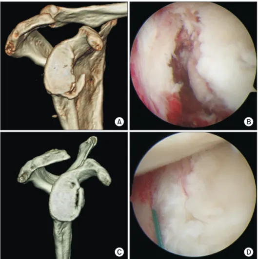
Fig. 2. Case 1. (A) Preoperative computed tomography (CT) scan shows a 9×19 mm osseous fragment and glenoid bone loss in the inferior glenoid circle of 28%. (B) Ar- throscopic appearance of the glenoid frac- ture (C) CT scan at the 3 months follow-up reveals articular congruence and union of the fragment. (D) Arthroscopic appearance of a completed bony Bankart repair.
A B
C D
Fig. 3. Case 2. (A) Preoperative computed tomography (CT) scan shows a 4.8×16.8 mm osseous fragment with 21% glenoid bone loss in the inferior glenoid circle. (B) The periosteum was detached from the bone fragment. (C) A CT scan at the 3 months follow-up reveals articular congruence and union of the fragment. (D) Arthroscopic appearance of the completed bony Bankart repair.
hension test was remarkably positive. Preoperatively, the Rowe score was 45, the ASES score was 39 and the UCLA score was 15. During arthroscopy, the periosteum was detached from the bony fragment, a Bigliani type II lesion was present (Fig. 3B), the rotator cuff was not repairable, and a bony spur was observed on the subacromial surface. Sutures were passed through the labroligamentous complex with periosteum. Articular congru- ence was maintained between the fragment and the glenoid after applying tension to the sutures. We inserted the knotless anchor completely into the pilot hole while applying constant tension (Fig. 3D). Additionally, only arthroscopic subacromial decompression was performed.
A CT scan at the 3 months follow revealed articular congru- ence and union of the fragment (Fig. 3C). The patient showed mild discomfort because of the rotator cuff injury at the 39 months follow-up, but had excellent range of motion without instability. The anterior apprehension test was negative and the Rowe score was 90, while the ASES score had improved to 95 and the UCLA score had improved to 30. Patient satisfaction at the final follow-up was 8 points.
3) Case 3
A 79-year-old woman presented with left shoulder pain after injury and was found to have her first episode of dislocation.
The shoulder was highly unstable after reduction. The anterior apprehension test result was remarkably positive, and the Rowe score, ASES score and UCLA score was 0, 27, and 7 preopera- tively.
A 3D-CT scan revealed 25% glenoid bone loss, a length to diameter ratio of 0.81, and a comminuted and shallow bony fragment (Fig. 4A). MRI showed a massive full thickness rota- tor cuff tear. The shallow bony fragment was comminuted on arthroscopy; however, the labroligamentous complex was not detached from the bony fragment, and a Bigliani type I lesion was present (Fig. 4B). Arthroscopic reduction and internal fixa- tion were performed using the MPS technique (Fig. 4D).
A CT scan at the 3 months follow-up revealed good reduc- tion and union of the fragment; however, the proximal fragment was tilted (Fig. 4C). The patient had nearly full range of motion with no instability or pain at the 12 months follow-up, as well as a negative anterior apprehension test and a Rowe score or 90.
Additionally, the ASES score had improved to 92 and the UCLA
A B
C D
Fig. 4. Case 3. (A) Preoperative computed tomography (CT) scan shows glenoid bone loss of 25%, and a comminuted and shallow bony fragment. (B) Arthroscopic appearance of the glenoid fracture. (C) A CT scan at the 3 months follow-up reveals permissible reduction and union of the fragment. (D) Arthroscopic appearance of the completed bony Bankart repair.
score had improved to 32. The patient satisfaction at the final follow-up was 9 points.
Discussion
Among various joints, the shoulder has the largest arc of mo- tion. Balance between bone structure and soft tissue results in a stable shoulder. In particular, the osseous contour of the glenoid affects instability of the shoulder during midrange motion more than that of the soft tissue.4) A neglected bony Bankart lesion is a major cause of recurrent shoulder instability following Bankart repair.
Rowe and Zarins24) were the first to report bony Bankart le- sions associated with classic Bankart lesions; however, other studies have also shown a relationship between the amount of glenoid bone loss and recurrence.22,25) Moreover, Burkhart and De Beer25) reported the importance of restoring the anterior- inferior glenoid. Therefore, many surgeons have explored new modalities to improve bony fragment fixation and decrease the recurrence of dislocations.
Open surgical treatment of a glenoid fossa fracture usually al- lows for anatomic restoration of the joint.9,14) However, this often requires an extensive approach and muscular dissection with the risk of stiffness, muscular weakness, and pain.
Many surgeons are interested in arthroscopic techniques as they allow for better evaluation of the intra-articular lesion and associated pathological findings. Smaller bony lesions may be amenable to arthroscopic treatments. However, larger bony lesions often require open surgery to prevent recurrent instabil- ity.26) Many surgeons have reported that shoulders with a large anterior-inferior osseous defect of the glenoid are not suitable for arthroscopic stabilization because of unacceptably high failure rates.25,27)
Several arthroscopic procedures have been developed and continue to be developed. Cameron7) described successful ar- throscopic reduction and fixation of an anteroinferior osseous fragment with a partially threaded 3.5 mm cancellous screw inserted through the subscapularis tendon fibers. Although fixa- tion with cannulated screws is feasible for larger fragments, there are some limitations. Specifically, placing cannulated screws can be technically challenging and may result in splitting the bony fragment. Inserting a cannulated screw through the subscapularis tendon entails a greater risk because the axillary and musculocu- taneous nerve runs just inferior to the subscapularis tendon.
Bauer et al.6) described arthroscopic repair using the transgle- noid technique, which is easier for achieving reduction and maintaining bony lesions because the reduction is performed by suturing the labroligamenotus complex without handling the bony fragment. The major drawback of this technique is the pos- terior fixation method, which is not firm because it is performed far from the lesion and the sutures are tied to muscle fascia.
Suture fixation on the fascia of the infraspinatus muscle is not as accurate as direct fixation with a screw or pin. Posterior-fixation has the risk of suprascapular nerve injury, and the posterior su- ture knot and posterior muscle dissection result in discomfort or limitations during internal rotation.18) Therefore, Marcacci et al.28) described a modification of the original Caspari technique in which the anchor is made directly on the bone of the scapular spine. However, this technique also requires an additional pos- terior incision and muscle dissection.
Surgeons have developed new techniques using the suture anchor as suture-anchor implants have evolved. Porcellini et al.12) reported an operative technique utilizing suture anchors for bony Bankart lesions. Since then, several arthroscopic tech- niques using suture anchors have been reported to treat bony Bankart lesions.12,16,29) However, there are some limitations to these techniques. For example, the suture anchor technique does not provide sufficient contact area for the fractured frag- ment because the suture crosses the surface of the fracture, and the bony fragment may tilt because of the one-point fixation.
To avoid the disadvantages of such a one-point fixation, Millett et al.15) reported the “bony Bankart bridge” procedure for ar- throscopic reduction and suture anchor fixation of bony Bankart fragments. This technique provides secure two-point fixation and compression of the fracture without tilting the bony frag- ments. However, this has a few disadvantages, mainly regarding the high technical demand and the suture crossing the articular surface.
Many authors have reported that suture anchor techniques for Bankart lesions are superior to the transglenoid techniques with regards to patient satisfaction, functional recovery, compli- cations, and recurrent instability.30,31) However, no studies have compared the transglenoid technique with the suture anchor technique for fixing a bony Bankart lesion. The transglenoid technique is useful for reducing bony fragments and anatomical restoration of the glenohumeral ligament and provides an easier way to control the amount of capsular plication.6) It was initially hypothesized that if the drawback of posterior fixation could be removed, the transglenoid technique would be as effective as the suture anchor technique for bony Bankart repair. Thus, we returned to the previous transglenoid technique and modified the fixation method using a knotless suture. The posterior fixa- tion method was changed (the transglenoid suture technique of Caspari) to anterior fixation using a knotless anchor (3.5 mm Pushlock; Arthrex). Thus, our technique does not have the draw- back of previous transglenoid techniques. Although our study comprised a small patient population with a short follow-up, the MPS technique provided good restoration of stability with high patient satisfaction and low complication rates.
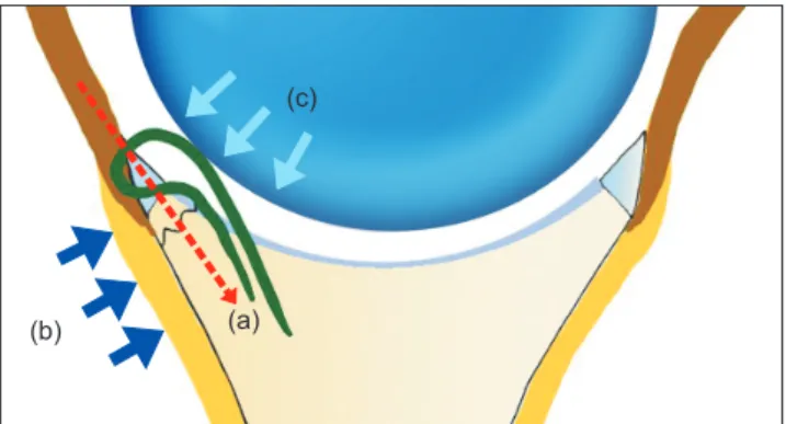
A follow-up CT scan was performed three months postopera- tively in all patients. The bony fragments were well united, and congruence of the articular surface was maintained. Unlike static
suture anchor fixation using the one-point fixation technique, the dynamic compression force of the MPS technique was ap- plied to the fracture site (Fig. 5). Although the pull-out strength was lower than fixation with several suture anchors, the knotless anchor did not pull out in any case. Therefore, we assume that the knotless anchor supplied sufficient fixation power until the fragment united.
The remnant fragment was smaller than the glenoid bone loss in two patients, and glenoid bone loss was >25% of the width of the inferior glenoid. The treatment goal for a bony Bankart lesion is different than that for a non-bony Bankart lesion. Fujii et al.5) reported that all bony fragments in bony Bankart lesions were viable in a histological study and suggested that these frag- ments could be used to treat a fractured glenoid defect. The primary treatment goal for a bony Bankart lesion should be fixa- tion of the fragment on the glenoid rim, regardless of remnant fragment size. In patients with bone losses >25% of the diam- eter of the inferior glenoid without a remnant fragment, many surgeons recommend a glenoid reconstruction procedure, such as an autobone graft, an allobone graft, the Bristow procedure, or the Latarjet procedure.23,25) We recommend fixing the frag- ment and a labroligamentous complex repair in cases of glenoid bone loss >25% of the diameter of the inferior glenoid if a bony fragment remains. The total surface of the inferior glenoid can- not be restored by fixing the remnant fragment, but the width of the inferior glenoid can be partially restored. One of two pa- tients presented with a redislocation because of a sports injury 3.6 years after the operation; however, this patient did not present with a dislocation before the re-injury.
In case 2, the labroligamentous complex and the periosteum were avulsed from osseous fragment, resulting in medial dis- placement and inferior shifting; however, the condition of the tissues was good. The surrounding labroligamentous complex and periosteum was mobilized and prepared. The suture ma-
terials were passed through the labroligamentous complex and the periosteum. When tension was applied to loaded sutures, sutured tissues were not torn and covered the osseous fragment.
If the condition of the labroligamentous complex and the perios- teum were poor, the arthroscopic MPS technique could not be performed.
When arthroscopic reduction using the suture anchor is performed, reducing and fixing large, long, or comminuted frag- ments is difficult and requires many anchors. For example, the
“double-row suture bridge” technique requires several anchors (mean, 5; range, 4–8).15) Furthermore, the larger the fragment, the more difficult it is to insert medial-low anchors. However, our technique requires only a knotless anchor, and is not affect- ed by the size of the bony lesion because it is an indirect fixation using ligamentotaxis. The osseous fragment can be anatomically reduced and fixed firmly using this technique, even for relatively large or comminuted bony fragments.
The MPS technique has additional theoretical advantages.
Specifically, it provides sufficient contact area of the osseous fragment because the sutures are not located on the fracture sur- face. Additionally, the pulling force of multiple sutures becomes a dynamic compression force on the osseous fragment, and the osseous fragment is supported by a labroligamentous complex like a buttress plate, and is compressed by the humeral head (Fig.
5). Thus, articular congruence is maintained by the combination of these forces.
Finally, our technique and the suture anchor technique dif- fer in the anchor insertion point. Our technique is an attractive option for revising a bony Bankart lesion or for a failed Bankart repair with a neglected bony fragment using suture anchors.
However, the failure strength of the knotless anchor (Pushlock 3.5 mm; Arthrex) used with our method has not been assessed.
Thus, further study is needed to determine cyclic load failure strength and pullout failure strength of knotless anchors loaded with multiple sutures. Additionally, our study was limited by the relatively small number of patients, which was attributable to the infrequent nature of this injury.
Conclusion
The arthroscopic MPS technique for bony Bankart lesions with large or comminuted osseous fragments provides relatively easy reduction and safe and stable fixation of osseous fragments.
Therefore, use of the arthroscopic MPS technique for anterior instability with a glenoid rim fracture resulted in good restoration of stability with high patient satisfaction and low complication rates. Larger patient populations with a longer follow-up are needed to draw more definite conclusions.
(b) (a)
(c)
Fig. 5. Theoretical basis for the multiple pulled suture technique. (a) The fracture site is compressed by the coupled forces (vector) of the pulled sutures and the reaction. (b) The anterior labroligamentous complex supports the bony fragment like a buttress plate. (c) The humeral head maintains articular congruence by compression and contact.
References
1. Bankart AS. Recurrent or habitual dislocation of the shoulder- joint. Br Med J. 1923;2(3285):1132-3.
2. Griffith JF, Antonio GE, Yung PS, et al. Prevalence, pattern, and spectrum of glenoid bone loss in anterior shoulder disloca- tion: CT analysis of 218 patients. AJR Am J Roentgenol. 2008;
190(5):1247-54.
3. Sugaya H, Moriishi J, Dohi M, Kon Y, Tsuchiya A. Glenoid rim morphology in recurrent anterior glenohumeral instability. J Bone Joint Surg Am. 2003;85(5):878-84.
4. Yamamoto N, Muraki T, Sperling JW, et al. Stabilizing mecha- nism in bone-grafting of a large glenoid defect. J Bone Joint Surg Am. 2010;92(11):2059-66.
5. Fujii Y, Yoneda M, Wakitani S, Hayashida K. Histologic analysis of bony Bankart lesions in recurrent anterior instability of the shoulder. J Shoulder Elbow Surg. 2006;15(2):218-23.
6. Bauer T, Abadie O, Hardy P. Arthroscopic treatment of glenoid fractures. Arthroscopy. 2006;22(5):569.e1-6.
7. Cameron SE. Arthroscopic reduction and internal fixation of an anterior glenoid fracture. Arthroscopy. 1998;14(7):743-6.
8. Kim SJ, Kim TW, Moon HK, Chang WH. A combined transgle- noid and suture anchor technique for bony Bankart lesions.
Knee Surg Sports Traumatol Arthrosc. 2009;17(12):1443-6.
9. Kavanagh BF, Bradway JK, Cofield RH. Open reduction and internal fixation of displaced intra-articular fractures of the gle- noid fossa. J Bone Joint Surg Am. 1993;75(4):479-84.
10. Kokubu T, Nagura I, Mifune Y, Kurosaka M. Arthroscopic bony bankart repair using double-threaded headless screw: a case report. Case Rep Orthop. 2012;2012:789418.
11. Millett PJ, Braun S. The “bony Bankart bridge” procedure: a new arthroscopic technique for reduction and internal fixation of a bony Bankart lesion. Arthroscopy. 2009;25(1):102-5.
12. Porcellini G, Campi F, Paladini P. Arthroscopic approach to acute bony Bankart lesion. Arthroscopy. 2002;18(7):764-9.
13. Sugaya H, Kon Y, Tsuchiya A. Arthroscopic repair of glenoid fractures using suture anchors. Arthroscopy. 2005;21(5):635.
14. Aulicino PL, Reinert C, Kornberg M, Williamson S. Displaced intra-articular glenoid fractures treated by open reduction and internal fixation. J Trauma. 1986;26(12):1137-41.
15. Millett PJ, Horan MP, Martetschläger F. The “bony Bankart bridge” technique for restoration of anterior shoulder stability.
Am J Sports Med. 2013;41(3):608-14.
16. Porcellini G, Paladini P, Campi F, Paganelli M. Long-term out- come of acute versus chronic bony Bankart lesions managed arthroscopically. Am J Sports Med. 2007;35(12):2067-72.
17. Anderl W, Kriegleder B, Heuberer PR. All-arthroscopic im-
plant-free iliac crest bone grafting: new technique and case report. Arthroscopy. 2012;28(1):131-7.
18. Savoie FH 3rd, Miller CD, Field LD. Arthroscopic reconstruc- tion of traumatic anterior instability of the shoulder: the Cas- pari technique. Arthroscopy. 1997;13(2):201-9.
19. Lee BI, Choi HS, Min KD, et al. A pulled sutures technique for bony Bankart lesion. Eur J Orthop Surg Traumatol. 2014;24(4):
641-5.
20. Burkhart SS, Debeer JF, Tehrany AM, Parten PM. Quantifying glenoid bone loss arthroscopically in shoulder instability. Ar- throscopy. 2002;18(5):488-91.
21. Chuang TY, Adams CR, Burkhart SS. Use of preoperative three-dimensional computed tomography to quantify glenoid bone loss in shoulder instability. Arthroscopy. 2008;24(4):376- 82.
22. Gerber C, Nyffeler RW. Classification of glenohumeral joint instability. Clin Orthop Relat Res. 2002;(400):65-76.
23. Bigliani LU, Newton PM, Steinmann SP, Connor PM, Mcllveen SJ. Glenoid rim lesions associated with recurrent anterior dislo- cation of the shoulder. Am J Sports Med. 1998;26(1):41-5.
24. Rowe CR, Zarins B. Recurrent transient subluxation of the shoulder. J Bone Joint Surg Am. 1981;63(6):863-72.
25. Burkhart SS, De Beer JF. Traumatic glenohumeral bone defects and their relationship to failure of arthroscopic Bankart repairs:
significance of the inverted-pear glenoid and the humeral en- gaging Hill-Sachs lesion. Arthroscopy. 2000;16(7):677-94.
26. Bushnell BD, Creighton RA, Herring MM. Bony instability of the shoulder. Arthroscopy. 2008;24(9):1061-73.
27. Itoi E, Lee SB, Berglund LJ, Berge LL, An KN. The effect of a glenoid defect on anteroinferior stability of the shoulder af- ter Bankart repair: a cadaveric study. J Bone Joint Surg Am.
2000;82(1):35-46.
28. Marcacci M, Zaffagnini S, Petitto A, Neri MP, Iacono F, Visani A. Arthroscopic management of recurrent anterior dislocation of the shoulder: analysis of technical modifications on the Cas- pari procedure. Arthroscopy. 1996;12(2):144-9.
29. Sugaya H, Moriishi J, Kanisawa I, Tsuchiya A. Arthroscopic os- seous Bankart repair for chronic recurrent traumatic anterior glenohumeral instability. Surgical technique. J Bone Joint Surg Am. 2006;88 Suppl 1 Pt 2:159-69.
30. van Oostveen DP, Schild FJ, van Haeff MJ, Saris DB. Suture anchors are superior to transglenoid sutures in arthroscopic shoulder stabilization. Arthroscopy. 2006;22(12):1290-7.
31. Kandziora F, Jäger A, Bischof F, Herresthal J, Starker M, Mit- tlmeier T. Arthroscopic labrum refixation for post-traumatic anterior shoulder instability: suture anchor versus transglenoid fixation technique. Arthroscopy. 2000;16(4):359-66.