Korean J Gastroenterol Vol. 59 No. 1, 44-47 http://dx.doi.org/10.4166/kjg.2012.59.1.44
CASE REPORT
Korean J Gastroenterol, Vol. 59 No. 1, January 2012 www.kjg.or.kr
혈변을 동반한 대장의 원발성 아밀로이드증 1예
권용환, 김지연, 김지훈, 박현우, 양해민, 전성우, 김성국
경북대학교 의학전문대학원 내과학교실
A Case of Primary Colon Amyloidosis Presenting as Hematochezia
Yong Hwan Kwon, Ji Yeon Kim, Ji Hun Kim, Hyun Woo Park, Hae Min Yang, Seong Woo Jeon and Sung Kook Kim Department of Internal Medicine, Kyungpook National University School of Medicine, Daegu, Korea
Amyloidosis is characterized by a deposition of insoluble fibrils in various organs and tissues. Amyloid deposition, in the gastro- intestinal track, provokes a dysfunction of the organ, due to an accumulation of fibrils, and causes a variety of clinical symptoms and endoscopic findings. Primary amyloidosis in the gastrointestinal tract is rarely reported in Korea. We experienced a case of recurrent intestinal bleeding, in a 59-year-old female patient with primary amyloidosis. A colonoscopy revealed the presence of multiple large circular ulcers. In the entire colon, diffuse nodular lesions with edema and bleeding were found. A colonoscopic biopsy established the diagnosis of amyloidosis, to the exclusion of other disease components. We concluded that the patient had localized amyloidosis. Though a definitive therapeutic strategy has not been established for localized gastrointestinal amyloi- dosis, the patient has been successfully treated with a high-dose of steroids and azathioprine. (Korean J Gastroenterol 2012;59:44 -47)
Key Words: Amyloidosis; Gastrointestinal hemorrhage; Colon
Received August 15, 2011. Revised September 19, 2011. Accepted September 19, 2011.
CC This is an open access article distributed under the terms of the Creative Commons Attribution Non-Commercial License (http://creativecommons.org/licenses/
by-nc/3.0) which permits unrestricted non-commercial use, distribution, and reproduction in any medium, provided the original work is properly cited.
교신저자: 전성우, 700-021, 대구시 중구 삼덕동 2가 50, 경북대학교병원 소화기내과
Correspondence to: Seong Woo Jeon, Department of Gastroenterology, Kyungpook National University Hospital, 50 Samdeok-dong 2-ga, Jung-gu, Daegu 700-021, Korea. Tel: +82-53-420-5515, Fax: +82-53-426-8773, E-mail: [email protected]
Financial support: None. Conflict of interest: None.
서 론
아밀로이드증은 저분자량의 비용해성 원섬유가 신체 내 다 양한 조직에서 세포 외 결합을 일으켜 생기는 질환이다. 현재 까지 발생기전은 알려져 있지 않으나, 아밀로이드증이 면역체 제의 이상과 명확한 연관성이 있는 것으로 알려져 있다.1-3 아 밀로이드증의 종류로는 다발골수종이나 형질세포 이상 등의 상태에서 발생하는 원발성 아밀로이드증(primary amyloido- sis, AL), 류마티스 관절염과 같은 만성 염증성 질병 및 오랜 기간 동안의 혈액투석 치료에서 발생하는 이차성 또는 반응성 아밀로이드증(secondary or reactive amyloidosis), 가족성 아밀로이드증(familial amyloidosis), 노인성 전신성 아밀로 이드증(senile systemic amyloidosis) 등으로 분류할 수 있 다.4-6 국내에서 이차성 아밀로이드증에 있어 위장관 침범은
흔히 발견되고 있으나,7 국소적으로 소화기계를 침범하여 내 시경검사에 의한 조직 생검으로 확진된 국소형 위장관 아밀로 이드증은 보고되는 경우가 드물다.8 이에 저자들은 출혈을 주 소로 내원한 환자에서 대장을 침범한 국소형 아밀로이드증을 진단하고 치료한 1예를 경험하였기에 문헌고찰과 함께 보고 한다.
증 례
59세 여자 환자가 내원 1개월 전부터 반복되는 혈변, 복통 및 전신 권태감으로 타 병원에서 시행한 대장내시경에서 대장 및 직장 전반에 걸친 다발성 궤양 및 발적 등의 소견으로 내원 하였다. 내원 당시 발열 등의 증상은 없었고, 오심 및 식욕 부진이 심한 상태로 내원 3일 전부터 거의 먹지 못하는 상태
Kwon YH, et al. A Case of Primary Colon Amyloidosis Presenting as Hematochezia
45
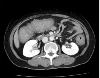
Vol. 59 No. 1, January 2012 Fig. 1. Abdominal CT findings. It demonstrated diffuse mural thic-
kening in the ascending colon, transverse colon and descending colon.
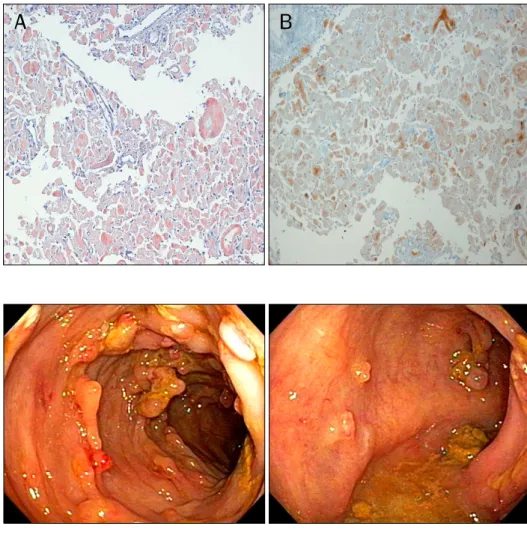
Fig. 2. Colonoscopic findings. (A) It demonstrated diffuse nodular le- sions with bleeding and friability at the ascending colon. (B) It demonstrated multiple irregular large ulcers with nodular lesions at the descending colon.
였으나, 3개월 간 몸무게 감소 소견은 보이지 않았다. 환자는 가정주부였으며, 술, 담배 및 기타 다른 약물의 복용력은 없었 다. 과거 30년 전 담낭염으로 담낭 제거 수술 시행한 병력 및, 급성 충수돌기염으로 충수돌기절제술을 시행한 병력이 있 었다. 그 외 고혈압, 결핵, 간염 및 류마티스 관절염 등의 병력은 없었고, 가족력에서도 특이사항은 없었다. 내원 당시 환자는 만성 병색을 띠었고, 혈압은 110/71 mmHg, 맥박수는 99회/분, 체온은 36.2oC였다. 흉부 청진에서 심잡음은 없었으 며, 양측 폐야에서 수포음도 없었다. 복부 진찰에서 하복부에 압통은 있었으나 반발통은 없었으며, 간이나 비장 종대의 소 견은 없었고, 직장 수지검사에서 종괴는 만져지지 않았으나 혈변을 확인하였다. 말초혈액검사에서 백혈구 4,940/mm3, 혈 색소 10.9 g/dL, 적혈구 용적률 33.7%, 혈소판 401,000/mm3 로 빈혈 소견을 확인하였고, 생화학검사에서 총단백은 5.5 g/dL, 알부민 2.7 g/dL로 저알부민혈증이 동반되었으며, 소변 검사에서 단백뇨 소견은 보이지 않았다. 혈액 요소 질소 7.5
mg/dL, 크레아티닌 0.47 mg/dL로 이상 소견은 보이지 않았 다. 면역혈청검사에서 류마티스 인자 및 학행항체, 항중성구 세포질 항체는 음성이었으며, Immunoglobulin G 865 mg/dL, Immunoglobulin A 266 mg/dL, Immunoglobulin M 76 mg/dL, C3/C4 137.4/34.8 mg/dL로 정상 소견을 보였 다. 형질세포(plasma cell) 이상으로 인한 면역글로불린 관련 질환을 감별하게 위한 소변과 혈액에서의 단백질 전기영동 (protein electrophoresis)과 면역 전기영동(immunoelec- trophoresis)에서 알부민 2.53 g/dL, alpha 1 globulin 0.49 g/dL, alpha 2 globulin 1.16 g/dL, beta globulin 1.14 g/dL 로 증가 소견을 보였으나, kappa free light chain 19.20 mg/dL, lambda free light chain 21.40 mg/dL, kappa/
lambda ratio 0.89로 정상 소견이었다. 골수 검사에서도 이상 소견은 보이지 않았다. 심전도에서 특이 소견 없었으며, 단순 흉부방사선촬영 결과에서도 특이 소견 없었다. 심장 초음파 검사에서 심근 비대나 심낭 삼출액은 없었다. 간헐적 복부통 증 및 혈변에 대한 감별을 위해 시행한 복부전산화단층촬영에 서는 대장 및 직장에 걸쳐 전반적인 점막 및 점막 하 부종 소견을 확인하였다(Fig. 1). 상부위장관내시경검사에서 출혈 이나 궤양이 없는 표재성 위염을 확인하였고, 십이지장에 특 별한 이상 소견 없었으나 무작위 조직검사를 시행하였으며, 조직검사에서 만성 염증 외 특이 소견은 보이지 않았다. 대장 내시경에서 항문, 직장 및 전 대장에 걸쳐 발적과 부종 및 출 혈을 동반한 결절성 종괴가 관찰되었다(Fig. 2). 대장내시경 소견 및 지속되는 하복부 불쾌감과 설사 등의 비특이적 증상 과 함께 반복적 혈변 등의 임상경과를 바탕으로 위장관을 침 범한 아밀로이드증을 의심하여, 상행결장, 횡행결장과 하행결 장의 결절에 각각 2개의 조직검사를 시행하였으며, 근위 및 원위부 직장 부위의 결절에 각각 2개의 조직검사와 항문 부위 에도 2개의 조직검사를 시행하였다. 조직의 Congo-red 염색 결과 고유층에 분홍색의 균질적 무형질의 아밀로이드가 침착 되어 있었으며, 편광현미경 검사로 아밀로이드 침착을 확인하
46
권용환 등. 혈변을 동반한 대장의 원발성 아밀로이드증 1예The Korean Journal of Gastroenterology
Fig. 3. Microscopic findings (Congo- red stain, ×100). (A) Congo-red stain revealed pink red deposits in the lamina propria of the colon. (B) The yellow-green birefringence of the de- posits was observed by polarizing mi- croscope.
Fig. 4. Colonoscopic findings. Follow- up colonoscopy demonstrated impro- vement of diffuse nodular lesions and ulceration at the entire colon.
였다(Fig. 3). 지속적 설사와 복통 및 반복적인 혈변의 원인으 로 장 아밀로이드증으로 진단하였다. 치료는 methylpredni- solone 1,000 mg으로 3일 간 정맥 투여한 후, prednisolone 1 mg/kg을 기준으로 매일 경구 50 mg을 경구 투여하였으며, azathioprine 50 mg을 스테로이드와 병용 투여하였다. 두 약 제로 치료 후 시행한 대장내시경에서, 전 대장에 걸쳐 발적과 부종 및 출혈을 동반한 결절성 종괴 및 염증 소견은 거의 소실 된 상태를 보였다(Fig. 4).
고 찰
아밀로이드증은 원섬유 구성 단백의 특성에 따라 분류되어, amyloid light chain protein으로 구성된 원발성(AL)과 결핵, 골수염, 기관지 확장증, 류마티스양 관절염 등에 속발하는 속발 성 아밀로이드증(AA), 가족성 아밀로이드증(ATTR, AApoAI, AGel 등), 국소형 아밀로이드증(Acal, AIAPP, AANF 등)으로 구분된다.9 이 중 원발성 아밀로이드증은 그 빈도가 전체 아밀 로이드증에 있어 10-20% 정도로 보고되는데,1 아밀로이드증 의 소화관 침범은 원발성 아밀로이드증의 약 70%, 그리고 속
발성 아밀로이드증 환자의 40-50%에서 발생한다.10,11 위장관 을 침범하는 아밀로이드증에서 나타날 수 있는 임상증상은 침 범된 장기에 따라 조금씩 다를 수 있으나, 대개는 장폐색, 궤 양, 흡수장애, 단백질 소실, 설사 등으로 나타날 수 있으며, 일부에서는 장관 내 결핵이나 육아종성 장염 등의 기저질환이 동시에 동반될 수 있다. 일반적인 위장관 아밀로이드증의 내 시경 소견은 점막 주름의 비후와 무름이며 미란, 궤양, 점막하 혈종 등 다양한 비특이적 소견 등을 보일 수 있다.12 위장관 아밀로이드증은 이런 임상증상 및 내시경적 소견만으로는 진 단하기에 한계가 있어 진단에 있어 조직학적으로 아밀로이드 소섬유를 확인하여 특징적인 소견인 Congo-red 염색 후 편광 현미경검사로 초록색의 이중 굴절상을 확인하여야 한다.13 조 직학적으로 아밀로이드증이 진단된 경우 다음으로 아형에 대 한 검사를 하여야 한다. AL형의 경우 주로 면역 글로불린의 경쇄로 구성되기 때문에 혈장이나 소변의 단백 전기영동검사 를 시행하여 단일 클론 면역 글로불린의 유무를 판단하고, 비 특이적인 소견을 보일 경우 골수검사를 시행하며, 만약 골수 검사에서 이상을 발견할 수 없을 때는 다른 아형을 고려해야 한다.5 이번 환자의 증례에서는 대장내시경을 통한 조직검사
Kwon YH, et al. A Case of Primary Colon Amyloidosis Presenting as Hematochezia
47
Vol. 59 No. 1, January 2012
및 전신성 아밀로이드증을 감별 진단하기 위한 골수검사와 혈 액 및 소변 전기영동검사, 자가면역질환 등에 의한 이차성 아 밀로이드증을 감별하기 위해 시행한 혈청학적 검사에서 특이 소견이 없었던 점 등을 고려하여, 대장만을 침범한 국소형 아 밀로이드증으로 진단하였다. 아밀로이드증의 치료는 아밀로 이드의 침착을 막고 흡수를 촉진시키는 데 있으나, 아직까지 만족스럽지 못하기 때문에 예후가 불량한 편이다. 위장관 출 혈을 동반한 아밀로이드증의 경우 출혈부위가 국소적일 때는 수술 치료 후 호전될 수 있다.14 약물적 치료로 mephalan과 prednisone의 병합 요법,15 colchicine,15 dimethylsulfoxide,16 D-penicillamine,17 epsilon-aminocaproic acid18 등이 출혈을 치료하는 데 유용하다고 알려져 있다. 최근에는 TNF-α 차단제 인 etanercept와 같은 생물학제제가 치료 약물로 시도되고 있다.19 이번 증례에서도 methylprednisolone 1,000 mg의 고용량 스테로이드 치료 후 환자는 복부통증, 압통 및 혈변 등의 임상적 증상의 개선을 보였으며, 고용량의 스테로이드 치료를 지속적으로 유지하기 어려우므로 스테로이드 감량 후 염증성 사이토카인을 적절하게 억제할 수 있는 면역 억제제 중 비교적 부작용이 적고 투여가 간편한 azathioprine을 스테 로이드를 감량하며 동시 투여하였다. 이후 스테로이드는 완전 히 중단한 상태에서 azathioprine 75 mg 단독유지요법을 시 행하며 추적 관찰한 대장내시경 소견도 호전을 보였으며, 임 상적으로 증상의 재발을 보이지 않고 있다.
저자들은 임상적으로 반복적 혈변과 복통을 주 증상으로 내원한 환자에서 위, 대장내시경을 통한 조직검사, 복부전산 화단층촬영 및 골수검사 등을 통하여 국소형 아밀로이드증을 진단 및 치료한 증례를 경험하였기에 문헌고찰과 함께 보고한 다.
REFERENCES
1. Scott PP, Scott WW Jr, Siegelman SS. Amyloidosis: an overview.
Semin Roentgenol 1986;21:103-112.
2. Kyle RA, Bayrd ED. Amyloidosis: review of 236 cases. Medicine (Baltimore) 1975;54:271-299.
3. Glenner GG, Ein D, Terry WD. The immunoglobulin origin of amyloid. Am J Med 1972;52:141-147.
4. Gertz MA, Lacy MQ, Dispenzieri A, Hayman SR. Amyloidosis. Best
Pract Res Clin Haematol 2005;18:709-727.
5. Falk RH, Comenzo RL, Skinner M. The systemic amyloidoses. N Engl J Med 1997;337:898-909.
6. Gertz MA, Comenzo R, Falk RH, et al. Definition of organ involve- ment and treatment response in immunoglobulin light chain amyloidosis (AL): a consensus opinion from the 10th Interna- tional Symposium on Amyloid and Amyloidosis, Tours, France, 18-22 April 2004. Am J Hematol 2005;79:319-328.
7. Moon W, Lee OY, Cho YJ, et al. The endoscopic findings and clin- ical characteristics of gastrointestinal amyloidosis. Korean J Gastrointest Endosc 2005;31:216-220.
8. Leem JM, Choi JH, Park NG, et al. A case of gastrointestinal amy- loidosis presenting with hematochezia. Korean J Gastrointest Endosc 2002;25:38-42.
9. Levy DJ, Franklin GO, Rosenthal WS. Gastrointestinal bleeding and amyloidosis. Am J Gastroenterol 1982;77:422-426.
10. Barth WF, Glenner GG, Waldmann TA, Zelis RF. Primary amyloidosis. Ann Intern Med 1968;69:787-805.
11. Chitkara NL, Chugh TD, Chhuttani PN, Chugh KS. Secondary amyloidosis. Indian J Pathol Bacteriol 1965;8:285-293.
12. Patel SA, al-Haddadin D, Schopp J, Cantave I, Duarte B, Watkins JL. Gastrointestinal manifestations of amyloidosis: a case of di- verticular perforation. Am J Gastroenterol 1993;88:578-582.
13. Puchtler H, Sweat F, Levine M. On the binding of congo red by amyloid. J Histochem Cytochem 1962;10:355-364.
14. Usui M, Matsuda S, Suzuki H, Hirata K, Ogura Y, Shiraishi T.
Gastric amyloidosis with massive bleeding requiring emergency surgery. J Gastroenterol 2000;35:924-928.
15. Kyle RA, Gertz MA, Greipp PR, et al. A trial of three regimens for primary amyloidosis: colchicine alone, melphalan and pre- dnisone, and melphalan, prednisone, and colchicine. N Engl J Med 1997;336:1202-1207.
16. Takahashi A, Matsumoto J, Nishimura S, et al. Improvement of endoscopic and histologic findings of AA-type gastrointestinal amyloidosis by treatment with dimethyl sulfoxide and prednisolone. Gastroenterol Jpn 1985;20:143-147.
17. Cohen HJ, Lessin LS, Hallal J, Burkholder P. Resolution of primary amyloidosis during chemotherapy. Studies in a patient with nephrotic syndrome. Ann Intern Med 1975;82:466-473.
18. Redleaf PD, Davis RB, Kucinski C, Hoilund L, Gans H.
Amyloidosis with and unusual bleeding diathesis; observations on the use of epsilon amino caproic acid. Ann Intern Med 1963;58:347-354.
19. Nakamura T, Higashi S, Tomoda K, Tsukano M, Baba S. Efficacy of etanercept in patients with AA amyloidosis secondary to rheu- matoid arthritis. Clin Exp Rheumatol 2007;25:518-522.