ISSN 0378-6471 (Print)⋅ISSN 2092-9374 (Online)
http://dx.doi.org/10.3341/jkos.2015.56.9.1359
Original Article
베바시주맙과 라니비주맙에 반응 없는 삼출성 나이관련황반변성에서 애플리버셉트 주입술의 효과
Intravitreal Aflibercept for Neovascular Age-Related Macular Degeneration Resistant to Bevacizumab and Ranibizumab
김종하1⋅조남천1,2⋅김우진1,2
Jong Ha Kim, MD1, Nam Chun Cho, MD, PhD1,2, Woo Jin Kim, MD1,2
전북대학교 의학전문대학원 안과학교실1, 전북대학교 임상의학연구소-전북대학교병원 의생명연구원2 Department of Ophthalmology, Chonbuk National University Medical School1, Jeonju, Korea
Research Institute of Clinical Medicine of Chonbuk National University-Biomedical Research Institute of Chonbuk National University Hospital2, Jeonju, Korea
Purpose: To evaluate outcomes of intravitreal aflibercept in cases resistant to bevacizumab and ranibizumab in neovascular age-related macular degeneration.
Methods: Twenty patients with neovascular age-related macular generation who were resistant to treatment with bevacizumab and ranibizumab were evaluated. After switching to aflibercept the best corrected visual acuity (BCVA) and central retinal thick- ness (CRT) were compared at baseline and at 1 month after injection. Additionally, changes in the intraretinal fluid, subretinal flu- id and pigment epithelial detachment were evaluated.
Results: The mean BCVA was 0.83 ± 0.56 log MAR and the mean CRT was 294.20 ± 12.99 μm before aflibercept treatment.
After switching to aflibercept the mean BCVA was 0.86 ± 0.61 log MAR with no statistical difference (p = 0.406) and the mean CRT was decreased to 232.45 ± 12.05 μm (p = 0.011). After 1 month of aflibercept injections, a reduction of intraretinal fluid in 4 eyes (80%), reduction of subretinal fluid in 11 eyes (78.6%) and reduction of pigment epithelial detachment in 5 eyes (50%) were observed. Increases in fluid or new lesions were not observed.
Conclusions: Aflibercept injection appears beneficial in patients with neovascular age-related macular generation who are re- sistant to bavacizumab and ranibizumab treatment.
J Korean Ophthalmol Soc 2015;56(9):1359-1364
Key Words: Aflibercept, Neovascular age-related macular degeneration, Treatment-resistant
■Received: 2015. 5. 15. ■ Revised: 2015. 6. 19.
■Accepted: 2015. 8. 14.
■Address reprint requests to Woo Jin Kim, MD
Department of Ophthalmology, Chonbuk National University Hospital, #20 Geonji-ro, Deokjin-gu, Jeonju 54907, Korea Tel: 82-63-250-1965, Fax: 82-63-250-1960
E-mail: [email protected]
ⓒ2015 The Korean Ophthalmological Society
This is an Open Access article distributed under the terms of the Creative Commons Attribution Non-Commercial License (http://creativecommons.org/licenses/by-nc/3.0/) which permits unrestricted non-commercial use, distribution, and reproduction in any medium, provided the original work is properly cited.
나이관련황반변성은 서구에서 65세 이상의 노인에서 실 명을 유발하는 중요한 원인이 되는 질환으로,1-3 최근 아시
아 국가에서도 노령 인구에서 시력저하와 실명의 중요한 원인으로 보고되고 있다.4 최근 우리나라에서도 노령화와 함께 유병률이 크게 증가하고 있다.
최근까지 나이관련황반변성에 동반된 맥락막신생혈관의 치료에는 혈관내피성장인자(vascular endothelial growth fac- tor, VEGF)를 억제시키는 베바시주맙과 라니비주맙이 사용 되었으며, 유리체내 베바시주맙, 라니비주맙과 주사치료로 나이관련황반변성 환자들에게 시력소실을 예방하여 평균 시력의 호전을 보였다.5,6
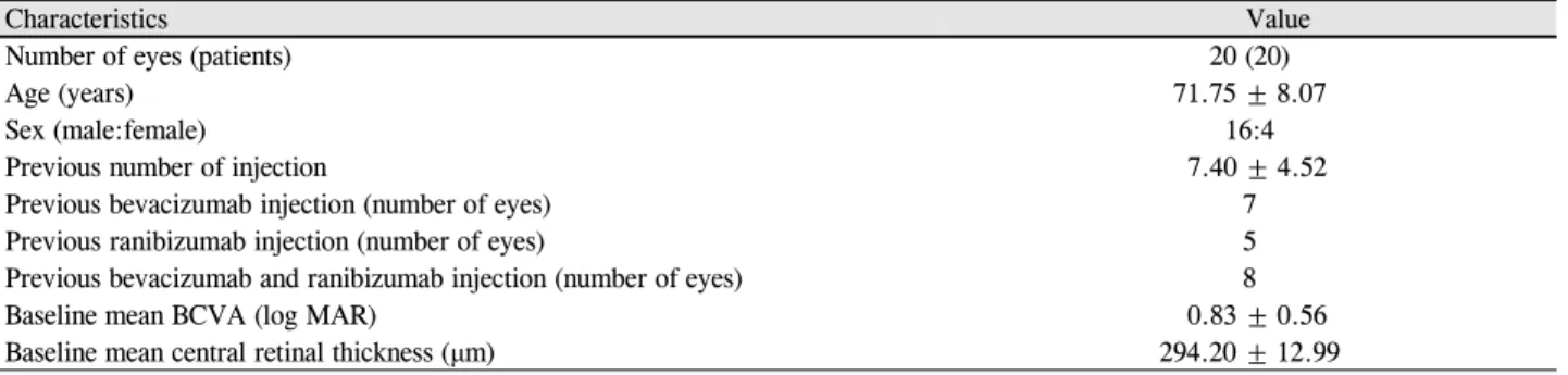
Table 1. Dermographic features and patient’s characteristics at baseline
Characteristics Value
Number of eyes (patients) 20 (20)
Age (years) 71.75 ± 8.07
Sex (male:female) 16:4
Previous number of injection 7.40 ± 4.52
Previous bevacizumab injection (number of eyes) 7
Previous ranibizumab injection (number of eyes) 5
Previous bevacizumab and ranibizumab injection (number of eyes) 8
Baseline mean BCVA (log MAR) 0.83 ± 0.56
Baseline mean central retinal thickness (μm) 294.20 ± 12.99
Values are presented as mean ± SD unless otherwise indicated.
SD = standard deviation; BCVA = best corrected visual acuity; log MAR = logarithm of the minimum angle of resolution.
이후 애플리버셉트(Eylea®, Regeneron, Tarrytown, New York, NY, USA and Bayer HealthCare, Berlin, Germany)는 수용성 재조합 데코이(decoy) 혈관내피성장인자 수용체로 immunoglobulin G-1 (IgG1)의 불변 fragment crystallizable (Fc) 부위에 융합된 VEGF 수용체-1, 2의 주요 세포 외 VEGF 결합 도메인을 포함하도록 설계되었다.7-11 VEGF-A 만 억제하는 베바시주맙과 라니비주맙과는 달리 애플리버 셉트는 VEGF-B와 placental growth factor (PlGF)에도 결합 하여 시험관내 실험에서 베바시주맙과 라니비주맙에 비해 서 더 높은 결합친화도를 보인다.12-14
국내에서는 아직 애플리버셉트를 사용한 연구결과가 보 고되지 않았기에 삼출성 나이관련황반변성 환자들 중에 기 존에 베바시주맙과 라니비주맙 치료를 시행하였지만 이에 반응하지 않는 환자를 대상으로 애플리버셉트로 교체하여 유리체강내 주입술을 시행하고 단기적 임상결과를 알아보 고자 하였다.
대상과 방법
2014년 10월부터 2015년 3월까지 기존에 나이관련황반 변성으로 진단 받고 유리체강내 베바시주맙과 라니비주맙 주입술을 시행 받은 환자에서 애플리버셉트로 교체하여 유 리체강내 주입술을 시행 받은 환자를 대상으로 후향적 의 무기록 분석을 시행하였다.
대상은 수차례 유리체강내 베바시주맙과 라니비주맙 주 입술을 시행 받고 이에 반응하지 않아 CIRRUSTM HD-OCT 4000 (Carl Zeiss, Jena, Germany)을 이용하여 빛간섭단층촬 영에서 망막하액, 망막내액 또는 망막색소상피박리가 유지 되는 환자로 총 20명 20안의 데이터를 분석에 사용하였다.
환자를 대상으로 연령, 성별, 기존에 유리체강내 베바시 주맙과 라니비주맙 주입술을 시행 받은 횟수, 기존 및 주사 후 1개월째 최대 교정시력과 기존 및 주사 후 1개월째 중심 망막 두께를 측정하였다. 최대 교정시력은 통계분석을 위
해 logMAR 시력으로 변환하였다.
유리체강내 애플리버셉트 주입술은 수술실에서 염산프 로파라카인 0.5% (Alcain®, Alcon, Fort Worth, TX, USA)로 점안마취 후 5% 포비돈 아이오다인 용액을 이용하여 결막 낭을 소독한 후 30게이지 주사바늘을 이용하여 각막윤부에 서 3.5 mm 떨어진 평면부를 통해 애플리버셉트 0.5 mg을 유리체강내로 주입하였다.
통계학적 분석은 SPSS version 18.0 software package (SPSS Inc. Chicago, IL, USA)를 사용하여 Paired t-test를 이용하였고 p값이 0.05 미만인 경우를 통계학적으로 의의 가 있는 것으로 판단하였다.
결 과
기존에 베바시주맙이나 라니비주맙 치료를 받은 환자에 서 해부학적으로 반응이 좋지 않았던 환자 20명 20안의 평 균 연령은 71.75 ± 8.07세였으며, 남자는 16명, 여자는 4명 이었다. 유리체강내 애플리버셉트 주입술 전 대상안의 평 균 최대 교정시력은 0.83 ± 0.56 logMAR였으며 평균 중심 망막 두께는 294.20 ± 12.99 μm였다. 기존에 베바시주맙과 라니비주맙의 유리체강내 총 주사 횟수는 7.40 ± 4.52회였 다(Table 1). 빛간섭단층촬영과 안저검사에서 망막내액 5 안, 망막하액 14안, 그리고 망막색소상피박리 10안이 관찰 되었으며 망막내액과 망막색소상피박리를 동반한 경우 1 안, 망막하액과 망막색소상피박리를 동반한 경우 6안, 망막 내액과 망막하액 그리고 망막색소상피박리를 동반한 경우 1안이었다.
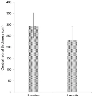
애플리버셉트로 전환하여 유리체강내 주입술을 시행하고 1 개월째 대상안의 평균 최대 교정시력은 0.86 ± 0.61 logMAR 로 애플리버셉트로 전환하기 전 시력과 통계적으로 유의 하게 변화하지 않았다(p=0.406). 하지만 유리체강내 애플 리버셉트 주입술을 시행하고 1개월째 평균 중심 망막 두께 는 232.45 ± 12.05 μm로 전환 이전에 비해 통계적으로 유
Table 2. Comparison of visual acuity, central macular thickness between at baseline and at 1 month after intravitreal aflibercept in-
jectionBaseline At 1 month p-value*
Visual acuity (log MAR) 0.83 ± 0.56 0.86 ± 0.61 0.406
Central retinal thickness (μm) 294.20 ± 12.99 232.45 ± 12.05 0.011
Values are presented as mean ± SD unless otherwise indicated.
SD = standard deviation.
*Paired t-test.
Table 3. Optical coherence tomography analysis after intravitreal aflibercept injection at 1 month
Number of eyes Resolution (%) Unchanged (%) Worse (%)
IRF 5 4 (80) 1 (20) 0 (0)
SRF 14 11 (78.6) 3 (21.4) 0 (0)
PED 10 5 (50) 5 (50) 0 (0)
IRF = intraretinal fluid; SRF = subretinal fluid; PED = pigment epithelial detachment.
Figure 2. A patient treated with 10th intravitreal bevacizumab and 9th intravitreal ranibizumab injection. Optical coherence tomog-
raphy (OCT) showed persistent intraretinal (IRF), subretinal fluid (SRF) and pigment epithelial detachment (PED) (A). 1 month af- ter the first intravitreal aflibercept injection, OCT shows complete absorption of IRF, SRF with decreased PED (B).0 50 100 150 200 250 300 350 400
Baseline 1 month
Central retinal thickness (µm)
Figure 1. Change in central retinal thickness from baseline to
1 month after intravitreal aflibercept injection who were re- sistant to treatment with bevacizumab and ranibizumab in neo- vascular age-related macular degeneration (Paired t-test, p = 0.011).의한 감소가 관찰되었다(p=0.011) (Table 2) (Fig. 1).
유리체강내 애플리버셉트 주입술을 시행하고 1개월째 망 막내액의 감소를 보인 경우는 4안(80%)이었으며 완전 흡수 를 보인 경우는 1안이 관찰되었다. 망막하액의 감소를 보인 경우는 11안(78.6%)이었으며 완전 흡수를 보인 경우는 9안 이 관찰되었다. 망막색소상피박리의 감소를 보인 경우는 5 안(50%)이 관찰되었다. 망막내액과 망막색소상피박리가 동 반된 1안에서 망막내액은 감소, 망막색소상피박리는 변화 가 없었으며, 망막하액과 망막색소상피박리가 동반된 6안 에서 망막하액의 감소를 보인 경우는 5안(83.3%), 망막색소 상피박리의 감소를 보인 경우는 4안(66.7%)이었다. 망막내 액과 망막하액 그리고 망막색소상피박리가 동반된 1안에서 망막내액, 망막하액, 망막색소상피박리 모두 감소하였다.
유리체강내 애플리버셉트 주입술 후 망막내액, 망막하액, 그리고 망막색소상피박리가 증가하거나 새로 발생한 경우 는 없었다(Table 3) (Fig. 2). 유리체강내 애플리버셉트 주입 술 후 특별한 합병증 및 안내염이 발생한 환자는 없었다.
A B
고 찰
혈관내피세포성장인자는 신생혈관증식을 유발하는 중요 한 역할을 하는 인자로 밝혀지면서 이에 대한 항혈관내피 세포성장인자가 나이관련황반변성에서 맥락막신생혈관 치 료에 이용되고 있다.15 기존에 두 가지 혈관내피세포성장인 자 억제제로 베바시주맙은 혈관내피성장인자에 대한 인간 화 단일 클론 항체(full-length monoclonal antibody), 라니비 주맙은 항원결합분절(Fab)로만 구성된 인간화 단일 콜론 항체로 사용되어 왔다.16 애플리버셉트는 수용성 재조합 데 코이(decoy) 혈관내피성장인자 수용체로 VEGF-A만 억제 하는 베바시주맙과 라니비주맙과는 달리 애플리버셉트는 VEGF-B와 placental growth factor (PlGF)에도 결합하여 시 험관 내 실험에서 베바시주맙과 라니비주맙에 비해서 더 높은 결합친화도를 보인다.12-14
본 연구에서는 기존에 베바시주맙과 라니비주맙 치료를 시행하였지만 이에 반응하지 않는 환자를 대상으로 하여 애플리버셉트로 전환하여 유리체강내 주입술을 시행하였다.
1개월째 경과관찰을 한 결과, 평균시력의 호전은 없었지만, 평균 중심 망막 두께는 애플리버셉트 치료 전(294.20 ± 12.99 μm)에 비해 유의하게 감소하여(232.45 ± 12.05 μm, p=0.011) 해부 학적 호전이 있음을 관찰할 수 있었다. 기존 치료에 반응하 지 않는 한 가지 원인으로는 베바시주맙과 라니비주맙을 반복적으로 사용하면서 생긴 과내성으로 생각되며 다른 항 혈관내피세포성장인자인 애플리버셉트로 전환을 하면서 해부학적 호전을 보였을 것으로 생각한다. 과내성은 항혈 관내피세포성장인자의 반복적인 치료에 의해 약제에 대한 반응이 저하되는 현상으로 섬유화, 광수용체층과 망막색소 상피층의 만성적인 변화, 대식세포의 이동과 활성의 증가 로 인한 VEGF의 발현 증가, 그리고 VEGF 수용체의 발현 증가를 포함하는 신생혈관막의 구조적인 변화에 의한 것으 로 알려져 있다.17,18 Gasperini et al18은 베바시주맙이나 라 니비주맙에 반응이 약화된 25명의 환자를 대상으로 다른 혈관내피세포성장 억제제를 투여하였을 때 81%에서 망막 하액의 감소를 보였다고 보고하여 애플리버셉트에 대한 해 부학적 호전을 뒷받침한다.
또한 애플리버셉트는 VEGF-A뿐만이 아닌 VEGF-B와 PlGF에도 결합하여 더 높은 결합친화도를 보여 베바시주맙 과 라니비주맙에 반응하지 않는 환자에서 해부학적 호전을 보인 원인으로 생각해볼 수 있다. Holash et al9의 연구에서 애플리버셉트의 VEGF에 대한 결합친화도(Kd=0.5 ρM)가 베바시주맙(Kd=58 ρM)과 라니비주맙(Kd=46 ρM)에 비해 상당히 크다고 보고하였다.
본 연구에서 애플리버셉트로 전환하고 1개월째 망막내액
의 감소를 보인 경우 4안(80%), 망막하액의 감소를 보인 경 우 11안(78.6%), 망막색소상피박리의 감소를 보인 경우 5 안(50%)이 관찰되었으며 망막내액과 망막색소상피박리가 동반된 1안에서 망막내액은 감소, 망막색소상피박리는 변 화가 없었으며, 망막하액과 망막색소상피박리가 동반된 6 안에서 망막하액의 감소를 보인 경우 5안(83.3%), 망막색소 상피박리의 감소를 보인 경우 4안(66.7%)이었으며, 망막내 액과 망막하액 그리고 망막색소상피박리가 동반된 1안에서 망막내액, 망막하액, 망막색소상피박리 모두 감소하였다.
유리체강내 애플리버셉트 주입술 후 망막내액, 망막하액, 그리고 망막색소상피박리가 증가하거나 새로 발생한 경우 는 없었다. Cho et al19은 베바시주맙과 라니비주맙에 반응 하지 않는 환자 28명을 대상으로 애플리버셉트로 전환하고 1개월째 망막하액의 감소를 보인 경우 19안(86%), 망막내 액의 감소를 보인 경우 7안(86%), 망막색소상피박리의 감 소를 보인 경우 6안(50%)으로 보고하였다. Ho et al20은 치 료에 반응하지 않는 삼출성 나이관련황반변성 환자 96명을 대상으로 애플리버셉트 3회 주사 후 망막내액의 감소를 보 인 경우 18안(58%), 망막하액의 감소를 보인 경우 29안 (59%), 망막색소상피박리의 감소를 보인 경우가 20안(27%) 으로 보고하였다. 기존의 연구와 본 연구의 결과가 크게 차 이가 없음을 확인할 수 있었으며 망막하액과 망막내액의 감소된 빈도에 비해 망막색소상피박리의 감소 빈도가 적은 경향도 일치하였다. 나이관련황반변성에서 망막색소상피박 리는 브루크막과 맥락막의 수리전도도의 감소, 맥락막과 망막색소상피 사이의 수분 교환 능력의 감소로 발생하며, 망막하와 망막색소상피하 공간에 인터루킨, 보체 등의 사 이토카인들과 활성화된 대식세포에서 분비된 혈관내피세 포성장인자들에 의해 투과성의 변화가 생겨 망막하액과 망 막내액에 비해 망막색소상피박리의 감소가 애플리버셉트 치료에 대한 반응이 좋지 못한 것으로 생각된다.21-23
연구의 한계점으로 본 연구가 후향적 연구이며 대상 환 자군이 20명으로 비교적 적으며 애플리버셉트로 전환 후 1 개월째 단기 효과에 국한되어 있다. 추후 장기간의 추적관 찰을 통해 평균시력의 변화, 해부학적인 호전의 지속 여부 의 비교가 필요할 것이다.
결론적으로, 베바시주맙과 라니비주맙에 반응하지 않는 삼 출성 나이관련황반변성 환자에서 애플리버셉트는 혈관내피 세포성장인자 억제제의 다른 대안 치료제로 유용하며 치료 계획 수립에 있어 많은 도움을 줄 수 있을 것으로 생각한다.
REFERENCES
1) Friedman DS, O'Colmain BJ, Muñoz B, et al. Prevalence of age-re-
lated macular degeneration in the United States. Arch Ophthalmol 2004;122:564-72.
2) Augood C, Fletcher A, Bentham G, et al. Methods for a pop- ulation-based study of the prevalence of and risk factors for age-re- lated maculopathy and macular degeneration in elderly European populations: the EUREYE study. Ophthalmic Epidemiol 2004;
11:117-29.
3) Resnikoff S, Pascolini D, Etya'ale D, et al. Global data on visual impairment in the year 2002. Bull World Health Organ 2004;
82:844-51.
4) Au Eong KG. Age-related macular degeneration: an emerging challenge for eye care and public health professionals in the Asia Pacific region. Ann Acad Med Singapore 2006;35:133-5.
5) Frampton JE. Ranibizumab: a review of its use in the treatment of neovascular age-related macular degeneration. Drugs Aging 2013;30:331-58.
6) Schouten JS, La Heij EC, Webers CA, et al. A systematic review on the effect of bevacizumab in exudative age-related macular degeneration. Graefes Arch Clin Exp Ophthalmol 2009;247:1-11.
7) Heier JS, Brown DM, Chong V, et al. Intravitreal aflibercept (VEGF trap-eye) in wet age-related macular degeneration. Ophthalmology 2012;119:2537-48.
8) Browning DJ, Kaiser PK, Rosenfeld PJ, Stewart MW. Aflibercept for age-related macular degeneration: a game-changer or quiet ad- dition? Am J Ophthalmol 2012;154:222-6.
9) Holash J, Davis S, Papadopoulos N, et al. VEGF-Trap: a VEGF blocker with potent antitumor effects. Proc Natl Acad Sci U S A 2002;99:11393-8.
10) Papadopoulos N, Martin J, Ruan Q, et al. Binding and neutraliza- tion of vascular endothelial growth factor (VEGF) and related li- gands by VEGF Trap, ranibizumab and bevacizumab. Angiogenesis 2012;15:171-85.
11) Dixon JA, Oliver SC, Olson JL, Mandava N. VEGF Trap-Eye for the treatment of neovascular age-related macular degeneration.
Expert Opin Investig Drugs 2009;18:1573-80.
12) Stewart MW, Rosenfeld PJ. Predicted biological activity of intra- vitreal VEGF Trap. Br J Ophthalmol 2008;92:667-8.
13) Chappelow AV, Kaiser PK. Neovascular age-related macular de- generation: potential therapies. Drugs 2008;68:1029-36.
14) Rudge JS, Thurston G, Davis S, et al. VEGF trap as a novel anti- angiogenic treatment currently in clinical trials for cancer and eye diseases, and VelociGene- based discovery of the next generation of angiogenesis targets. Cold Spring Harb Symp Quant Biol 2005;
70:411-8.
15) Ferrara N. Vascular endothelial growth factor: basic science and clinical progress. Endocr Rev 2004;25:581-611.
16) Ferrara N, Damico L, Shams N, et al. Development of ranibizu- mab, an anti-vascular endothelial growth factor antigen binding fragment, as therapy for neovascular age-related macular degeneration.
Retina 2006;26:859-70.
17) Binder S. Loss of reactivity in intravitreal anti-VEGF therapy: ta- chyphylaxis or tolerance? Br J Ophthalmol 2012;96:1-2.
18) Gasperini JL, Fawzi AA, Khondkaryan A, et al. Bevacizumab and ranibizumab tachyphylaxis in the treatment of choroidal neo- vascularisation. Br J Ophthalmol 2012;96:14-20.
19) Cho H, Shah CP, Weber M, et al. Aflibercept for exudative AMD with persistent fluid on ranibizumab and/or bevacizumab. Br J Ophthalmol 2013;97:1032-5.
20) Ho VY, Yeh S, Olsen TW, et al. Short-term outcomes of aflibercept for neovascular age-related macular degeneration in eyes pre- viously treated with other vascular endothelial growth factor inhibitors. Am J Ophthalmol 2013;156:23-8.e2.
21) Moore DJ, Hussain AA, Marshall J. Age-related variation in the hydraulic conductivity of Bruch's membrane. Invest Ophthalmol Vis Sci 1995;36:1290-7.
22) Hageman GS, Luthert PJ, Victor Chong NH, et al. An integrated hypothesis that considers drusen as biomarkers of immune-medi- ated processes at the RPE-Bruch's membrane interface in aging and age-related macular degeneration. Prog Retin Eye Res 2001;20:
705-32.
23) Oh H, Takagi H, Takagi C, et al. The potential angiogenic role of macrophages in the formation of choroidal neovascular membranes.
Invest Ophthalmol Vis Sci 1999;40:1891-8.
= 국문초록 =
베바시주맙과 라니비주맙에 반응 없는 삼출성 나이관련황반변성에서 애플리버셉트 주입술의 효과
목적: 유리체강내 베바시주맙과 라니비주맙 주입술에 반응하지 않는 삼출성 나이관련황반변성에서 유리체강내 애플리버셉트 주입술 의 효과를 알고자 하였다.
대상과 방법: 나이관련황반변성으로 진단 받고 유리체강내 베바시주맙과 라니비주맙 주입술을 시행 받고 반응이 없는 환자에서 애플 리버셉트로 교체하여 유리체강내 주입술을 시행 받은 20명의 환자를 대상으로 주입술 후 1개월째 시력과 중심 망막두께의 변화를 조사하였다. 또한 망막하액, 망막내액, 망막색소상피박리의 변화를 조사하였다.
결과: 유리체강내 애플리버셉트 주입술 전 대상안의 평균 최대 교정시력은 0.83 ± 0.56 logMAR였으며 평균 중심 망막 두께는 294.20
± 12.99 μm였다. 애플리버셉트로 전환하여 유리체강내 주입술을 시행하고 1개월째 대상안의 평균 최대 교정시력은 0.86 ± 0.61 logMAR로 기존 시력과 통계적으로 유의하게 변화하지 않았고(p=0.406), 평균 중심 망막 두께는 232.45 ± 12.05 μm로 전환 이전에 비해 통계적으로 유의한 감소가 관찰되었다(p=0.011). 유리체강내 애플리버셉트 주입술을 시행하고 1개월째 망막내액의 감소를 보인 경우 4안(80%), 망막하액의 감소를 보인 경우 11안(78.6%), 망막색소상피박리의 감소를 보인 경우 5안(50%)이 관찰되었으며 망막내 액, 망막하액 및 망막색소상피박리가 증가하거나 새로 발생한 경우는 없었다.
결론: 유리체강내 베바시주맙과 라니비주맙 주입술에 반응하지 않는 삼출성 나이관련황반변성 환자에서 애플리버셉트는 유용한 치료 제임을 확인할 수 있었다.
<대한안과학회지 2015;56(9):1359-1364>