The Effect of Acitretin to the Expression of Vascular Endothelial Growth Factor in Psoriasis
Chi Yeon Kim, Seong Min Kim and Gun-Do Kim1*
Department of Dermatology, School of Medicine, Gyeongsang National University, Gyeongsang Institute of Health Science, 92 Chilam-Dong, Chinju 660-702, Korea
1Department of Microbiology, College of Natural Sciences, Pukyong National University, Busan 608-737, Korea Received January 18, 2009 /Accepted March 17, 2009
Psoriasis is a well known disorder of keratinization. In this disease, several reports revealed that der- mal micro vessels are increased and angiogenic factors such as vascular endothelial growth factor (VEGF) and basic fibroblast growth factor (bFGF) are over-expressed. Angiogenesis may play an im- portant role in the progression of psoriasis. Acitretin is widely used as an anti-psoriatic drug because of its potent action on keratinocyte growth and differentiation, but its effects on angiogenesis are uncertain. The goal of this immunohistochemical study was to investigate the effects of acitretin on the expression of VEGF in psoriatic lesions of the skin. We compared the expression levels of VEGF between pre- and post- acitretin treated skin - 10 psoriatic skin lesions and 3 normal (control) skins.
The expressions of VEGF in psoriatic skin lesions were significantly higher than in normal control skin. The expressions of VEGF in psoriatic skin lesions post-treatment were lower than those pre-treatment. Acitretin revealed inhibitory effects on angiogenesis by reducing the expression of an- giogenic factors such as VEGF in psoriatic skin lesions. We suggest that acitretin may be useful in therapeutic approaches to psoriasis management, possibly related to angiogenesis.
Key words : Acitretin, angiogenesis, psoriasis, vascular endothelial growth factor
*Corresponding author
*Tel:+82-51-629-5618, Fax:+82-51-629-5619
*E-mail : [email protected]
Introduction
Psoriasis is a papulosquamous disease with the incidence of 1–2% of population worldwide, and its characteristic is etythema accompanying slivery scales and it is a chronic re- current disases. Histologically, its characteristics are the rete ridge elongation together with proliferation in the under- neath area, and the elongation of the dermal papillar and edema. In the dermal papillar area, Malpighii's layer is thinner than normal, the appearance of spongiotic pustules, the loss of granular layer, abnormal parakeratosis, the for- mation of Munro's microabscess, and the number of blood vessles is increased in pathologic as well as non-pathologic areas [7,25]. In addition, in psoriasis lesions, vascular endo- thelial cells proliferate resulting in the expansion of the pat- tern of capillary blood vessels or the appearance of im- mature blood vessels with a pattern of abnormally twisted, and the permeability of blood vessel is increased and thus inflammatory cells can be accumulated [4]. The etiology of psoriasis has not been elucidated yet, however, the hypoth-
esis that it is caused by the alteration of keratinocytes, blood vessels and the interaction of inflammatory cells has been accepted currently [1]. The theory that the alteration of the dermis in response to stimulations such as antigens, toxins, and trauma proceeded, and as its secondary re- sponse, the alteration of the epidermis such as the abnormal proliferation and differentiation of keratinocytes, etc. is prevalent [6]. In response to stimulation, the release of Interleukin (IL) -2 and Tumor necrosis factor α (TNF-α) from dendritic cells in the dermis is increased, con- sequently, T cells and endothelial cells are activated. The ac- tivation of these cells induces the release of IL-8 and stim- ulates the proliferation of keratinocytes [16]. In such man- ners, as the causality of psoriasis, the alteration of the der- mis appears to affect the alteration of the epidermis. In ad- dition, normal human keratinocytes in tissue culture ex- press angiogenetic factor vascular endothelial growth factor (VEGF)/vascular permeability factor (VPF) [8], and the stimulation of keratinocytes with transforming growth fac- tor α (TGF-α) that is increased in psoriasis increases VEGF/VPF. The increased VEGF reacts with its receptor on endothelial cells in the dermis and increases vascular per- meability, which induces the invasion of a number of neu-
trophils to psoriasis lesions, and the proliferation of kerati- nocytes is induced again [11,15]. Hence, in regard to the ab- normal proliferation of keratinocytes in psoriasis, the abnor- mal formation of the blood vessel by angiogenetic factor vascular endothelial growth factor (VEGF)/vascular perme- ability factor (VPF) is considered to mediate such effect, al- though its mechanism is not elucidated yet.
Acitretin, a derivative of Retinoic acid (RA), is ther- apeutics of psoriasis. RA is a subsnce that controls cell growth and differentiation, it induces the proliferation and differentiation of epithelial cells and mesenchymal cells, and it is involved in the apoptosis of tumor cells [5]. RA was confirmed to suppress the formation of blood vessel induced in tumor tissues [18], and it was reported to sup- press the reaction of angiogenic factors with endothelial cells, or it suppresses the increased phenomenon of migra- tion of endothelial cells induced by tumor cell culture me- dia [24]. As RA controls the proliferation and differ- entiation of keratinocytes, it has been applied as a ther- apeutic of proriasis, and among the effects on cultured ker- atinocytes, it is known to have a function to suppress the angiogenic factor VEGF [10,30], however, in regard to the expression of VEGF in psoriasis lesions, the study on its modulation is not sufficient.
VEGF is a protein that induces the formation of blood vessel by stimulating the proliferation and migration of vascular endothelial cells. The fact that the formation of blood vessel in the dermis in response to the increased VEGF in the blood or in tissues influences the develop- ment of new psoriasis lesiona and mediates its ex- acerbation [13,14], which suggests that therapeutics that suppresses the formation of blood vessel in response to VEGF may be effective as a therapeutic of psoriasis.
Acitretin that has been used primarily for the control of the differentiation of keratinocytes previously not only de- creases the expression of VEGF in cultured keratinocytes [28], but if it decreased the expression of VEGF after ad- ministered to psoriasis lesions, which may be taken as the suggestion of the possibility as a therapeutics of psoriasis mediated by a mechanism different from the suppressor of the formation of blood vessels. Hence, in our study, by comparing with normal, the expression pattern of VEGF that is a factor inducing the formation of blood vessel was assessed, and the effect of the psoriasis therapeutics aci- tretin on psoriasis lesions before after its administration was assessed by immunochemical staining.
Materials and Methods The study population
In 10 patients diagnosed as psoriasis in the department of dermatology from January 2003 and March 2004, tissue biopsy of the lesion area was performed, and age-matching 5 healthy individuals without dermatological diseases were selected as controls. The patients were not treated locally or systemically during the past 2 months minimum. After the treatment with 10 mg Acitretin (Neotigason®, Roche, Korea), tissue biopsy was performed on the improved lesions.
Immunohistochemical test
Immunohistochemical test was performed by using the immunoperoxidase technique (ABC technique). The skin of each patients was fixed in formalin and subsequently pre- pared as a paraffin block, prepared as secial sections 6 µm in thickness, attached to slide glasses, and tissue staining was performed. Initially, paraffin tissue sections were depar- affined by treating with xylene for 3 times, and to remove endogenous peroxidase, treated with 0.3% H2O2 (Sigma, St.
Louis, MO, USA) diluted with methanol for 30 minutes, and washed with 0.02 M phosphate buffered saline (PBS). Next, to perform the antigen retrieval step, tissue slides were add- ed to a beaker filled with Citrate buffer, pH 6.0 (Sigma), treated with microwave processing for 10 minutes, and washed with PBS buffer. Subsequently, the samples were treated with 10 % normal goat serum to block non-specific reactions, and incubated with primary antibody overnight in a refrigerator. After the treatment with primary antibody, tissue sections were washed with PBS sufficiently, and in- cubated with the secondary antibody biotinylated goat an- ti-mouse immunoglobulin (Vector laboratories, Inc., Burlingame, CA) diluted to 1:200 or biotinylated goat anti- rabbit immunoglobulin (Vector laboratories) for 30 minutes, and washed. Next, the sections were treated with Avidin- peroxidase (Vector laboratory, Inc.) for 30 minutes and stained with PBS containing 0.15% H2O2 and 0.01 % 3-3' dia- minobenzidine (Sigma) for 5 minutes, washed with distilled water, counterstained with Harris hematoxylin for 1 mi- nutes, and sealed with Permount. As control, the skin of nor- mal individuals or the skin treated with a similar experiment procedure was used. To assess the distribution of the angio- genic factor VEGF in the lesion of proriasis patients before and after treatment and in the skin of normal individuals, anti-VEGF polyclonal antibody (Abcam, Cambridge, UK)
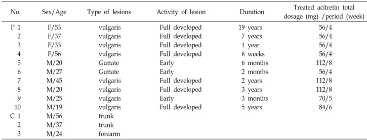
Table 1. Clinical characteristics of psoriasis patients (P) and normal control skin (C)
No. Sex/Age Type of lesions Activity of lesion Duration Treated acitretin total
dosage (mg) /period (week) P 1
2 3 4 5 6 7 8 9 10 C 1 2 3
F/53 F/37 F/33 F/56 M/20 M/27 M/45 M/20 M/25 M/19 M/56 M/37 M/24
vulgaris vulgaris vulgaris vulgaris Guttate Guttate vulgaris vulgaris vulgaris vulgaris trunk trunk forearm
Full developed Full developed Full developed Full developed Early
Early
Full developed Full developed Early
Full developed
19 years 7 years 1 year 6 weeks 6 months 2 months 2 years 3 years 3 months 5 years
56/4 56/4 56/4 56/4 112/8 56/4 112/8 112/8 70/5 84/6 staining was performed.
The evaluation of the result of staining
The expression of fluorescent VEGF in the epidermis, the dermis, vascular endothelial cells and endocrine glands was assessed, and the intensity of staining was classified to 4 grades, from 0 to 3, according to their staining level and evaluated by 3 investigators using a light microscope (under 100× magnification). The cases without staining was classi- fied as 0, locally moderately stained was 1 point, locally strongly stained/diffused moderately was 2 point, and dif- fused strongly was 3 points and graded [12]. The alteration of VEGF before and after the treatment of proriasis was ana- lyzed by Student’s t-test, and the significance was examined based on p<0.05.
Results Clinical characteristic
The age of patients was diverse, from 19 years to 56 years, and the mean age was 33.5 years. Regarding the ratio of male and female, male was 6 cases and female was 4 cases, and the disease duration varied from 6 weeks to 19 years.
The activity of lesion areas was classified based on histo- logical findings as early lesions and sufficiently advanced lesions. The histological findings of early lesion was defined as dyskeratosis with papilla in some area, the decrease of granular layer, the moderate level of acanthosis, the ex- pansion of capillary blood vessel in the dermal papilla, and the invasion of lymphocytes and some neutrophils in the vicinity of blood vessel. Sufficiently advanced lesions was
defined as the typical psoriasis histological findings the elon- gation of the rete ridge regularly, the thinness of Malpighii’s layer in the dermal papilla, the appearance of spongiotic pustures, the loss or reduction of the granular layer, para- keratosis, Munro's microabscess, twisted capillary blood ves- sels, etc. Clinically, among 10 patients, 8 patients were diag- nosed as psoriasis vulgaris and 2 patients were diagnosed as guttate psoriasis. In the patient group, an evident differ- ence of staining according to the gender, age, clinical pattern, the disease duration, the activity in lesions, etc. was not detected. Acitretin was administered total 20 mg per day, divided to 2 times per day, the administered dose was from 56 mg to 112 mg, and the average treatment dosage was 77±25.8 mg. The treatment period was from 4 weeks to 8 weeks, and the average treatment duration was 5.5±1.8 weeks. A specific medical history was not detected, and dur- ing the treatment, except xerotic cheilitis, a specific side ef- fect was not reported (Table 1).
Immunohistochemical test
In the immunohistochemical staining against VEGF, in the normal skin used as negative control, VEGF was detected at the basal layer in epidermal with the intensity 1 whereas in psoriasis lesions, in the entire epidermis layer, the in- tensity 2 was detected. In the vascular epithelial cells in the dermis and secretory glands, the staining intensity was sig- nificantly increased in comparison with control group, the staining intensity was decreased after the treatment with aci- tretin, particularly, the reduction was evident in the entire epidermis (p<0.01), and it was decreased significantly in se- cretory glands (p<0.05). The staining intensity in vascular
Fig. 1. Representative photomicrographs of VEGF immunohisto- chemistry in the normal skin (a), psoriatic lesional skin (b), and acitretin treated psoriatic skin (c). VEGF im- munostaining intensity score of epidermis and dermal endothelial cells was higher (b) than (a) and was de- creased in (c) ×200.
Fig. 2. VEGF immunostaining intensity score in normal skin (Nl), psoriatic lesional skin (PLS), and acitretin treated psoriatic lesional skin (APLS). VEGF immunostaining score was higher PLS than APLS and Nl in keratinocyte (KC), gland (Gl), endothelial cell (EC). Staining results suggested keratinocyte derived VEGF decreased by treated with acitretin(Stain intensity scored as: 0=no stain; 1= weak; 2=moderate; 3=intense, *p<0.01, ‡ p<0.05, Student's t-test).
endothelial cells in the dermis was also decreased. However, it was not statistically significant (Fig. 1, 2).
Discussion
Psoriasis is a benign proliferative dermatosis and a chron- ic recurrent disease with the characteristic of the excess pro- liferation and differentiation of the epidermis, the invasion of inflammatory cells widely in the epidermis and the der- mis, and the alteration of the vascular vessel in the dermis [7,25]. In psoriasis, it has been reported that the change of vascular vessles such as the elongation of the capillary blood vessel in the dermal papilla, the increase of the permeability of blood vessel, the increase of blood flow, etc. are involved in the cause of psoriasis [1,4]. Hence, the abnormal pro- liferation of blood vessel may be considered as a causality of psoriasis, and such abnormality of the blood vessel was considered to mediate the effect on the alteration of the der-
mis such as abnormal proliferation of keratocytes [6,16].
The alteration of blood vessel observed in psoriasis is ap- peared due to the imbalance of the factors controlling the formation of the blood vessels resulting in the proliferation of vascular endothelial cells, migration and the continuous formation of the blood vessels [8,14]. Besides VEGF, angio- genic factors increased in psoriasis are TGF-alpha, IL-8, PD-ECGF, bFGF, HGF/SF, angiopoietin, etc. [17,19,20]. It has been reported that the release of these factors is increased whereas the release of Thrombospondin-1 that is known to suppress the formation of the blood vessel is decreased [27].
It has been reported that in keratinocytes excessively prolif- erated, the expression of VEGF is increased, and the amount of VEGF released is higher than normal resulting in the ab- normal proliferation of the blood vessel [9].
Vascular Endothelial Cell Growth Factor (VEGF) released by keratinocytes is a mitototic accelerator specific to endo- thelial cells, and involved in the development of the neo- vasculature such as the proliferation and migration of endo- thelial cells, the reformation of extracellular matrix, the for- mation of capillary blood vessels, and others [26]. VEGF is synthesized not only by normal cells for example keratino- cytes but also by transformed cells, which plays an im- portant role in the formation of neovasculature such as the development of the coronary vascular system, physiological phenomenon of normal vascular system, rheumatoid arthri- tis, cancer, etc [23]. Considering that VEGF has a signal se- quence that could be expressed extracellularly in contrast to bFGF and other angiogenic factor and thus it can induce the growth of vascular endothelial cells and if it were in- creased, it could form a structure similar to the blood vessel [26], it increases the vascular permeability [2], etc. and it can induce all procedures prerequisite for the formation of neo- vasculature, it shows that among angiogenic factors, VEGF is a protein that particularly plays a central role.
According to the study that used mice with the modified VEGF gene [23], it was confirmed that the administration of VEGF increases the density of subcutaneous blood ves- sels, capillary blood vessels with many folds and branches were formed, and the permeability of endothelial cells was increased. VEGF released acts as a cell proliferation factor specific to endothelial cells and thus increases the pro- liferation of endothelial cells [9], or it has been reported that it enhances the expression of the fibronectin receptor on vas- cular endothelial cells alphaV beta3 and the collagen re- ceptors alpha1 beta 1 and alpha 2 beta 1, and stimulates
the migration of endothelial cells resulting in increasing the formation of blood vessels [29]. In regard to psoriasis, con- sidering the reports that in the papillary layer of the vascular endothelial cells, the expression of the VEGF receptor kdr and flt-1 was increased [8], it may be conceivable that in psoriasis, VEGF is released by keratinocytes and acts on the receptor on vascular endothelial cells resulting into the stim- ulation of the proliferation of blood vessels, and in addition, VEGF released in response to the stimulation in the dermis acts directly as a proliferation factor of endothelial cells in the vascular endothelial cells, stimulates the formation of the blood vessel, and increases the permeability of blood vessel, which facilitates the migration of inflammatory cells to the dermis, and thus it stimulates the proliferation of keratino- cytes in the dermis. Our results showed that the expression of VEGF in psoriasis lesions was enhanced not only in kera- tinocytes but also in vascular endothelial cells and secretory glands, and based on the results, in psoriasis, the expression of VEGF is considered to be an important etiological factor.
Acitretin is Retinoic acid (RA) that is a metabolite of Vitamin A, it controls the proliferation and differentiation of keratinocytes, it has an anti-inflammatory function, and it has been used for dermatological diseases showing para- keratosis such as psoriasis, lamellar ichthyosis, ichthyosis vulgaris, pityriasis rubra pilaris, lichen planus, bullous ich- thyosiform erythroderma, etc [21]. According to the tissue culture study using keratinocytes reported by Weninger et al. [30], in the culture of keratinocytes, the treatment with 10-6 M all-trans RA, 13-cis RA or all-trans retinol decreases the amount of VEGF released by 50~60%.
In our study, taken into account that during the normal- ization of the differentiation of keratinocytes, it was ob- served that the release of VEGF is reduced, VEGF in psor- iasis lesions is altered before and after acitretin admin- istration, and in the epidermis and dermis of psoriasis, the release of VEGF that was increased in vascular endothelial cells and secretory glands was reduced. In regard to the eti- ology of psoriasis, the excess proliferation of keratinocytes is a major causality, and the effect of RA on its treatment is well known. In our study, considering that the formation of blood vessels is an important factor of the etiology psor- iasis and the importance of VEGF as angiogenic factor perti- nent to it, the enhancement of the expression of VEGF in psoriasis lesions was evaluated. In addition, efforts have been made to confirm that after the administration of the psoriasis therapeutic acitretin, the reduction of VEGF in the
epidermis and the dermis of psoriasis lesions where the ex- cessive proliferation of the epidermis was observed, and to suggest the suppressive effect on the proliferation of blood vessels that as a therapeutic action of acitretin. Regarding RA suppressing the formation of blood vessels, it has been reported that in vitro culture, by controlling the transcription of cells through the nuclear receptor RAR and the receptor RXR, it suppresses the proliferation of endothelial cells or decreases the synthesis of enzymes degrading extracellular matrix by endothelial cells resulting in the suppression of neovascularization, and in vivo, it is known to suppress the neovascularization and the vascular permeability [3,22,30], nevertheless, regarding acitretin, a RA, it is rare to assess its effect on the reduction of the vascularization or the change of angiogenic factors in psoriasis lesions. In our study as well as previous reports, we believe that in psoriasis patients administered acitretin as a therapeutic to suppress the formation of blood vessels, even before the thickened epidermis becoming normal, it blocks the steps that released VEGF acting on vascular endothelial cells directly, or di- rectly controls the blood vessel formation genes in vascular endothelial cells, and thus inhibits the formation of blood vessels. In our study, we encountered several problems in the assessment of the change of VEGF in psoriasis lesion before and after acitretin treatment. First, it was believed that the thickness of sections might affect the intensity of staining. Although an identical tome was used, the thickness may vary, consequently, it was believed that it might affect the intensity of staining. Second, the duration of the storage of tissues varied, and thus the fixation state of tissues may affect staining. In the cases of vascular endothelial cells, as it was damaged during the process of tissue section and preparation, it was difficult to evaluate. Hence, it is believed that additional studies of the quantitative analysis of the change of VEGF in tissues before and after the treatment with acitretin are required.
Hence, it suggests the possibility that the therapeutic ef- fect of acitretin as an inhibitor of the formation of blood vessel may be anticipated. In addition, in the treatment of psoriasis, besides the therapeutic approach of treating the excessive proliferation of keratin layer, the suppression of the formation of blood vessel may be an approach. In regard to the mechanism of acitretin suppressing the formation of blood vessel induced by VEGF, it is believed that in depth researches on various signal transduction systems in vitro and in vivo are required, and the result of our research may
serve as the basic information for future studies.
Based on research results, the fact known presently, be- sides the action of acitretin to normalize the differentiation of keratinocytes showing excessive proliferation in psoriasis lesions, by decreasing the expression of VEGF in keratino- cytes, vascular endothelial cells in the dermis and secretory glands, it was found that it functions to suppress the for- mation of blood vessels.
In our study, the results of immunohistochemical staining showed that in psoriasis lesion, the expression of the angio- genic factor VEGF was increased in the epidermis of psor- iasis lesions, vascular endothelial cells and secretory glands.
In addition, after the administration of acitretin that has been widely used as psoriasis therapeutic for controlling the dif- ferentiation of keratinocytes, the expression of VEGF in the lesions was decreased. Hence, in psoriasis, as therapeutic re- actions of acitretin for psoriasis, not only the suppression of the formation of blood vessels could be anticipated, fur- thermore, it suggests that it may be possible to apply it as a therapeutic for other diseases with the enhanced formation of blood vessels, and continuous studies on it may be required.
Acknowledgment
This work was supported by the Pukyong National University research fund in 2004.
References
1. Anderson, T. F. and J. Woorhees. 1986. Psoriasis. pp. 67-84, In Thiers, B. and W. Dobson (eds.), Pathogenesis of skin disease. McGraw-Hill book, New York.
2. Berse, B., L. F. Brown, L. Van De Water, H. F. Dvorak, and D. R. Senger. 1992. Vascular permeability factor (vascular endothelial growth factor) gene is expressed differentially in normal tissues, macrophages, and tumors. Mol. Biol. Cell 3, 211-220.
3. Braunhut, S. J. and M. A. Moses. 1994. Retinoids modulate endothelial cell production of matrix-degrading proteases and tissue inhibitors of metalloproteinases (TIMP). J. Biol.
Chem. 269, 13472-13479.
4. Braverman, I. M. and A. Keh-Yen. 1986. Three-dimensional reconstruction of endothelial cell gaps in psoriatic vessels and their morphologic identity with gaps produced by the intradermal injection of histamine. J. Invest. Dermatol. 86, 577-581.
5. Camacho, L. H. 2003. Clinical applications of retinoids in cancer medicine. J. Biol. Regul. Homeost. Agents. 17, 98-114.
6. Christopher, F. and G. G. Krueger. 1987. Psoriasis. pp.
461-491, In Fitzpatrick, T. B., A. Z. Eisen, and D. Wolff (eds.), Dermatology in general medicine, 3rd ed, MaGraw-Hill Book, New York.
7. Christophers, E., R. Parzefall, and O. Braun-Falco. 1973.
Initial events in psoriasis: quantitative assessment. Br. J.
Dermatol. 89, 327-334.
8. Detmar, M., L. F. Brown, K. P. Claffey, K. T. Yeo, O. Kocher, R. W. Jackman, B. Berse, and H. F. Dvorak. 1994.
Overexpression of vascular permeability factor/vascular endothelial growth factor and its receptors in psoriasis. J.
Exp. Med. 180, 1141-1146.
9. Detmar, M., K. T. Yeo, J. A. Nagy, L. Van de Water, L.
F. Brown, B. Berse, B. M. Elicker, S. Ledbetter, and H. F.
Dvorak. 1995. Keratinocyte-derived vascular permeability factor (vascular endothelial growth factor) is a potent mi- togen for dermal microvascular endothelial cells. J. Invest.
Dermatol. 105, 44-50.
10. Diaz, B. V., M. C. Lenoir, A. Ladoux, C. Frelin, M.
Demarchez, and S. Michel. 2000. Regulation of vascular en- dothelial growth factor expression in human keratinocytes by retinoids. J. Biol. Chem. 275, 642-650.
11. Dvorak, H. F., L. F. Brown, M. Detmar, and A. M. Dvorak.
1995. Vascular permeability factor/ vascular endothelial growth factor, microvascular hyperpermeability, and angiogenesis. Am. J. Pathol. 146, 1029-1039.
12. Edward, R. S., N. Mark, C. W. James, K. S. Andres, L.
Samuel, and H. Meenhard. 1999. Vascular endothelial growth factor is a marker of tumor invasion and metastasis in squamous cell carcinomas of the head and neck. Clin.
Cancer Res. 5, 775-782.
13. Ferrara, N. 1999. Role of vascular endothelial growth factor in the regulation of angiogenesis. Kidney Int. 56, 794-814.
14. Folkman, J. and M. Klagsburn. 1987. Angiogenic factors.
Science 235, 442-447.
15. Gille, J., M. Khalik, V. Konig, and R. Kaufmann. 1998.
Hepatocyte growth factor/scatter factor (HGF/SF) induces vascular permeability factor (VPF/VEGF) expression by cultured keratinocytes. J. Invest. Dermatol. 111, 1160-1165.
16. Gottlieb, A. B. 1997. Immunopathogenesis of psoriasis. Arch.
dermatol. 133, 781-782.
17. Ishikawa, F, K. Miyazono, and U. Hellman. 1989.
Identification of anangiogenic activity and the cloning and expression of platelet derived endothelial cell growth factor.
Nature 338, 557-562.
18. Kini, A. R., L. A. Peterson, M. S. Tallman, and M. W.
Lingen. 2001. Angiogenesis in acute promyelocytic leuke- mia: induction by vascular endothelial growth factor and inhibition by all-trans retinoic acid. Blood 97, 3919-3924.
19. Koch, A. E., P. J. Polverini, S. L. Kunkel, L. A. Harlow, L.
A. DiPietro, V. M. Elner, S. G. Elner, and R. M. Strieter.
1992. Interleukin-8 as a macrophage-derived mediator of angiogenesis. Science 58, 1798-1801.
20. Kuroda, K., A. Sapadin, T. Shoji, R. Fleischmajer, and M.
Lebwohl. 2001. Altered expression of angiopoietins and Tie2 endothelium receptor in psoriasis. J. Invest. Dermatol. 116,
713-720
21. Lachgar, S., M. Charveron, Y. Gall, and J. L. Bonafe. 1999.
Inhibitory effects of retinoids on vascular endothelial growth factor production by cultured human skin keratinocytes. Dermatology 199, 25-27.
22. Lansink, M, P. Koolwijk, V. van Hinsbergh, and T. Kooistra.
1998. Effect of steroid hormones and retinoids on the for- mation of capillary-like tubular structures of human micro- vascular endothelial cells in fibrin matrices is related to ur- okinase expression. Blood 92, 927-938.
23. Larcher, F., R. Murillas, M. Bolontrade, C. J. Conti, and J.
L. Jorcano. 1998. VEGF/VPF overexpression in skin of transgenic mice induces angiogenesis, vascular hyper- permeability and accelerated tumor development. Oncogene 17, 303-311.
24. Lingen, M. W., P. J. Polverini, and N. P. Bouck. 1996.
Retinoic acid induces cells cultured from oral squamous cell carcinomas to become anti-angiogenic. Am. J. Pathol. 149, 247-258.
25. Mordovtsev, V. N. and V. I. Albanova. 1989. Morphology of skin microvasculature in psoriasis. Am. J. Dermatopathol.
11, 33-42.
26. Neufeld, G., T. Cohen, S. Gengrinovitch, and A. Poltorak.
1999. Vascular endothelial growth factor (VEGF) and its receptors. FASEB J. 13, 9-22.
27. Nickoloff, B. J., R. S. Mitra, J. Varani, V. M. Dixit, and P.
J. Polverini. 1994. Aberrant production of interleukin-8 and thrombospondin-1 by psoriatic keratinocytes mediates angiogenesis. Am. J. Pathol. 44, 820-828.
28. Pal, S., M. L. Iruela-Arispe, V. S. Harvey, H. Zeng, J. A.
Nagy, H. F. Dvorak, and D. Mukhopadhyay. 2000. Retinoic acid selectively inhibits the vascular permeabilizing effect of VPF/VEGF, an early step in the angiogenic cascade.
Microvasc. Res. 60, 112-120.
29. Senger, D. R., K. P. Claffey, J. E. Benes, and C. A. Perruzzi, A. P. Sergiou, and M. Detmar. 1997. Angiogenesis promoted by vascular endothelial growth factor: regulation through alpha1beta1 and alpha2beta1 integrins. Proc. Natl. Acad. Sci.
USA 94, 13612-13617.
30. Weninger, W., M. Rendl, M. Mildner, and E. Tschachler.
1998. Retinoids downregulate vascular endothelial growth factor/vascular permeability factor production by normal human keratinocytes. J. Invest. Dermatol. 111, 907-911.
초록:건선(psoriasis)에서 혈관내피 성장인자(VEGF)에 대한 acitretin의 효과
김지연․김성민․김군도1*
(경상대학교 의과대학 피부과학교실 & 의학연구소, 1부경대학교 자연과학대학 미생물학과)
건선(psoriasis)은 각화(keratinization)의 장애에 의한 질병으로 잘 알려져 있다. 이 질병에있어서, 몇몇 보고에 따르면 피부 미세혈관이 증가되고, 혈관 신생 유도 인자들인 내피 성장 인자(VEGF) 및 섬유아세포 성장 인자(bFGF) 가 과발현된다고 보고 되어져 있다. 신생혈관(angiogenesis)은 건선의 진행성에 있어 중요한 역할을 할지도 모른다.
Acitretin은 keratinocyte 성장과 분화에 그것의 잠재적인 활동 때문에 anti-psoriatic 약으로 널리 이용된다. 그러나 신생혈관에 대한 효과는 여전히 불명확하다. 본 면역혈청학적 연구 목표는 건선염 피부에 있는 VEGF의 발현에 acitretin의 효과를 조사하기 위한 것이었다. 우리는 10명의 건선염 피부환자와 대조군으로 3명의 정상인 피부에 acitretin의 치료 전과 후의 VEGF의 발현량을 비교하였다. 치료 전 대조군인 정상인 피부에서보다 건선염 피부환자 에서 VEGF의 발현은 명백히 높은 수준이었다. 건선염 피부환자에서 VEGF의 발현량은 acitretin 치료 후 치료 전과 비교시 감소되었다. Acitretin은 건선 피부에서 VEGF와 같은 혈관 신생 유도 인자의 발현을 감소 시켜 신생혈 관형성에 있어 억제의 효과가 있음을 나타낸다. 우리는 신생혈관 관점에서 acitretin이 건선에 대한 치료 수단이 될 수 있다는 것을 제안한다.