3)
접수일자: 2004년 7월 23일 통과일자: 2005년 3월 4일
책임저자: 권수경, 인제대학교 의과대학 동래백병원 내과
갑상샘 종양에서 Vascular Endothelial Growth Factor (VEGF) 발현의 의의
인제대학교 의과대학 내과학교실, 고신대학교 의과대학 내과학교실1, 병리학교실2
권수경․최영식1․박요한1․장희경2
Meanings of Expression of Vascular Endothelial Growth Factor in Thyroid Tumors
Su Kyoung Kwon, Young Sik Choi1, Yo Han Park1, Hee Kyung Jang2
Department of Internal Medicine, Inje University College of Medicine, Department of Internal Medicine, Kosin University College of Medicine1, Department of Pathology, Kosin University College of Medicine2
ABSTRACT
Background: Angiogenesis is essential for tumor growth and metastasis. Vascular endothelial growth factor (VEGF), also known as vascular permeability factor (VPF), is an angiogenic factor that plays important roles in tumor growth. Angiogenesis studies on VEGF deal with various types of malignant tumors, but little is known about the role or significance of VEGF in human thyroid neoplasms. Therefore, this study was performed to determine whether the VEGF expression in different histological types of thyroid tumors is altered and to see if there was a relationship between the expression of VEGF and either metastasis or the invasiveness of thyroid carcinomas.
Methods: Forty-two cases that underwent thyroidectomy at Kosin Medical Center, between March, 1999 and February, 2000, were included in this study. Of the 42 cases, 27 were malignant (26 papillary carcinoma, 1 Hürthle cell carcinoma) and 15 were benign lesions. The expression of VEGF was determined by immunohistochemistry using paraffin embedded thyroid tissue blocks, and was quantified as negative (absent), + (1~24%), ++ (25~49%), +++ (50~74%) and ++++ (≥75%), according to the extent of positive cells.
Results: VEGF was stained with red-brown colored granules in the cytoplasm of the thyroid tumor epithelium and was expressed in 27 of the 42 cases (+ 1, ++ 8, +++ 5, ++++ 13). Most malignant tumors (24 of 27 cases) were stained with VEGF, but only 3 of the 15 benign tumors cases were stained (P <0.001). When the VEGF expression was divided into ++ or below and +++ or above groups, the expression of VEGF was much more extensive in the malignant than benign tumors (P <0.001). Of the 27 malignant tumors cases, lymph node metastasis and/or invasion was noted in 13. VEGF expression was more extensive in malignant tumors with lymph node metastasis and/or invasion than in those without (P <0.001).
Conclusion: In this study, the rate and extent of VEGF expression were greater in the malignant than the benign thyroid tumors, and also the extent of VEGF expression was the extent of VEGF greater in the malignant tumors with lymph node metastasis and/or invasion than those without (J Kor Soc Endocrinol 20:134
~141, 2005).
ꠏꠏꠏꠏꠏꠏꠏꠏꠏꠏꠏꠏꠏꠏꠏꠏꠏꠏꠏꠏꠏꠏꠏꠏꠏꠏꠏꠏꠏꠏꠏꠏꠏꠏꠏꠏꠏꠏꠏꠏꠏꠏꠏꠏꠏꠏꠏꠏꠏꠏꠏꠏꠏꠏꠏꠏꠏꠏꠏꠏꠏꠏꠏꠏꠏꠏꠏꠏꠏꠏꠏꠏꠏꠏꠏꠏꠏꠏꠏꠏꠏꠏꠏꠏꠏꠏꠏꠏꠏꠏꠏꠏꠏꠏꠏꠏꠏꠏꠏ Key Words: Vascular endothelial growth factor, Thyroid tumor, Metastasis, Invasion
서 론
신생혈관형성은 종양의 성장과 전이에 필수적인 과정이 다[1]. 포유동물은 생존을 위해 산소와 영양분의 공급이 반 드시 필요하고 혈관으로부터 100~200 μm을 벗어나면 확산 에 의한 산소 공급이 한계에 달하므로, 종양세포 역시 혈관 의 생성 없이는 수 cm이상의 성장이나 타장기로의 전이를 일으킬 수는 없다[2]. 발생 초기의 종양은 무혈관 상태이나 크기가 1~2 mm3 이상이 되면 종양세포나 종양세포 주위에 침윤된 대식세포 혹은 섬유모세포 등에서 혈관형성을 자극 하는 물질을 분비하여 미세혈관을 증식시킨다[3,4]. 증식된 미세혈관은 종양세포에 영양을 공급하고, 혈관내피세포는 basic fibroblast growth factor (bFGF), insulin like growth factor-2, platelet derived growth factor (PDGF) 등과 같은 성장인자들을 분비하여 종양을 성장시키고[5], urokinase, collagenase, plasminogen activator 등의 분해효소를 생성 하여 종양세포가 주위조직으로 침윤하는데 기여한다[6,7].
혈관형성인자로서 지금까지 밝혀진 것으로는 vascular endo- thelial growth factor (VEGF)-A, VEGF-B, VEGF-C, VEGF- D, acidic fibroblast growth factor (aFGF), bFGF, trans- forming growth factor-α (TGF-α), TGF-β, hepatocyte gro- wth factor (HGF), tumor necrosis factor-α (TNF-α), angio- genin, interleukin-8, PDGF, placental growth factor 등이 있다[8~10]. 그 중에서도 VEGF는 혈관내피세포에 선택적 으로 작용하여 강력한 유사분열을 일으키는 물질로서 생리 적 또는 병적 상태 모두에서 신생 혈관 형성의 가장 핵심적 인 인자이다. Vascular permeability factor (VPF)로 명명하 기도 하며, 1989년에 처음 발견되었고 아미노산 수에 따라 VEGF-121, -165, -189, -206의 네 종류가 존재한다[10~
12]. 주로 산소압 저하에 의해 발현이 증가되나 epidermal growth factor (EGF), TGF-β, keratinocyte growth factor 등의 cytokine과 p53 등의 종양억제유전자에 의해서도 발현 이 조절된다[13~15]. 발현된 VEGF는 혈관내피세포 표면에 있는 flt-1이나 KDR과 같은 수용체와 결합하여 내피세포의 증식 및 내피세포에서 urokinase, collagenase, serine protease 등의 발현을 유도하여 세포외 기질을 분해시켜 혈 관내피세포나 종양세포의 침윤을 용이하게 한다. 이러한 VEGF의 특성으로 인해 유방, 방광, 뇌, 소화기계, 난소 등 의 악성종양에서 VEGF 발현이 이들 악성종양의 국소침윤 과 전이에 관계한다는 연구가 광범위하게 시행되어 왔으나 [16~19], 갑상샘 종양에서의 VEGF 발현에 대한 연구는 드 물고 보고자들 간에 상반된 결과를 제시하기도 하였다 [20,21].
이에 저자들은 양성과 악성 갑상샘 종양에서 VEGF 발현 양상의 차이를 살펴보고, VEGF의 발현과 임상적 예후인자 와의 연관성을 살펴보고자 본 연구를 시행하였다.
대상 및 방법 1. 대상
1999년 3월부터 2000년 2월까지 고신 대학교 병원에서 갑상샘 종양으로 갑상샘 절제술을 시행한 환자의 조직 중 양성종양 15예, 악성종양 27예 (유두상암종 26예, Hürthle 세포암 1예)를 대상으로 시행하였으며, 종양의 분류는 세계 보건기구 기준 (WHO, 1998)[22]에 따라 분류하였다
2. 방법
1) 면역조직학적 검사
조직을 10% 중성 완충포르말린으로 고정하여 파라핀 포 매괴를 만들어 4~5 μm 두께로 박절한 후 probe on plus 슬 라이드 (Fisher Scientific)에 부착시켰다. 56℃에서 6시간 이 상 건조하고 100% xylene으로 5분간 2회 처리하여 파라핀 을 제거한 후 100%, 95%, 90%, 80%, 75% 알코올로 처리 하고 증류수로 함수시켰다. 파라핀 고정으로 조직내 감추어 진 항원을 노출시키기 위해 펩신으로 처리하였고, 조직내 내인성 과산화효소의 작용을 억제시키기 위해 3% 과산화수 소로 6분간 처리 후 완충액(10X Immunoassay buffer, Bio- meda)으로 세척하였다. 배경의 비특이적인 결합을 막기 위 해 정상 면양 혈청을 가하여 6분간 반응시킨 후 일차항체 (Santa Cruz #SC-152)를 처리하여 실온에서 40분간 반응시 켰으며, 이차항체인 biotin과 결합한 항생쥐 면양 혈청을 13 분간 반응시킨 후 각각 완충액으로 세척하였다. Avidin과 결합한 과산화효소복합체를 가하여 15분간 반응시키고 다 시 완충액으로 세척한 다음 3-amino-9-ethylcarbazole (AEC) 로 발색시켰다. 마지막으로 Mayer's hematoxylin으로 대조 염색하고 봉입하였다.
2) 면역조직학적 염색의 판독
한 명의 병리과 전문의가 환자의 임상 정보를 전혀 모르 는 상태에서 반복하여 면역조직화학염색 결과를 판독하였 다. VEGF 염색의 성적은 갑상샘 여포세포의 세포질에서 발현되는 적갈색의 염색 범위에 따라 음성, 1-24%를 +, 25-49%를 ++, 50-74%를 +++, 75% 이상일 경우를 ++++
(Fig. 1)로 판정하였다.
3) 통계 분석
통계처리는 SPSS (ver 10.0) 통계프로그램을 사용하였으 며 갑상샘 양성종양과 악성종양에서의 VEGF 발현의 차이 와 전이나 주위 조직에 침윤의 유무에 따른 VEGF 발현의 차이는 χ2 test를 이용하여 검정하였고 P값이 0.05미만인 경 우 통계적으로 유의한 것으로 판정하였다.
결 과 1. VEGF의 발현양상
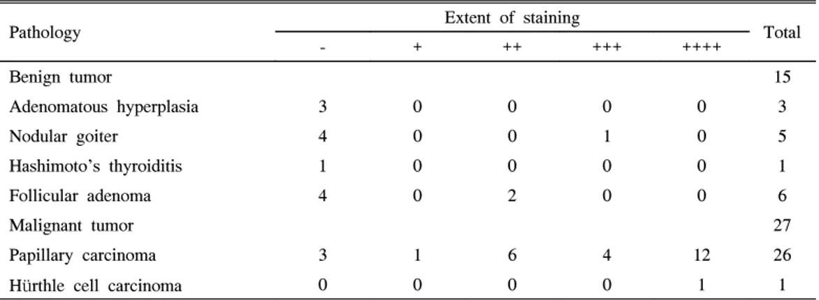
VEGF는 주로 종양세포의 세포질 내에서 적갈색의 과립 상으로 염색되었으며 염색의 강도는 부위에 따라 다양하였 다. 총 42예 중 27예에서 VEGF가 발현되었는데 양성종양 은 여포상 선종 2예, 결절성 갑상선종 1예로 모두 3예에서,
악성종양은 유두상암종 23예, Hürthle 세포암 1예로 모두 24예에서 VEGF발현을 보였다. 발현의 정도는 양성종양에 서는 3예 중 ++ 2예, +++ 1예이었고, 악성종양은 24예 중 + 1예, ++ 6예, +++ 4예, ++++ 13예였다. 양성종양 12예와 악성종양 3예에서는 발현되지 않았으며 ++++로 나타난 것 은 모두 악성종양이었다 (Table 1).
악성종양과 양성종양에서의 VEGF의 발현정도를 ++이하 군과 +++이상 군으로 나누어 비교하였을 때 악성 종양은 27예 중 ++이하가 10예, +++이상이 17예였고, 양성종양은 15예 중 ++이하가 14예, +++이상이 1예로 악성종양이 양 성종양에 비해 의미있게 높게 발현되었다 (P < 0.001) (Fig.
2).
2. 악성종양에서 전이 및 주위조직 침습 유무에 따른 VEGF의 발현 양상
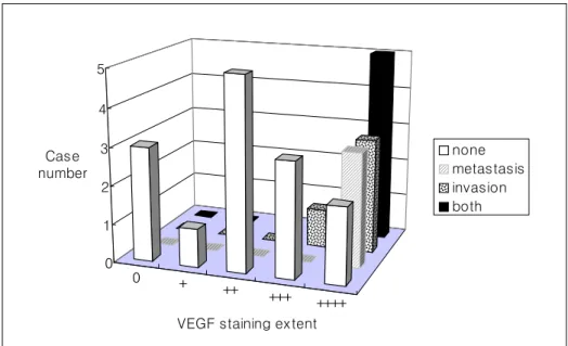
악성종양에서 림프절 전이나 주위 조직에 침습이 있었던 경우는 27예 중 13예였고 이 중 3예는 림프절 전이만, 4예 는 주위 조직에 침습만, 6예는 전이와 침습이 모두 있었다.
VEGF의 발현 정도는 전이나 침습이 없었던 14예 중 음성 은 3예, + 1예, ++ 5예, +++ 3예, ++++ 2예였으며, 전이나 침습이 있었던 13예 중 음성인 경우는 없었으며, ++ 1예, Fig. 1. Immunohistochemical staining for vascular endothelial growth factor (VEGF) in thyroid tumors
A: Negative control of VEGF staining (X200).
B: Papillary carcinoma, weakly positive (+) immunoreactivity of VEGF staining was observed (X200).
C: Strong positive (++++) immunoreactivity was observed in the cytoplasm of papillary carcinoma epithelial cell (X200).
B C
A
A
BB CA A
Table 1. Expression of Vascular Endothelial Growth Factor (VEGF) in the Thyroid Tumors
Pathology Extent of staining
Total
- + ++ +++ ++++
Benign tumor 15
Adenomatous hyperplasia 3 0 0 0 0 3
Nodular goiter 4 0 0 1 0 5
Hashimoto's thyroiditis 1 0 0 0 0 1
Follicular adenoma 4 0 2 0 0 6
Malignant tumor 27
Papillary carcinoma 3 1 6 4 12 26
Hürthle cell carcinoma 0 0 0 0 1 1
Fig. 2. Difference of expression of VEGF between benign and malignant thyroid tumors
0 2 4 6 8 10 12 14 16 18
++이하 +++이상
VEGF staining extent
Case Number
benign tumor malignant tumor
0 2 4 6 8 10 12 14 16 18
++이하 +++이상
VEGF staining extent
Case Number
benign tumor malignant tumor
+++ 1예, ++++ 11예였다 (Fig. 3).
전이나 주위조직으로 침윤이 있는 경우와 없는 경우의 VEGF 발현 정도를 ++이하 군과 +++이상 군으로 나누어 비교하였을 때 전이나 침윤이 있는 군은 13예 중 ++이하가 1예, +++이상이 12예였고, 전이나 침윤이 없는 군은 14예 중 ++이하가 9 예, +++이상이 5예로 전이나 침윤이 있는 군이 비전이 비침윤 군에 비해 VEGF의 발현 정도가 유의 하게 높았다 (P < 0.001) (Fig. 4).
고 찰
신생혈관형성은 이미 존재하는 혈관으로부터 새로운 혈 관이 생성되는 것으로 배아 발생, 상처 복구 및 생식주기 등 의 생리적 과정에 필수적인 요소로 정상에서는 혈관 형성인 자와 혈관 형성 억제인자 사이의 균형에 의해 엄격히 조절
된다[9]. 일반적으로 성숙한 혈관내피세포는 휴면상태에 있 지만 자극에 의해 다시 세포 주기로 들어갈 수 있다. 혈관 형성인자와 혈관 형성 억제인자 사이의 균형이 깨어지면 (angiogenic switch) 종양에서는 지속적인 혈관 신생이 일어 나고[23], 생성된 종양 내의 신생혈관들은 종양세포의 성장 및 침윤과 전이에 기여하게 된다. 신생혈관의 형성이 악성 종양의 성장 이외에도 전이 및 침윤성과 관계가 있다는 것 은 유방, 피부, 소화기계 및 자궁경부암 등을 대상으로 시행 한 여러 연구에서 밝혀졌다[24~26].
종양에서의 혈관 형성인자 및 혈관 형성 억제인자간의 불균형에 대한 다양한 보고가 있지만 그 중에서도 가장 분 명한 것은 VEGF 발현의 극적인 증가이다. Ferrara 등[27]
은 VEGF의 발현에 관한 연구에서 갑상선, 폐, 유방, 소화기 계, 신장, 방광, 난소 및 자궁경부에서 발생하는 암종, 수막 종, 혈관 육종 등에서 VEGF mRNA가 과발현됨을 보고하 였고, Wong 등[28]은 대장 및 직장 종양을 대상으로 한 연 구에서 VEGF의 발현이 정상보다는 선종에서, 그리고 선종 보다는 악성종양에서 증가한 것을 보고하였다. 뿐만 아니라 유방암, 소화기계암 등에서 VEGF의 발현이 종양의 악성도, 임상적 병기, 림프절 전이 및 예후와도 상관 관계가 있다는 보고가 있다[29~31]. 따라서 여러 고형종양에서 VEGF의 발현이 증가되어 있고 과발현된 VEGF는 종양의 성장, 주 위 조직으로의 침윤 및 전이에 관여하여 불량한 예후를 초 래한다고 할 수 있겠다.
갑상샘암은 다른 조직에 발생한 암에 비해 비교적 천천 히 자라므로 대부분 예후가 양호하지만, 일부에서는 주위 조직을 침범하거나 원격 전이를 일으켜 치명적인 결과를 초 래하기도 한다. 일반적으로 갑상샘암은 나이가 40세 이상, 주위 조직으로 침범한 경우, 전이된 경우, 조직학적으로 미 Fig. 3. Expression of VEGF according to metastasis and/ or invasion in the thyroid tumor
0 + ++ +++ ++++
0 1 2 3 4 5
Case number
VEGF staining extent
none metastasis invasion both
Fig. 4. VEGF overexpression in the thyroid malignancies between metastasis or invasion and those without 0
2 4 6 8 10 12 14
++이하 +++이상
VEGF staining extent
Case Number none
metastasis or invasion
분화암일 때 예후가 좋지 않은 것으로 알려져 있으나, 이와 같은 양상을 보이지 않는 경우에서도 나쁜 경과를 보이는 경우가 있다. 이러한 경우에 이를 미리 알아 낼 수 있는 종 양지표가 있다면 임상적으로 많은 도움을 얻을 수 있을 것 이다.
갑상샘 조직에서의 VEGF 발현에 관한 연구를 보면 종양 세포뿐 아니라 그레이브스병, 하시모토 갑상샘염 및 정상 갑상샘 조직에서도 정도의 차이는 있으나 발현된다고 한다 [32,33]. 갑상샘 종양에서의 VEGF 발현에 관한 저자들의 연구에서 VEGF의 발현은 양성종양에서 보다 악성종양에서 과발현되어 (P <0.001) Soh 등[34]이나, Bunone 등[35], Fenton 등[36]의 연구 결과와 일치한다. 하지만 최근 Jubb 등[37]은 세포주와 신선동결 인체조직을 이용하여 갑상샘 종양에서의 VEGF 발현을 유전자배열 및 조직배열법을 이 용하여 검사 하였을 때 갑상샘 종양에서 정상조직에 비하여 의미있게 VEGF의 발현이 감소하였다는 상반된 보고를 하 였다. 이에 관하여서는 향후 보다 대규모의 검체를 이용한 연구가 필요 할 것으로 생각된다. 악성 갑상샘 종양에서 VEGF의 발현과 예후 인자와의 상관관계를 보면 Bunone 등[35]은 갑상샘의 악성종양에서 전이가 있는 경우가 없는 경우에 비해 VEGF의 발현이 더 높았다고 보고하였고 Fenton 등[36]은 VEGF의 발현이 유두상암종과 여포상암 등의 악성종양에서 양성종양보다 높고 종양의 크기와 상관 이 있으며, 재발한 유두상암종에서 특히 과발현 된다고 하 였다. 또한 갑상샘 유두상암종에서의 VEGF 발현에 관한 여러 연구에서 전이가 있거나 국소 또는 원격 재발한 경우 에 VEGF의 발현이 높아 갑상샘 악성종양, 특히 유두상암 종에서 VEGF의 발현이 예후인자로서 의미가 있음을 시사 하였다[38~40]. 상기한 결과들은 악성종양에서는 림프절 전 이나 주위조직에 침윤이 있는 경우에 종양이 갑상샘에 국한 되어 있는 것보다 VEGF의 발현이 더 강하였다는 저자들의 연구 결과와 일치한다고 할 수 있겠으며, VEGF가 임상적 인 예후인자로 유의한 의미를 가지며 종양의 공격성향이나 전이.능력을 예측할 수 있는 유용한 수단이 될 수 있고 나아 가 종양 치료의 표적으로서의 의의를 가질 수 있음을 시사 한다고 할 수 있겠다. 그러나 갑상샘 종양에서의 VEGF 과 발현이 주위조직 침윤, 전이 또는 병기 등과 유의한 상관관 계를 보이지 않는다는 보고들도 있어 이에 대한 추가적인 연구가 필요할 것으로 생각된다[34,41].
VEGF의 발현이 갑상샘암의 분화에도 영향을 미친다는 보고들이 있는데, Viglietto 등[20]은 미분화 갑상샘암의 경 우 정상 갑상샘에 비해 20배 정도 과발현된다고 하였으며, 더 나아가 Katoh 등[21]은 VEGF가 저등급암에서 고등급암 으로 이행하는데 관여한다고 하였다. 이러한 연구 결과들은 갑상샘암의 증식, 전이 및 분화에 혈관형성인자인 VEGF가 일부 관여함을 시사해준다. 그러나 Huang 등[41]은 갑상샘
유두상암종에서 과발현되었던 VEGF가 미분화 갑상샘암에 서는 감소되어 있음을 보고하여 VEGF는 발암과정의 초기 에 관여하고 미분화 암종으로 이행하면서 발현도가 떨어진 다고 하였다.
갑상샘 종양에서의 VEGF의 발현에 관한 본 연구는 비록 유두상암종과 Hürthle 세포암 외에 다른 종류의 갑상샘암은 연구에 포함되어 있지 않았고, 분화가 서로 다른 암에 관한 연구는 시행하지 못하였지만, 갑상샘 종양에서 VEGF의 발 현이 양성종양보다 악성종양에서 높으며, 악성종양 중에서 도 주위조직으로 침윤이 있거나, 림프절 전이가 있는 경우 높음을 알 수 있었으며, 이는 VEGF의 발현정도가 갑상샘종 양의 예후인자로 의미가 있음을 나타낸다고 할 수 있겠다.
향후 갑상샘의 다른 암종 및 분화 정도가 서로 다른 암 사이 의 VEGF의 발현에 대한 연구를 통해 갑상샘암의 분화에 미 치는 VEGF의 영향에 대한 연구도 필요할 것으로 생각된다.
요 약
연구배경: 갑상샘암은 다른 조직에 발생한 암에 비해 비 교적 천천히 자라므로 대부분 예후가 양호하지만, 일부에서 는 광범위한 주위 조직으로의 침윤 혹은 원격전이로 치명적 인 결과를 초래할 수 있다. 갑상샘암의 침윤 및 전이에는 종 양세포의 분화도, 혈관, 면역계 및 결체조직 간의 여러 중요 한 상호작용이 관여한다. 이 중 종양의 신생혈관형성은 종 양의 국소적 증식이나 전이에 필수적이다. 신생혈관형성 인 자 중 vascular endothelial growth factor (VEGF)가 가장 강력한 인자로 알려져 있으나 갑상샘암에서의 VEGF에 관 한 연구는 드물다. 본 연구는 갑상샘 종양에서의 VEGF 발 현과 임상소견과의 연관성을 살펴보고자 하였다.
대상 및 방법: 1999년 3월부터 2000년 2월까지 고신대학 교 복음병원에서 갑상샘 종양으로 갑상샘 절제술을 시행한 환자의 조직 중 양성종양 15예, 악성종양 27예 (유두상암종 26예, Hürthle 세포암 1예)로 총 42예를 대상으로 VEGF에 대해 면역조직화학검사를 시행하였으며, VEGF 염색의 성 적은 적갈색의 염색범위에 따라 음성, + (1~24%), ++
(25~49%), +++ (50~ 74%), ++++ (75% 이상)로 판정하였 다.
결과: 1) 악성종양 27예 중 13예에서 주위 림프절 전이나 주위조직에 침윤이 있었으며, 나머지 14예는 전이나 주위조 직에 침윤 없이 종양이 갑상샘에 국한되어 있었다.
2) VEGF는 주로 종양세포의 세포질 내에서 적갈색의 과 립상으로 염색되었으며 염색의 강도는 부위에 따라 다양하 였다. 총 42예 중 27예에서 VEGF가 발현되었으며, + 1예, ++ 8예, +++ 5예 및 ++++ 13예였다. 양성종양 15예 중 12 예와 악성종양 27예 중 3예에서 발현되지 않아서, 양성종양 에서 악성종양보다 VEGF의 발현이 낮았다 (P <0.001). 양
성종양과 악성종양에서의 VEGF의 발현을 ++이하 군과 +++이상 군으로 나누어 비교하였을 때 악성종양에서 양성 종양에 비해 의미있게 과발현되었다 (P <0.001).
3) 악성종양 27예 중 림프절 전이 또는 주위조직에 침윤 이 있었던 13예는, 림프절 전이나 주위조직에 침윤이 없는 14예에 비해 VEGF의 발현이 높았다 (P < 0.001).
결론: 본 연구에서 갑상샘 종양에서 VEGF의 발현은 양 성종양보다 악성종양에서 높으며, 악성종양 중에서도 주위 조직으로 침윤이 있거나, 림프절 전이가 있는 경우에 VEGF 의 발현이 더 높음을 알 수 있었다.
참 고 문 헌
1. Folkman J: What is the evidence that tumors are angiogenesis dependent?. J Natl Cancer Inst 82:4-6, 1990
2. Carmeliet P, Jain RK: Angiogenesis in cancer and other disease. Nature 407:249-257, 2000
3. Turner HE, Harris AL, Melmed S, Wass JA: Angio- genesis in endocrine tumors. Endocr Rev 24:600-632, 2003
4. Moscatelli DA, Gross J, Rifkin D: Angiogenesis factors stimulate plasminogen activator and coll- agenase production by capillary endothelial cells. J Pathol 170(Suppl):338a, 1993
5. Weidner N, Semple JP, Welch WR, Folkman J:
Tumor angiogenesis and metastasis correlation in invasive breast carcinoma. N Engl J Med 324:1-8, 1991
6. Pepper MS, Ferrara N, Orci L, Montesano R: Vas- cular endothelial growth factor (VEGF) induces plas- minogen activators and plasminogen activator inhi- bitor-1 in microvascular endothelial cells. Biochem Biophys Res Commun 181:902-906, 1991
7. Mandriota SJ, Seghezzi G, Vassalli JD, Ferrara N, Wasi S, Mazzieri R, Mignatti P, Pepper MS: Vascular endothelial growth factor increases urokinase receptor expression in vascular endothelial cells. J Biol Chem 270:9709-9716, 1995
8. Semenza GL: Angiogenesis in ischemic and neo- plastic disordes. Annu Rev Med 54:17-28, 2003 9. Folkman J: The role of angiogenesis in tumor growth.
Semin Cancer Biol 3(2):65-71, 1992
10. Leung DW, Cachianes G, Kuang WJ, Goeddel DV, Ferrara N: Vascular endothelial growth factor is a secreted angiogenic mitogen. Science 246:1306-1309,
1989
11. Tischer E, Mitchell R, Hartman T, Silva M, Gos- podarowicz D, Fiddes JC, Abraham JA: The human gene for vascular endothelial growth factor. Multiple protein forms are encoded through alternative exon splicing. J Biol Chem 266:11947-11954, 1991 12. Houck KA, Ferrara N, Winer J, Cachianes G, Li B,
Leung DW: The vascular endothelial growth factor family: identification of a fourth molecular species and characterization of alternative splicing of RNA.
Mol Endocrinol 5:1806-1814, 1991
13. Frank S, Hubner G, Breier G, Longaker MT, Green- halgh DG, Werner S: Regulation of vascular endo- thelial growth factor expression in cultured keratino- cytes. Implications for normal and impaired wound healing. J Biol Chem 270:12607-12613, 1995 14. Dameron KM, Volpert OV, Tainsky MA, Bouck N:
Control of angiogenesis in fibroblasts by p53 regu- lation of thrombospondin-1. Science 265:1582-1584, 1994
15. Kieser A, Weich HA, Brandner G, Marme D, Kolch W: Mutant p53 potentiates protein kinase C induction of vascular endothelial growth factor expression. Onco- gene 9:963-969, 1994
16. Kolch W, Martiny-Baron G, Kieser A, Marme D:
Regulation of the expression of the VEGF/ VPS and its receptors: role in tumor angiogenesis. Breast Cancer Res Treat 36:139-155, 1995
17. Dvorak HF, Brown LF, Detmar M, Dvorak AM:
Vascular permeability factor/vascular endothelial growth factor, microvascular hyperpermeability, and angiogenesis. Am J Pathol 146:1029-1039, 1995 18. Senger DR, Van de Water L, Brown LF, Nagy JA,
Yeo KT, Yeo TK, Berse B, Jackman RW, Dvorak AM, Dvorak HF: Vascular permeability factor (VPF, VEGF) in tumor biology. Cancer Metastasis Rev 12:
303-324, 1993
19. Brown LF, Berse B, Jackman RW, Tognazzi K, Manseau EJ, Senger DR, Dvorak HF: Expression of vascular permeability factor (vascular endothelial growth factor) and its receptors in adenocarcinomas of the gastrointestinal tract. Cancer Res 53:4727-4735, 1993 20. Viglietto G, Maglione D, Rambaldi M, Cerutti J,
Romano A, Trapasso F, Fedele M, Ippolito P, Chiappetta G, Botti G, et al: Upregulation of vascular endothelial growth factor (VEGF) and down-
regulation of placenta growth factor (PlGF) associ- ated with malignancy in human thyroid tumors and cell lines. Oncogene 11:1569-1579, 1995
21. Katoh R, Miyagi E, Kawaoi A, Hemmi A, Ko- miyama A, Oyama T, Shibuya M: Expression of vascular endothelial growth factor (VEGF) in human thyroid neoplasms. Hum Pathol 30:891-897, 1999 22. Hedinger C, Williams ED, Sobin LH: Histological
typing of thyroid tumors. WHO 2nd Ed. Springer Verlag, Berlin, 1998
23. Hanahan D, Folkman J: Patterns and emerging mechanism of angiogenic switch during tumor- igenesis. Cell 86:353-364, 1996
24. Staibano S, Boscaino A, Salvatore G, Orabona P, Palombini L, De Rosa G: The prognostic significance of tumor angiogenesis in nonaggressive and aggre- ssive basal cell carcinoma of the human skin. Hum Pathol 27:695-700, 1996
25. Weidner N, Folkman J, Pozza F, Bevilacqua P, Allred EN, Moore DH, Meli S, Gasparini G: Tumor angiogenesis: a new significant and independent prognostic indicator in early-stage breast carcinoma. J Natl Cancer Inst 84:1875-1887, 1992
26. Bremer GL, Tiebosch AT, van der Putten HW, Schouten HJ, de Haan J, Arends JW: Tumor angiogenesis: an independent prognostic parameter in cervical cancer. Am J Obstet Gynecol 174:126-131, 1996
27. Ferrara N, Davis-Smyth T: The biology of vascular endothelial growth factor. Endocr Rev 18:4-25, 1997 28. Wong MP, Cheung N, Yuen ST, Leung SY, Chung
LP: Vascular endothelial growth factor is up- regulated in the early pre-malignant stage of colo- rectal tumour progression. Int J Cancer 81:845-850, 1999
29. Toi M, Hoshina S, Takayanagi T, Tominaga T:
Association of vascular endothelial growth factor expression with tumor angiogenesis and with early relapse in primary breast cancer. Jpn J Cancer Res 85:1045-1049, 1994
30. Maeda K, Chung YS, Ogawa Y, Takatsuka S, Kang SM, Ogawa M, Sawada T, Sowa M: Prognostic value of vascular endothelial growth factor expression in gastric carcinoma. Cancer 77:858-863, 1996 31. Takahashi Y, Kitadai Y, Bucana CD, Cleary KR, Ellis
LM: Expression of vascular endothelial growth factor
and its receptor, KDR, correlates with vascularity, metastasis, and proliferation of human colon cancer.
Cancer Res 55:3964-3968, 1995
32. Nagura S, Katoh R, Miyagi E, Shibuya M, Kawaoi A:
Expression of vascular endothelial growth factor (VEGF) and VEGF receptor-1 (Flt-1) in Graves disease possibly correlated with increased vascular density. Human path 32:10-17, 2001
33. Klein M, Picard E, Vignaud JM, marie B, Bresler L, Toussaint B, Weryha G, Duprez A, Leclere J:
Vascular endothelial growth factor gene and protein:
strong expression in thyroiditis and thyroid carcinoma.
J Endocrinol 161:41-49. 1999
34. Soh EY, Duh QY, Sobhi SA, Young DM, Epstein HD, Wong MG, Garcia YK, Min YD, Grossman RF, Siperstein AE, Clark OH: Vascular Endothelial Growth Factor Expression Is Higher in Differentiated Thyroid Cancer than in Normal or Benign Thyroid. J Clin Endocrinol Metab 82:3741-3747, 1997
35. Bunone G, Vigneri P, Mariani L, Buto S, Collini P, Pilotti S, Pierotti MA, Bongarzone I: Expression of angiogenesis stimulators and inhibitors in human thyroid tumors and correlation with clinical patho- logical features. Am J Pathol 155:1967-1976, 1999 36. Fenton C, Patel A, Dinauer C, Robie DK, Tuttle RM,
Francis GL: The expression of vascular endothelial growth factor and the type 1 vascular endothelial growth factor receptor correlate with the size of papillary thyroid carcinoma in children and young adults. Thyroid 10:349-357, 2000
37. Jubb AM, Pham TQ, Hanby AM, Frantz GD, Peale FV, Wu TD, Koeppen HW, Hillan KJ: Expression of vascular endothelial growth factor, hypoxia inducible factor 1alpha, and carbonic anhydrase IX in human tumours. J Clin Pathol 57:504-512, 2004
38. Lennard CM, Patel A, Wilson J, Reinhardt B, Tuman C, Fenton C, Blair E, Francis GL, Tuttle RM: Inten- sity of vascular endothelial growth factor expression is associated with increased risk of recurrence and decreased disease-free survival in papillary thyroid cancer. Surgery 129:552-558, 2001
39. Klein M, Vignaud JM, Hennequin V, Toussaint B, Bresler L, Plénat F, Leclère J, Duprez A, Weryha G.:
Increased Expression of the Vascular Endothelial Growth Factor Is a Pejorative Prognosis Marker in Papillary Thyroid Carcinoma. J Clin Endocrinol
Metab 86:656-658, 2001
40. Kilicarslan AB, Ogus M, Arici C, Pestereli HE, Cakir M, Karpuzoglu G: Clinical importance of vascular endothelial growth factor (VEGF) for papillary thy-
roid carcinomas. APMIS 111:439-443, 2003
41. Huang SM, Lee JC, Wu TJ, Chow NH: Clinical Relevance of Vascular Endothelial Growth Factor for Thyroid Neoplasms. World J surg 25:302-306, 2001