Inhibition of Neointima Formation and Migration of Vascular Smooth Muscle Cells by Anti-vascular Endothelial Growth Factor Receptor-1 (Flt-1) Peptide in Diabetic Rats
Min Seop Jo, M.D.*, Ki Dong Yoo, M.D.**, Chan-Beom Park, M.D.*, Deog Gon Cho, M.D.*, Kue Do Cho, M.D.*, Ung Jin, M.D.*, Kun Woong Moon, M.D.**, Chul Min Kim, M.D.**,
Sun Hee Lee, M.D.*, Young Pil Wang, M.D.*
Background: Vascular endothelial growth factor (VEGF) plays an important role in angiogenesis, including stimulating the proliferation and migration of vascular smooth muscle cells (VSMCs). It has been known that diabetes is associated with accelerated cellular proliferation via VEGF, as compared to that under a normal glucose concentration. We investigated the effects of selective blockade of a VEGF receptor by using anti-Flt-1 peptide on the formation and hyperplasia of the neointima in balloon injured-carotid arteries of OLETF rats and also on the in vitro VSMCs’ migration under high glucose conditions. Material and Method: The balloon-injury method was employed to induce neointima formation by VEGF. For 14 days beginning 2 days before the ballon injury, placebo or vascular endothelial growth factor receptor-1 (VEGFR-1) specific peptide (anti-Flt-1 peptide), was injected at a dose of 0.5 mg/kg daily into the OLETF rats. At 14 days after balloon injury, the neointimal proliferation and vascular luminal stenosis were measured, and cellular proliferation was assessed by counting the proliferative cell nuclear antigen (PCNA) stained cells. To analyze the effect of VEGF and anti-Flt-1 peptide on the migration of VSMCs under a high glucose condition, transwell assay with a matrigel filter was performed. And finally, to determine the underlying mechanism of the effect of anti-Flt-1 peptide on the VEGF-induced VSMC migration in vitro, the expression of matrix metalloproteinase (MMP) was observed by performing reverse transcription-polymerase chain reaction (RT-PCR). Result: Both the neointimal area and luminal stenosis associated with neointimal proliferation were significantly decreased in the anti-Flt-1 peptide injected rats, (0.15±0.04 mm
2and 36.03±3.78%
compared to 0.24±0.03 mm
2and 61.85±5.11%, respectively, in the placebo-injected rats (p<0.01, respectively).
The ratio of PCNA(+) cells to the entire neointimal cells was also significantly decreased from 52.82±4.20% to 38.11±6.89% by the injected anti-Flt-1 peptide (p<0.05). On the VSMC migration assay, anti-Flt-1 peptide significantly reduced the VEGF-induced VMSC migration by about 40% (p<0.01). Consistent with the effect of anti-Flt-1 peptide on VSMC migration, it also obviously attenuated the induction of the MMP-3 and MMP-9 mRNA expressions via VEGF in the VSMCs. Conclusion: Anti-Flt-1 peptide inhibits the formation and hyperplasia of the neointima in a balloon-injured carotid artery model of OLETF rats. Anti-Flt-1 peptide also inhibits the VSMCs’
migration and the expressions of MMP-3 and MMP-9 mRNA induced by VEGF under a high glucose condition.
*가톨릭대학교 의과대학 흉부외과학교실
Department of Thoracic and Cardiovascular Surgery, College of Medicine, The Catholic University of Korea
**가톨릭대학교 의과대학 성빈센트병원 순환기내과학교실
Division of Cardiology, Department of Internal Medicine, St. Vincent's Hospital, College of Medicine, The Catholic University of Korea 논문접수일:2006년 12월 29일, 심사통과일:2007년 1월 26일
책임저자:이선희 (150-713) 서울시 영등포구 여의도동 62번지, 가톨릭대학교 의과대학 성모병원 흉부외과 (Tel) 02-3779-1020, (Fax) 02-3779-1181, E-mail: [email protected]
본 논문의 저작권 및 전자매체의 지적소유권은 대한흉부외과학회에 있다.
당뇨병 쥐에서 혈관내피 성장인자 수용체-1 차단 펩타이드를 이용한 신내막 형성과 혈관평활근세포 이동의 억제
조민섭*·유기동**·박찬범*·조덕곤*·조규도*·진 웅*·문건웅**·김철민**·이선희*·왕영필*
서 론
경피적 관상동맥성형술과 관상동맥 우회술 및 동맥내 막 절제술 시행 후에 발생하는 혈관재협착은 혈관손상에 대한 동맥의 치유 과정으로 일어나는 현상으로 관상동맥 재관류술(coronary revascularization)의 중요한 제한점이 되 고 있다[1]. 혈관재협착은 탄성반도(elastic recoil)와 혈관벽 재형성(vessel wall remodeling)에 의한 급성 또는 만성 혈 관 크기의 변화와 신내막 비대(neointimal hyperplasia) 등의 기전에 의해 발생하는 것으로 알려져 있다[2,3].
신내막은 중막에 있는 혈관평활근세포(vascular smooth muscle cells)가 증식하고 내막으로 이동하여 비대해지며 [4,5], 외막의 섬유모세포가 혈관평활근세포와 비슷한 근 섬유모세포로 형질전환된 후 증식하고 이동하여 신내막 형성에 참여한다[6].
혈관내피 성장인자(vascular endothelial growth factor, VEGF)는 혈관평활근세포의 증식과 이동의 촉진을 포함한 다양한 기전으로 혈관신생(angiogenesis)을 유도하는 성장 인자이다[7]. VEGF의 주요 수용체(vascular endothelial growth factor receptor, VEGFR)로는 VEGF 수용체-1 (VEGFR-1, Fms-like tyrosine kinase, Flt-1)과 VEGF 수용체-2 (VEGFR-2, fetal liver kinase, Flk-1)가 있다. VEGFR-1은 혈관내피세포 뿐만 아니라 혈관평활근세포, 대식세포 및 단핵구 등에 분포하여 혈관평활근세포 및 단핵구의 활성과 이동[8,9], 그리고 혈관투과성에 영향을 줄 수 있다[10]. VEGFR-2는 주로 혈관내피세포에 분포되어 있으며 이 수용체를 통하 여 VEGF에 의한 혈관신생(angiogenesis)이 일어난다[11].
혈관평활근세포의 내막내로의 이동과정 중에는 내탄력 층의 통과가 필수적인데 이는 기존의 성장인자들의 작용 으로는 불가능하며 단백분해물질인 기질금속단백분해효
소(matrix metalloprotenase, MMP)의 작용에 의한 것으로 알려져 있다. 실제로, VEGF에 의해서 MMP-1, 3, 9의 발현 이 촉진되는 것으로 관찰되어 VEGF가 MMP의 발현을 증 가시켜 혈관내피세포의 이동을 일으키는 것으로 생각되 고 있다[12-14].
당뇨병은 혈관합병증을 유발함으로써 동맥경화와 심혈 관협착에 의한 사망률을 증가시킨다. 정상 혈당상태에서 보다 고혈당 시에 세포의 증식이 더 촉진되는 것이 관찰 되어, 당뇨병 시에는 혈관평활근세포의 증식과 이동이 정 상상태에서 보다 더 촉진되어 혈관합병증을 일으키는 것 으로 보고되었다[15-17].
신내막 형성 과정에서 VEGFR-1을 선택적으로 차단할 경우 신내막 형성이 억제되는 것을 사전실험에서 확인하 였다. 이에 본 연구에서는 VEGFR-1의 선택적 차단 펩타 이드(항-Flt-1 펩타이드: anti-Flt-1 peptide)가 당뇨병쥐에서 의 고혈당환경에서도 신내막 형성을 억제하는지를 관찰 하였으며, VEGF에 의한 혈관평활근세포의 이동과 MMP 의 발현에 항-Flt-1 펩타이드가 미치는 영향을 알아보았다.
대상 및 방법
1) 대상동물 및 처치400∼450 g의 제2형 당뇨모델인 OLETF (Otsuka Ling- Evans Tokushima Fatty)계의 수컷 흰쥐를 대상으로 무작위 로 실험군(n=5)과 대조군(n=5)으로 나눴다. Xylazine (4.6 mg/kg)과 ketamine (70 mg/kg)을 복막 내 주사하여 마취한 후 경부의 정중앙을 절개하고 좌측 총경동맥 및 외경동맥 을 노출시켰다. 좌측 외경동맥을 통하여 2F Fogarty 풍선 도자를 대동맥궁까지 삽입한 후 총경동맥 기시부에서 약 간의 저항을 느낄 정도로(0.3∼0.4 mL) 공기를 넣어 풍선
Therefore, these results suggest that specific blockade of VEGFR-1 by anti-Flt-1 peptide may have therapeutic potential against the arterial stenosis of diabetes mellitus patients or that occurring under a high glucose condition.
(Korean J Thorac Cardiovasc Surg 2007;40:264-272) Key words: 1. Restenosis
2. Endothelial growth factors 3. Diabetes
4. Cell signaling proteins
5. Vascular smooth muscle cell
을 확장시키고 확장된 풍선도자를 회전시키면서 경동맥 분지부까지 잡아당겨 경동맥의 내막을 박리했다. 동일개 체에 이와 같은 술기를 3회 반복하고 풍선도자를 제거한 후 외경동맥을 결찰하고 절개부위를 봉합했다. 실험군은 풍선도자로 경동맥의 내막박리 2일 전부터 내막박리 후 2 주간 매일 Bae 등[18]이 합성하여 사용하였던 항-Flt-1 펩 타이드(Gly-Asn-Gln-Trp-Phe-Il: Peptron, Inc., Daejon, Korea) 를 0.5 mg/kg의 용량으로 완충액에 혼합하여 피하 주사했 다. 대조군은 위약을 투여했다.
2) 조직채취
경동맥의 내막박리 2주 후 다음과 같은 방법으로 조직 을 얻었다. 마취 후 우측 대퇴동맥을 박리하여 노출한 뒤 도관을 삽입하여 상행대동맥까지 전진시키고 4% 포르말 린 용액을 사용하여 90∼100 mmHg의 압력으로 5분간 관 류 고정했다. 관류 고정이 시작된 후 꼬리정맥을 통하여 과량의 thiopental sodium을 주사하여 안락사시켰다. 관류 고정 후 좌측 경동맥을 채취하여 4% 포르말린 용액에 24 시간 동안 더 고정한 후 파라핀에 포매하고 5μm의 절편 을 한 마리당 4개씩 만들어 조직형태학적 검사(histomor- phometric study)와 면역조직화학적 검사(Immunohistochemical study)를 시행하였다.
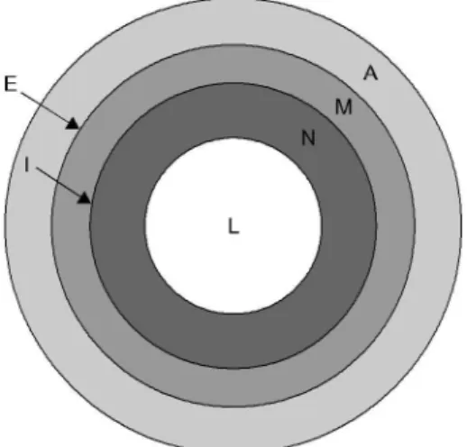
3) 조직형태학적 검사(Histomorphometric study) 좌측경동맥 절편을 Hematoxylin-Eosin 염색 후 100배 확 대 시야에서 디지털카메라로 촬영하고 AxioVision LE v.4.1 (Carl Zeiss Vision Gmbh, Germany)을 사용하여 신내 막과 중막의 경계, 즉 내탄력층이 이루는 면적에서 혈관 내강의 면적을 빼서 신내막의 면적을 구하고 중막과 외막 의 경계, 즉 외탄력층이 이루는 면적에서 내탄력층이 이루 는 면적을 빼서 중막의 면적을 구했다. 내강의 협착 정도는 (1-내강면적/내탄력층면적)×100으로 계산하였다(Fig. 1).
4) 면역조직화학 염색(Immunohistochemical stain) 세포의 증식을 평가하기 위하여 준비한 조직절편에서 파라핀 제거 후 인산완충식염수(Phosphate-buffered saline, PBS)로 세척하고 조직 내의 내인성 과산화효소를 제거하 기 위하여 실온에서 3% 과산화수소에 15분간 반응시겼다.
이어 PBS로 세척 후 비특이적 항원-항체 결합의 제거를 위하여 Rodent Block (UltraVision Mouse Tissue Detection System, LAB VISION, Fremont)으로 실온에서 60분간 반응 시킨 후 PBS로 세척하였다. 일차항체로 proliferative cell
nuclear antigen (NeoMarkers, Fremont, MS)을 실온에서 반 응시켰다. 세척 후 이차항체(UltraVision Mouse Tissue De- tection System, LAB VISION, Fremont)에 반응시키고 다시 PBS로 세척한 후 streptavidin peroxidase (Ultra Vision Mouse Tissue Detection System, LAB VISION, Fremont)로 biotin - avidin 특이결합을 유도하기 위하여 실온에서 반응시켰다.
10분 후 PBS로 세척하고 DAB (3,3'-Diaminobenzidine tetra- hydrochloride, Zymed Cap-Plus Detection Kit, San Francisco) 로 처리하여 발색시키고 증류수로 세척한 다음 Mayer's Hematoxylin 대조염색을 한 후 탈수과정 후 canada balsam 으로 봉입한 후 광학현미경으로 관찰하였다. 400배 시야 에서 네 방향에서 세포 수를 측정하여 전체 세포 수에 대 하여 양성반응을 보인 세포 수의 비율을 구했다.
5) 혈관평활근세포 배양 및 이동측정(Cell migration assay)
흰쥐의 흉부 및 복부 대동맥을 적출하여 분리한 평활근 세포주를 10% FBS (Gibco BRL, Life Technologies)가 포함 된 DMEM (Dulbeco’s minium essential media, Gibco BRL 12320 low glucose, Life Technologies)에 분주하여, 5% CO2
가 공급되는 37oC 배양기에서 3일간 배양 후 세포의 배지 를 교환하였다. 그 후 25 mmol/L의 glucose를 첨가한 배지 에서 배양하여 세포이동측정 실험에 사용하였다[19].
Fig. 1. Cross-sectional view of the carotid artery. A=Adventitia;
M=Media; N=Neointima; L=Lumen; E=External elastic lamina; I=
Internal elastic lamina. The area of the media represents the area surrounded by E minus the area surrounded by I, and the area of the neointima represents the area surrounded by I minus the area of the lumen. Luminal stenosis represents the ratio of the neointima area to the area surrounded by I.
세포의 이동능력은 pore size가 8μm인 polycarbonate mem- brane의 24-well의 Transwell plate (Corning Inc., Corning, NY)를 이용한 transwell assay로 측정했다[20,21]. 5×10⁴개 의 배양된 혈관평활근세포를 matrigel이 coating된 filter 상 층부에 분주하고 하층부에는 VEGF (50 ng/mL)를 단독 또 는 항-Flt-1 펩타이드(3μg/ml)와 함께 넣고 배양하였다. 16 시간 후에 filter를 통과하지 못한 세포를 제거하고, 통과한 세포를 2% paraformaldehyde 용액으로 고정한 후 hemato- xylin 용액으로 염색하여 광학현미경하에서 200배율로 관 찰하였다. 혈관평활근세포의 이동 정도는 임의로 지정된 5군데의 시야에서 관찰된 세포 수를 합산하여 VEGF를 첨 가하지 않고 배양한 세포가 이동한 정도에 대한 상대적인 비율로 나타냈다.
6) Reverse transcription-polymerase chain reaction (RT-PCR)
아무런 조작도 가하지 않은 고혈당 환경에서 배양된 혈 관평활근세포를 대조군으로 하고, 혈관평활근세포에 VEGF (50 ng/mL)를 첨가하거나 VEGF와 함께 항-Flt-1 펩타이드 (3μg/ml)를 첨가하여 실험에 사용하였다. 각각의 세포에 서 Trizol시약(Invitrogen, Carlsbad, CA)을 사용하여 총 RNA 를 분리하였다. 분리한 RNA에 200 unit의 reverse trans-
criptase와 500 ng의 random hexamer를 넣고 50 mM Tris- HCl (pH 8.3), 75 mM KCl, 3 mM MgCl2, 10 mM dithio- threitol, 그리고 1 mM dNTPs를 혼합한 다음 42oC에서 1시 간 동안 반응시켜 cDNA를 합성한 후 70oC에서 15분간 작 용시켜 반응을 중지시켰다.
각 중합효소연쇄반응은 3μL의 cDNA에 50 mM KCl, 10 mM Tris-HCl (pH 8.3), 1.5 mM MgCl2, 0.2 mM dNTPs, 2.5 units의 Taq DNA polymerase, 그리고 0.25μM의 각 primer (Table 1)를 혼합하여 DNA thermal cycler (Gene Amp9700, Applied Biosystems, Foster City, CA)에서 94oC에서 5분간 변성 후 94oC에서 30초, 55oC에서 30초, 72oC에서 30초간 25회 반복하여 반응을 시키고, 72oC에서 10분간 연장반응 을 시행하였다. 반응이 끝난 증폭산물은 ethidium-bromide 가 포함된 1.5% agarose gel에서 전기영동을 실시하였다.
7) 통계분석
통계분석은 SAS system (Ver. 8.02)을 사용하였으며, 각 실 험값은 평균±표준편차로 나타냈고, 두 군 간의 정량적인 비교는 Wilcoxon rank-sum test, 세 군 간의 비교는 ANOVA 로 분석 후 Sheffe's test로 사후검정하였으며, p<0.05인 경 우 유의한 것으로 판정하였다.
결 과
1) 조직형태학적 검사(Histomorphometric study) 경동맥 손상 2주 후에 정상적인 경동맥에 비하여 비대 해진 신내막 형성을 관찰할 수 있었다. 위약을 투여한 쥐 Table 1. Sequences of primers
Primer sequence Product size
Flt-1
Sense 5'-AAACCTCACAGCCACTCTCATCGT-3'
491 bp Antisense 5'-TGACCACACAGGACAGTTTCAGGT-3' Flk-1
Sense 5'-GCTTGCCTTATGATGCCAGCAAGT-3'
696 bp Antisense 5'-GGGCCAAGCCAAAGTCACAGATTT-3' MMP-2
Sense 5'-TGTTTACCATGGGTGGCAATGCTG-3'
583 bp Antisense 5'-TGTTTGCAGATCTCCGGAGTGACA-3'
MMP-3
Sense 5'-ATGTCCTCGTGGTACCCACCAAAT-3'
509 bp Antisense 5'-GACAGCATCCACCTTTGTGCCAAT-3'
MMP-9
Sense 5'-TTCTCGAATCACGGAGGAAGCCAA-3' 280 bp Antisense 5'-ATTTCCAGATACGTTCCCGGCTGA-3'
GAPDH
Sense 5'-ACCACAGTCCATGCCATCAC-3'
520 bp Antisense 5'-TCCACCACCCTGTTGCTGTA-3'
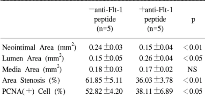
Table 2. Histomorphometric and immunohistochemical study indices 14 days after balloon injury
-anti-Flt-1 +anti-Flt-1
peptide peptide p
(n=5) (n=5)
Neointimal Area (mm2) 0.24±0.03 0.15±0.04 <0.01 Lumen Area (mm2) 0.15±0.05 0.26±0.04 <0.05 Media Area (mm2) 0.18±0.03 0.17±0.02 NS Area Stenosis (%) 61.85±5.11 36.03±3.78 <0.01 PCNA(+) Cell (%) 52.82±4.20 38.11±6.89 <0.05 Values are mean±SD. Both the neointimal area and luminal stenosis associated with neointimal proliferation were significantly decreased by treatment of anti-Flt-1 peptide. The media area is not statistically significant. The percentage of PCNA(+) cells in the anti-Flt-1 treated group was significantly smaller in number.
에서 형성된 신내막의 면적은 0.24±0.03 mm2였으나 항- Flt-1 펩타이드를 처리한 쥐는 0.15±0.04 mm2로 유의하게 작았으며(p<0.01, Table 2, Fig. 2) 신내막 형성에 따른 내 강의 협착 정도도 61.85±5.11%에서 항-Flt-1 처리에 의하 여 36.03±3.78%로 유의하게 협착이 억제되었다(p<0.01, Table 2, Fig. 2).
2) 면역조직화학 염색(Immunohistochemical stain) 신내막에서 증식하고 있는 세포를 측정하기 위하여 분 열하는 세포의 핵에서 발현되는 proliferating cell nuclear antigen (PCNA) 단백질의 발현을 면역조직화학 염색법으 로 관찰하고, 전체 세포 수에 대하여 PCNA(+)인 세포를 백분율로 구하였다. 경동맥 손상 후 비대해진 신내막의 PCNA(+) 세포는 52.82±4.20%였으나, 항-Flt-1 펩타이드 를 처리한 쥐의 PCNA(+) 세포는 38.11±6.89%로 유의(p<
0.05, Table 2, Fig. 3)하게 적었다. 즉, 항-Flt-1 펩타이드 처 리가 경동맥 손상에 의한 세포의 분열과 증식을 감소시킴 을 알 수 있다.
3) 혈관평활근세포의 이동
경동맥 손상에 의한 신내막 형성과 비대에 대한 항- Flt-1 펩타이드의 억제효과의 기전을 분석하기 위하여 혈 관평활근 세포의 이동에 대한 항-Flt-1 펩타이드의 효과를 분석하였다. 경동맥 손상 부위에서 증가되는 VEGF가 혈 관평활근세포의 이동을 촉진하는 것은 이미 밝혀진 바 [4,5], 이에 대한 항-Flt-1 펩타이드의 효과를 transwell assay 를 시행하여 알아보았다. 아무런 처리를 하지 않은 혈관 평활근 세포의 이동 정도를 1로 하였을 때, VEGF를 첨가 한 군에서는 2.58±0.38로 세포의 이동이 증가하였으며, VEGF와 항-Flt-1 펩타이드를 함께 첨가한 군에서는 상대 Fig. 2. Cross sectional view of ca- rotid arteries of OLETF rats. After 14 days of balloon-injury in the absence (A) or presence (B) of anti-Flt-1 pep- tide, cross-sectional view of carotid arteries was examined by hematoxylin- eosin staining as described in Ma- terials and Methods. While the for- mation and hyperplasia of neointima was remarkably observed in balloon- injured carotid artery (A), thickness of the neointima was significantly reduced in the presence of ant-Flt-1 peptide (B). N=Neointima; M=Media;
A=Adventitia (×100).
Fig. 3. Injured-carotid arteries were analyzed by immunohistochemistry as described in Materials and Meth- ods. Black arrow indicates PCNA (+) cells (×400).
적 이동 정도가 1.77±0.31로 VEGF만 첨가한 군보다 유의 하게 이동이 감소하였다(p<0.01, Fig. 4). 즉, 항-Flt-1 펩타 이드가 VEGF에 의한 혈관평활근 세포의 이동 촉진을 차 단하고 있음을 알 수 있다.
4) Reverse transcription-polymerase chain reaction (RT- PCR)에 의한 VEGF 수용체의 발현 조사 본 실험에서 사용한 혈관평활근세포에서 발현되는 VEGF 에 대한 수용체의 종류를 알아보기 위하여 VEGFR-1과 VEGFR-2에 대한 RT-PCR을 시행하였다. Fig. 5에서 볼 수 있듯이 Flt-1은 발현되고 있으나 Flk-1은 발현되지 않았다.
따라서, 위의 실험 결과에서 나타난 항-Flt-1 펩타이드의 효과가 VEGFR-1에 대한 특이한 차단 반응에 의한 것임을
시사 받을 수 있었다.
VEGF에 의한 혈관평활근세포의 이동에 대한 항-Flt-1 펩타이드의 억제 효과의 기전을 알아보기 위하여 VEGF 에 의해 증가하는 것으로 알려진 MMP의 발현 변화를 RT-PCR로 측정하였다.
고혈당 환경에서 배양된 혈관평활근 세포에 VEGF를 첨가하였을 때 MMP-3과 MMP-9의 발현이 크게 증가하였 고, VEGF와 항-Flt-1 펩타이드를 함께 투여한 세포에서는 VEGF 단독 처리한 세포보다 MMP3, 9의 발현이 억제되었 다(Fig. 6). 따라서 항-Flt-1 펩타이드는 VEGF에 의한 MMP-3 과 -9의 발현 증가를 감소시켜 VEGF에 의한 혈관평활근 세포의 이동을 억제함을 시사 받을 수 있다.
고 찰
혈관손상 후 일어나는 신내막의 형성은 손상에 대한 정 상적인 치유과정으로 이해되고 있다. 현재까지 밝혀진 신 내막 형성의 기전으로는 중막에 있는 혈관평활근세포의 활성화와 미성숙한 세포로의 형질전환을 동반한 증식, 중 Fig. 4. Relative migration of vascular smooth muscle cells (VSMCs).
Migration of VSMCs cultured under high glucose condition was estimated by transwell assay as described in Materials and Meth- ods. VSMC migration was induced by VEGF (50 ng/mL) treatment and anti-Flt-1 peptide significantly attenuated the VEGF-induced VSMC migration (p<0.01). Values are mean±SD. VEGF=Vascular endothelial growth factor.
Fig. 5. PCR amplification of VEGF receptors of cultured vascular smooth muscle cells (VSMCs) of the rat under high glucose condition. RT-PCR with total RNA purified from cultured VSMCs and specific primers against Flt-1 and Flk-1 was performed as described in Materials and Methods. The PCR products in agarose gel were visualized by ethidium bromide staining. VEGF=Vascular endothelial growth factor.
Fig. 6. PCR amplification of MMP-2, MMP-3 and MMP-9 from cultured VSMCs of the rat under high glucose condition. VSMCs were seeded in 6-well culture plates and treated with VEGF (50 ng/ml) in the absence or presence of anti-Flt-1 peptide (3μg/ml).
After 16 hours, total RNA was purified and RT-PCR against MMP-2, -3 and -9 was performed as described in Materials and Methods. The PCR products in agarose gel were stained with ethidium bromide. The expression of MMP-3 and MMP-9 was enhanced by VEGF treatment. Anti-Flt-1 peptide significantly attenu- ated the increase of expression of MMP-3 and MMP-9 by VEGF.
VSMCs=Vascular smooth muscle cells; VEGF=Vascular endothelial growth factor.
막으로부터 내탄력층을 통과하여 내막으로의 이동, 그리 고 혈관평활근세포의 내막에서의 증식 및 내막의 세포외 물질의 생성 및 축적 등이 있다[4]. 혈관평활근 세포의 비 정상적인 성장은 고혈압, 동맥경화, 당뇨병 등의 질환과 혈관이식이나 혈관성형술 등의 혈관수술 후에 나타나는 특징적인 소견이다[22]. 혈관평활근세포의 활성화는 세포 이동, 화학주성물질(chemoattractant)과 부착분자(adhesion mol- ecule)의 발현, 그리고 세포기질의 조절을 유발한다.
이러한 과정들은 재협착이나 동맥경화증의 시작과 진 행에 중요하다. 또한, 풍선 손상 모델의 혈관평활근세포에 서 혈관내피성장인자의 활성이 관찰되었으며 이는 VEGF 가 재협착 병변이 생기는 것에 중요한 역할을 한다는 것 을 의미한다[23]. VEGF는 혈관투과성을 증가시키고, 부착 분자와 단핵구 화학주성인자를 조절하여 내막세포를 활 성화하고, 혈관평활근세포의 증식과 이동을 유발하여 혈 관의 성장을 자극한다.
VEGF는 크게 VEGF-A, VEGF-B, VEGF-C, VEGF-D, VEGF-E, Placenta growth factor (PIGF) 등 6가지로 분류된 다. 이들의 아이소형(isoform)들은 각각 특징적인 VEGFR 에 결합하는데, VEGFR로는 VEGFR-1, VEGFR-2, VEGFR- 3, Neuropilin-1, Neuropilin-2, Heparan sulfate proteoglycan 등이 있다[24,25].
이들 중 VEGFR-1 (Fms-like tyrosine kinase, Flt-1)과 VEGFR-2 (Fetal liver kinase, Flk-1)가 주요작용을 하는데 Flt-1은 혈관내피세포 이외에 혈관평활근세포, 단핵구, 골 모세포 등에 존재하여 허혈상태, 염증, 종양 등에서 혈관 신생에 주요한 역할을 한다. Flk-1은 주로 혈관내피세포에 분포되어 혈관신생에 관여한다.
VEGF와 그 수용체에 대한 억제는 혈관의 과도한 성장 을 특징으로 하는 암 등의 질환에서 연구되고 있다[26].
VEGF의 수용체인 Flt-1의 차단은 혈관신생의 억제 이외에 도 염증성 질환이나 죽상경화증의 치료적인 방법으로 제 시될 수 있을 것이다.
VEGFR-1에 대한 단일클론 항체는 VEGF와 PIGF가 VEGFR-1에 결합하는 것을 막아서 종양의 혈관생성, 관절 염, 동맥경화를 억제한다고 보고된 바 있다[27].
Bae 등[18]은 항-Flt-1 펩타이드를 이용하여 VEGFR-1은 단순히 혈관내피세포의 증식을 유발하는 것이 아니라 혈 관평활근세포의 이동에도 관여한다고 주장하였다.
혈관평활근세포의 이동에 관여하는 여러종류의 성장인 자가 확인되었는데, Zempo 등[28]과 Bendeck 등[29]은 혈 관평활근세포의 내막 내로의 이동과정 중에는 내탄력층
의 통과가 필수적이며 이는 단백분해물질인 MMP와 연관 된다고 주장하였으며, 특히 MMP-2와 MMP-9의 과다발현 을 입증하였다. Mason 등[30]은 MMP-9의 과다발현이 혈 관평활근세포의 이동을 촉진한다고 주장하였다. 그리고 Wang 등[13]은 VEGF에 의해 MMP-1, MMP-3, MMP-9의 발현이 촉진되었는데 이는 VEGFR-1과 관여함을 밝혔다.
당뇨는 혈관합병증을 유발하여 동맥경화의 발생과 심 혈관질환을 악화시킬 수 있다. Natarajan 등[15]은 동맥경 화 발생의 주요한 원인으로 혈관평활근세포의 증식과 비 대를 주장하였고, 또한 동맥경화에서 관찰되는 신생혈관 의 형성의 원인으로 VEGF의 발현을 언급하였다. Aiello 등[16]은 증식성 당뇨병성 망막증과 같은 망막혈관 질환 에서 VEGF가 관여한다고 하였고, Sone 등[17]은 고혈당 환경의 망막표피세포에서 VEGF의 발현이 증가함을 관찰 하였다.
고혈당 환경에서의 본 연구에서는 항-Flt-1 펩타이드에 의해 신내막 형성이 억제되었다. 또한, VEGF에 의한 혈관 평활근세포의 이동과 MMP-3와 MMP-9의 발현이 항-Flt-1 펩타이드에 의해 유의하게 억제되었다. 이러한 결과들로 미루어, 항-Flt-1 펩타이드에 의한 VEGFR-1의 특이한 차 단은 VEGF에 의한 혈관평활근세포에서의 MMP-3와 -9을 비롯한 다양한 MMP의 발현증가를 감소시켜 혈관평활근 세포의 이동을 억제하여 신내막 형성을 방지할 수 있음을 예상할 수 있다. 본 연구에서는 Zempo 등[28]과 Bendeck 등[29]의 보고와는 달리 VEGF에 의해 MMP-2의 발현이 증가되지 않았는데, 이것이 분석방법의 차이인지 아니면 고혈당 환경에 의한 특이한 현상인지에 대해서는 더 깊은 실험 연구가 필요할 것으로 생각하나, 중요한 역할을 하 는 것으로 알려진 MMP-9을 포함한 MMP-3의 발현이 감 소되는 현상을 관찰한 것으로부터 MMP 활성의 억제를 통 하여 혈관평활근세포의 이동을 막는 것으로 추측된다. 또 한, 본 실험에서는 혈관평활근세포에서 VEGFR-1이 발현되 고 VEGFR-2가 발현되지 않는 것을 확인하였다. VEGFR-1 은 세포의 이동에 관여한 신호전달체계를 활성화하고, VEGFR-2는 세포의 증식과 성장을 일으키는 신호전달체 계를 활성화하는 것으로 보고되었는 바, VEGF는 주로 혈 관평활근세포에서 발현되는 VEGFR-1의 자극을 통하여 MMP의 활성화를 비롯한 다양한 기전으로 혈관평활근세 포의 이동을 촉진하며, 항-Flt-1 펩타이드는 VEGFR-1과 VEGF의 결합을 차단하거나 VEGFR-1의 활성화를 방해하 여 VEGFR-1 하위단계의 신호 전달을 막음으로써 혈관평 활근세포의 이동을 억제하고 신내막 형성을 방지하는 것
으로 생각된다. 그러나 이러한 가설은 VEGFR-1이 완전히 결손된 동물에서 검증되어야 할 것이다.
결 론
고혈당 환경에서 항-Flt-1 펩타이드가 혈관평활근세포의 이동의 억제를 통하여, 혈관손상 후 발생하는 신내막 형 성을 경감시킬 수 있을 것으로 생각된다. 항-Flt-1 펩타이 드는 비교적 쉽게 합성할 수 있으며, 면역원성이 낮기 때 문에 임상적으로 적용하기에 장점이 있다.
향후 이 연구를 토대로 경피적 관상동맥 성형술, 관상 동맥 우회술, 그리고 혈관내막절제술 등 관상동맥 재관류 술 후 발생하는 혈관재협착을 막기위한 치료제의 개발에 항-Flt-1 펩타이드가 중요한 단서를 제공할 수 있을 것으 로 생각되며, 이를 이용한 보다 깊은 연구가 수행되어야 할 것으로 생각한다.
참 고 문 헌
1. Schwartz RS, Huber KC, Murphy JG, et al. Restenosis and
the proportional neointimal response to coronary artery injury: results in a porcine model. J Am Coll Cardiol 1992;
19:267-74.
2. Kimura T, Kaburagi S, Tamura T, et al. Remodeling of
human coronary arteries undergoing coronary angioplasty or atherectomy. Circulation 1997;96:475-83.
3. Post MJ, de Smet BJ, Van der Helm Y, Borst C, Kuntz RE.
Arterial remodeling after balloon angioplasty or stenting in an atherosclerotic epxerimental model. Circulation 1997;96:
996-1003.
4. Owens Gk. Regulation and differentiation of vascular smooth
muscle cells. Physiol Rev 1995;75:487-517.
5. Sartore S, Franch R, Reelofs M, Chiavegato A. Molecular
and cellular phenotypes and their regulation in smooth muscle. Rev Physiol Biochem Pharmacol 1999;134:235-320.
6. Zalewski A, Shi Y. Vascular myofibroblasts: lessons from
coronary repair and remodeling. Arterioscler Thromb Vasc
Biol 1997;17:417-22.7. Ferrara N. Role of vascular endothelial growth factor in
regulation of physiological angiogenesis. Am J Physiol Cell
Physiol 2001;280:C1358-66.8. Barleon B, Sozzani S, Zhou D, Weich HA, Mantovani A, Marme D. Migration of human monocytes in response to
vascular endothelial growth factor (VEGF) is mediated via the VEGF receptor flt-1. Blood 1996;87:3336-43.
9. Clauss M, Weich H, Breier G, et al. The vascular endothelial
growth factor receptor FLT-1 mediates biological activities:
implications for a functional role of placenta growth factor
in monocyte activation and chemotaxis. J Biol Chem 1996;
271;17269-34.
10. Connolly DT, Olander JV, Heuvelman D, et al. Human
vascular permeability factor; isolation from U937 cells. J
Biol Chem 1989;264:200017-24.11. Neufeld G, Cohen T, Gengrinovitch S, Poltorak Z. Vascular
endothelial growth factor (VEGF) and its receptors. FASEB
J 1999;13:9-22.12. Newby AC. Dual role of matrix metalloproteinases (Matrixins)
in intimal thickening and atherosclerotic plaque rupture.
Physiol Rev 2005;85:1-31.
13. Wang H, Keiser JA. Vascular endothelial growth factor
upregulates the expression of matrix metalloproteinases in vascular smooth muscle cells; role of flt-1. Circ Res 1998;
83:832-40.
14. Newby AC. Matrix metalloproteinases regulate migration,
proliferation, and death of vascular smooth muscle cells by degrading matrix and non-matrix substrates. Cardiovasc Res
2006;69:614-24.15. Natarajan R, Bai W, Lanting L, Gonzales N, Nadler J.
Effects of high glucose on vascular endothelial growth factor expression in vascular smooth muscle cells. AJP-Heart
1997;273:2224-31.16. Aiello LP, Pierce EA, Foley ED, et al. Supression of retinal
neovascularization in vivo by inhbition of vascular endo- thelial growth factor (VEGF) using soluble VEGF-receptor chimeric proteins. Proc Natl Acad Sci USA 1995;92:10457-61.
17. Sone H, Kawakami Y, Okuda Y, et al. Vascular endothelial
growth factor is induced by long-term high glucose concentration and up-regulated by acute glucose deprivation in cultured bovine retinal pigmented epithelial cells.
Biochem Biophys Res Commun 1996;221:193-8.
18. Bae DG, Kim TD, Li G, Yoon WH, Chae CB. Anti-flt1
peptide, a vascular endothelial growth factor receptor 1- specific hexapeptide, inhibits tumor growth and metastasis.
Clin Cancer Res 2005;11:2651-61.
19. Peiro C, Lafuente N, Matesanz N, et al. High glucose
induces cell death of cultured human aortic smooth muscle cells through the formation of hydrogen peroxide. Br J
Pharmacol 2001;133:967-74.20. Hidaka Y, Eda T, Yonemoto M, Kamei T. Inhibition of
cultured vascular smooth cell migration by simbastatin (MK-733). Atherosclerosis 1992;95:87-94.
21. Liu S, Wang L, Wang W, et al. TSC-36/FRP inhibits
vascular smooth muscle cell proliferation and migration. Exp
Mol Pathol 2006;80:132-40.22. Grunwald J, Schaper W, Mey J, Hauss WH. Special char-
acteristics of cultured smooth muscle cell subtypes of hypertensive and diabetic rats. Artery 1982;11:1-14.
23. Ruef J, Hu ZY, Yin LY, et al. Induction of vascular
endothelial growth factor in balloon-injured babboon arteries:
a novel role for reactive oxygen species in atherosclerosis.
Circ Res 1997;81:24-33.
24. Kliche S, Waltenberger J. VEGF receptor signaling and
endothelial function. IUBMB Life 2001;52:61-6.
25. Suarez SC, Fjälman AZ, Hofer KB. The role of VEGF
receptors in angiogenesis; complex partnerships. Cell Mol
Life Sci 2006;63:601-15.26. Carmeliet P, Jain RK. Angiogenesis in cancer and other
diseases. Nature 2000;407:249-57.
27. Luttun A, Tjwa M, Moons L, et al. Revascularization of
ischemic tissues by PlGF treatment, and inhibition of tumor angiogenesis, arthritis and atherosclerosis by anti-Flt1. Nat
Med 2002;8:831-40.28. Zempo N, Kenagy RD, Au T, et al. Matrix metalloproteinases
of vascular wall cells are increased in balloon-injured rat carotid artery. J Vasc Surg 1994;20:209-17.
29. Bendeck MP, Zempo N, Clowes AW, Galardy RE, Reidy MA. Smooth muscle cell migration and matrix metallopro-
teinase expression after arterial injury in the rat. Circ Res
1994;75:539-45.30. Mason DP, Kenagy RD, Hasenstab D, Bowen-Popes DF, Seifert RA, Coats S. Matrix metalloproteinase-9 overexpression
enhances vascular smooth muscle cell migration and alters remodeling in the rat carotid artery. Circ Res 1999;85:
1179-85.
=국문 초록=
배경: 혈관내피 성장인자(vascular endothelial growth factor, VEGF)는 혈관평활근세포(vascular smooth muscle cell)의 증식과 이동을 촉진함으로써 혈관신생에 중요한 역할을 한다. 당뇨병은 VEGF의 발현 과 연관되어 정상 혈당상태에서 보다 세포의 증식을 더욱 촉진시킨다. 당뇨병쥐에서 VEGF 수용체의 선택적 차단이 손상된 혈관에서 신내막 형성과 혈관평활근세포의 이동에 미치는 영향에 대해 알아보 고자 했다. 대상 및 방법: 당뇨병 쥐의 경동맥 풍선손상 모델에서 위약을 투여하거나, 혈관내피 성장 인자 수용체-1(VEGFR-1)에 선택적으로 작용하는 항-Flt-1 펩타이드(anti-Flt-1 peptide; Gly-Asn-Gln-Trp- Phe-Ile)를 풍선손상 2일 전부터 0.5 mg/kg의 용량으로 2주간 매일 투여한 군으로 나누어 Hematoxylin- Eosin 염색을 하여 신내막의 형성정도와 혈관내강의 협착정도를 비교하였으며, proliferative cell nuclear antigen (PCNA)에 대한 면역조직화학염색법을 시행하여 세포의 증식정도를 관찰하였다. 혈관평활근 세포를 고혈당환경에서 배양하고 transwell assay를 시행하여 혈관평활근세포의 이동 정도를 측정하였 다. 고혈당 환경에서 자라고 있는 혈관평활근세포에 50 ng/mL의 VEGF를 단독 또는 3 ug/mL의 항- Flt-1 펩타이드와 함께 처리하고 일정시간이 지난 후 matrigel filter를 통과한 세포를 세어 아무런 처치 를 받지 않은 세포가 이동한 정도와 비교하였다. 또한, 혈관평활근세포에 세포이동 정도 측정 시와 같은 처리를 한 후, RNA를 분리하고 reverse transcription-polymerase chain reaction (RT-PCR)을 시행하 여 기질금속단백분해효소(matrix metalloprotenase, MMP)의 발현 정도를 관찰하였다. 결과: 신내막의 면적은 위약 투여 쥐는 0.24±0.03 mm2이었으나, 항-Flt-1 펩타이드의 처리에 의해 0.15±0.04 mm2로 유의하게 감소하였으며(p<0.01), 신내막 형성에 따른 내강의 협착 정도도 위약 투여 쥐는 61.85±
5.11%, 항-Flt-1 펩타이드 투여 쥐는 36.03±3.78%로 항-Flt-1 펩타이드 투여에 의하여 유의하게 감소 하였다(p<0.01). 신내막의 전체 세포수에 대한 PCNA(+)인 세포를 백분율로 구하였으며, 위약 투여 쥐와 항- Flt-1 펩타이드 투여 쥐에서 각각 52.82±4.20%, 38.11±6.89%로 나타나 항-Flt-1 펩타이드를 투여한 쥐에서 PCNA(+)인 세포가 유의하게 적음을 보이고 있다(p<0.05). 혈관평활근세포의 이동 정 도 측정에서는 항-Flt-1 펩타이드 처리에 의하여 VEGF에 의한 혈관평활근 세포의 이동이 유의하게 감소하였다(p<0.01). 또한, 항-Flt-1 펩타이드 처리에 의하여 VEGF에 의한 MMP-3와 MMP-9 mRNA의 발현 증가가 억제되었다. 결론: 항-Flt-1 펩타이드는 당뇨병쥐의 경동맥손상모델에서 신내막 형성을 억제하였으며, 고혈당 환경에서 배양된 혈관평활근세포의 이동과, MMP-3와 MMP-9의 활성을 억제하 였다.
중심 단어:1. 재협착
2. 혈관내피성장인자 3. 당뇨
4. 기질금속단백분해효소 5. 혈관평활근세포