의의
가톨릭대학교 의과대학 1내과학교실, 2흉부외과학교실
김승준1, 이정미1, 김진숙1, 강지영1, 이상학1, 김석찬1, 이숙영1, 김치홍1, 안중현1, 권순석1, 김영균1, 김관형1, 문화식1, 송정섭1, 박성학1, 문석환2, 왕영필2
Prognostic Value of Vascular Endothelial Growth Factor (VEGF) and Basic Fibroblast Growth Factor (bFGF) Expression in Resected Non-small Cell Lung Cancer
Seung Joon Kim, M.D.
1, Jung Mi Lee
1, Jin Sook Kim
1, Ji Young Kang, M.D.
1, Sang Hak Lee, M.D.
1, Seok Chan Kim, M.D.
1, Sook Young Lee, M.D.
1, Chi Hong Kim, M.D.
1, Joong Hyun Ahn, M.D.
1, Soon Seog Kwon, M.D.
1, Young Kyoon Kim, M.D.
1, Kwan Hyoung Kim, M.D.
1, Hwa Sik Moon, M.D.
1, Jeong Sup Song, M.D.
1, Sung Hak Park, M.D.
1, Seok Hwan Moon, M.D.
2, Yeong Pil Wang, M.D.
2Departments of
1Internal Medicine, and
2Thoracic and Cardiovascular Surgery, The Catholic University of Korea College of Medicine, Seoul, Korea
Background: Tumor angiogenesis plays an important role in tumor growth, maintenance and metastatic potential.
Tumor tissue produces many types of angiogenic growth factors. Vascular endothelial growth factor (VEGF) and basic fibroblast growth factor (bFGF) have both been implicated to have roles in tumor angiogenesis. In this study, the expression of tissue VEGF and bFGF from non-small cell lung cancer (NSCLC) patients were analyzed.
Methods: We retrospectively investigated 35 patients with a histologically confirmed adenocarcinoma or squamous cell carcinoma of the lung, where the primary curative approach was surgery. An ELISA was employed to determine the expression of VEGF and bFGF in extracts prepared from 35 frozen tissue samples taken from the cancer patients.
Results: VEGF and bFGF concentrations were significantly increased in lung cancer tissue as compared with control (non-cancerous) tissue. The VEGF concentration was significantly increased in T2 and T3 cancers as compared with T1 cancer. Expression of VEGF was increased in node-positive lung cancer tissue as compared with node-negative lung cancer tissue (p=0.06). VEGF and bFGF expression were not directly related to the stage of lung cancer and patient survival.
Conclusion: Expression of VEGF and bFGF were increased in lung cancer tissue, and the expression of VEGF concentration in lung cancer tissue was more likely related with tumor size and the presence of a lymph node metastasis than the expression of bFGF. However, in this study, expression of both VEGF and bFGF in tissue were not associated with patient prognosis. (Tuberc Respir Dis 2008;64:200-205)
Key Words: VEGF, bFGF, Angiogenesis, Lung cancer, Prognosis
Address for correspondence: Young Kyoon Kim, M.D.
Department of Internal Medicine, Kangnam St. Mary's Hospital, 505, Banpodong, Seocho-gu, Seoul 137-040, Korea Phone: 82-2-590-2756, Fax: 82-2-599-3589
E-mail: [email protected] Received: Nov. 15, 2007
Accepted: Feb. 4, 2008
서 론
폐암은 우리나라뿐만 아니라 전 세계적으로 악성 종양 관련 사망의 가장 중요한 원인을 차지하는 질환으로 잘
알려져 있다1. 폐암의 약 80% 정도를 차지하고 있는 비소 세포폐암에서 완치가 기대되는 치료방법은 아직까지 외 과적 절제술이지만 수술이 가능한 1, 2기의 경우에도 5년 생존율이 각각 약 60%와 40% 정도로 높지 않은 편이며 대부분의 환자가 진단 당시 3기 이상으로 진행된 상태여 서 예후가 더욱 좋지 않다2.
비소세포성 폐암에서 현재까지 잘 알려진 예후인자에 는 종양의 크기, 림프절의 침범 정도, 전이 여부 등에 따른 병기이지만 그 이외에도 종양유전자 및 종양억제유전자 의 발현 정도, 혈관신생 등이 알려져 있다. 혈관신생은 악
Table 1. The clinicopathological factors of the 35 patients with non-small cell lung cancer
Age (mean±SD) 60.8±9.5
Sex (male:female) 23:12
Histology
Adenocarcinoma 26
Squamous cell carcinoma 9 T factor
T1 10
T2 19
T3 6
N factor
N0 24
N1 7
N2 4
Stage
I 20
II 11
III 4
성종양의 성장 및 전이에 관여하는 중요한 인자로서 환자 의 예후와 관련된다고 알려져 있다3,4.
종양조직은 여러 종류의 혈관형성 촉진인자, 즉 혈관내피 세포성장인자(vascular endothelial growth factor, VEGF), 염기성섬유모세포성장인자(basic fibroblast growth factor, bFGF), 표피성장인자(epidermal growth factor), 인터루 킨-8 (interleukin-8), 전환성장인자(transforming growth factor-α, -β), 혈소판유래성장인자(platelet-derived growth factor), 간세포성장인자(hepatocyte growth fac- tor), 종양괴사인자(tumor necrosis factor) 등을 생성하는 데 이러한 성장인자들이 혈관신생, 화학주성, 세포분화, 조직복구 등에 관련하고 있다3,5,6.
여러 신생혈관형성 촉진인자 중 VEGF 및 bFGF는 선택 적으로 혈관내피세포의 증식을 유도하여 혈관형성에 중 요한 역할을 하는 것으로 알려져 있다3. 본 연구에서는 수 술적으로 절제한 폐암조직에서 VEGF 및 bFGF의 발현정 도를 임상적으로 환자의 병기 및 예후와 어떻게 관련되는 지 알아보고자 하였다.
대상 및 방법
1. 연구 대상
2000년 6월부터 2004년 6월까지 비소세포폐암으로 진단 되어 근치적 폐절제술을 시행받은 환자 중 조직의 고정 및 보관상태가 양호한 35예의 환자를 대상으로 임상기록 을 재검토하였으며 임상양상, 병기 및 생존기간을 조사하 였다. 수술 전 항암화학요법을 받았거나 수술로 완전 절 제되지 않은 경우는 대상에서 제외하였고 연구를 위해 환 자의 사전 동의 취득 및 가톨릭임상시험위원회의 승인을 받았다. 대조조직은 폐암수술을 받은 환자의 조직 중에서 종양조직을 제외하고 육안적으로 정상적인 형태를 보이 는 22예의 폐조직을 대상으로 하였다.
2. 조직 내 VEGF, bFGF 농도의 측정
VEGF 및 bFGF의 조직 내 농도는 상품화된 ELISA 키트 (Biosource, California, USA)를 이용하여 측정하였다. 우 선 동결된 조직을 빠르게 실온 하에서 녹이고, 조직무게의 4배의 단백효소억제제가 들어있는 RIPA buffer (lysis sol- ution)를 넣고 조직분쇄기를 이용하여 갈았다. 갈아 낸 조 직을 1.5 ml 튜브에 분주하고 12,000 rpm에서 15분간 원 심분리한 후 상층액 모았다. 조직 상층액을 각각 bio- tinylated anti-VEGF solution과 biotinylated anti-bFGF
solution을 넣고 한 시간 동안 결합시킨 후 streptavi- din-HRP working solution으로 효소결합을 한 후에 기질 로 발색반응을 시키고 450 nm에서 흡광도를 측정하였다.
3. 통계처리
통계방법은 SPSS 프로그램(version 10.0)을 이용하였 다. Independent sample T-test를 이용하여 각 군 간을 비교하였고, Kaplan-Meier법으로 생존곡선을 구하였는데 각 요소가 생존에 미치는 영향은 log-rank법으로 분석하 였다. 통계적 유의수준은 p값이 0.05 미만일 때로 하였다.
결 과
1. 환자 특성
환자의 평균연령은 61세였고 남자가 23예, 여자가 12예 였다. 환자의 조직병리학적 소견은 샘암종 26예, 편평상 피세포암종 9예였으며, 병기분포는 1 병기 20예, 2 병기 11예, 3 병기 4예였다(Table 1). 환자 중 3예는 수술 후 보조항암화학요법, 5예에서는 수술 후 방사선요법을 받았 다.
2. 대조조직 및 종양조직 내 VEGF, bFGF의 농도 비교
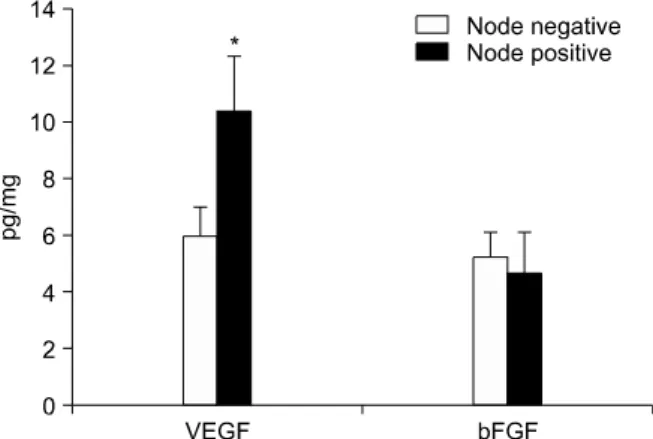
샘암종과 편평상피세포암종을 포함한 모든 종양조직을 대조조직과 비교해 보았을 때 종양조직이 대조조직보다 VEGF 및 bFGF의 농도가 유의하게 높았다(VEGFFigure 3. VEGF and bFGF concentrations in lung cancer tissue according to N stage. Compared with node neg- ative, node positive showed increased tendency of VEGF level in lung cancer tissue. *Increased tendency in node positive lung cancer tissue compared with node negative lung cancer tissue (p=0.06).
Figure 4. VEGF and bFGF concentrations in lung cancer tissue according to pathological stage. There was no sig- nificant difference according to pathological stage.
Figure 1. VEGF and bFGF concentrations in control and lung cancer tissue. Compared with control tissue, lung cancer tissue showed significantly increased VEGF and bFGF levels. *Significant difference between control and lung cancer tissue (p<0.01), †Significant difference be- tween control and lung cancer tissue (p<0.05).
Figure 2. VEGF and bFGF concentrations in lung cancer tissue according to T stage. Compared with T1, T2+T3 showed significantly increased VEGF level in lung cancer tissue. *Significant difference between T1 and T2+T3 (p
<0.05).
7.3±0.9 pg/mg vs 4.2±0.4 pg/mg, p<0.01; bFGF, 5.0±0.7 pg/mg vs 2.9±0.6 pg/mg, p<0.05)(Figure 1).
병리조직학적으로 샘암종과 편평상피세포암종을 나누어 보았을 때, 샘암종의 경우 종양조직과 대조조직 간에 VEGF의 농도가 유의한 차이를 보였으나(p<0.01), 편평 상피세포암종에서는 차이를 보이지 않았다.
3. 종양조직 내 VEGF, bFGF의 농도와 병기 비교
종양의 크기에 따라 T1 (n=10)과 T2+T3 (n=25)로 구분해 보았을 때 T2+T3의 종양조직이 T1의 종양조직보다 유 의하게 VEGF의 농도가 높았으나(8.5±1.1 pg/mg vs 4.3±1.1 pg/mg, p<0.05), bFGF는 차이를 보이지 않았 다(4.0±0.8 pg/mg vs 7.4±1.5 pg/mg, p>0.05)(Figure 2). 림프절의 전이 유무에 따라 구분해 보았을 때 림프절 전이가 있었던 경우가(n=11) 없었던 경우보다(n=24) 종 양조직에서 VEGF의 농도가 증가한 경향을 보였다(10.3±
2.0 pg/mg vs 5.9±0.9 pg/mg, p=0.06)(Figure 3). 병기 에 따라 구분해 보았을 때 각 군 간에 차이는 없었다
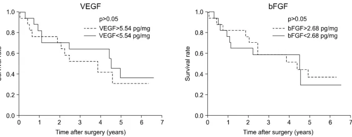
Figure 5. Kaplan-Meier survival curves of non-small cell lung cancer patients after curative surgery according to the median value of VEGF and bFGF concentrations.
(Figure 4).
4. 종양조직 내 VEGF, bFGF의 농도에 따른 생존율의 비교
대상환자의 조직 내 VEGF의 농도를 중앙값인 5.54 pg/mg을 기준으로 나누어 보았을 때 VEGF 저농도군에서 중앙생존기간은 4.5년, 고농도군은 3.9년으로 저농도군에 서 생존율이 증가하는 경향을 보였으나 통계적으로 유의 한 차이를 보이지는 않았으며(p=0.55), bFGF의 농도를 중앙값인 2.68 pg/mg을 기준으로 나누어 보았을 때 bFGF 저농도군에서 중앙생존기간은 4.5년, 고농도군은 4.4년으로 양군간에 차이를 보이지 않았다(p=0.79) (Figure 5).
고 찰
혈관신생은 기존에 존재하는 혈관으로부터 혈관내피세 포의 증식으로 새로운 혈관이 생성되는 것을 말한다. 정 상적인 혈관신생의 대부분은 임신과 관련해서 배아 시기 에 일어나지만 성인에서도 난소주기 또는 상처치유 동안 에 나타나며, 병적인 상태에서는 당뇨병성 망막증, 관절 염, 허혈 상태 등의 비종양성 질환과 악성종양에서 발생할 수 있다7,8.
1971년, Folkman9은 처음으로 종양이 성장하기 위해서 는 혈관신생이 필요하다는 가설을 세웠다. 그는 혈관신생 이 없으면 종양은 1∼2 mm3 이상의 크기로 성장할 수
없기 때문에 종양이 성장하기 위해서는 혈관신생을 촉진 하는 물질을 분비할 것이고 이를 이용하면 궁극적으로는 종양치료에 응용할 수 있을 것이라고 하였다. 당시에 Folkman은 주위의 학자들로부터 상당한 비판을 받았으나 결국 그의 가설은 증명이 되었고 1989년 혈관신생에 가장 중요한 물질인 VEGF가 Napoleone Ferrara에 의해 확인 되었다10.
VEGF는 VEGF-A, -B, -C, -D, -E, placental growth fac- tor로 여섯 가지 그룹이 지금까지 알려져 있고 그 중에서 VEGF-A가 혈관신생에 가장 주요한 물질이고 VEGF-C, D 는 림프관신생에 관여한다. VEGF의 신호전달은 VEGF 수 용체를 통하는데, VEGF가 혈관내피세포에 존재하는 VEGFR-1 (fms-like tyrosine kinase receptor, Flt-1)과 VEGFR-2 (kinase insert domain containing receptor, KDR)에 결합하여 혈관신생을 촉진시킨다. 반면에 VEGFR-3는 림프관에 존재한다. 혈관신생의 주된 작용은 VEGF-A가 VEGFR-2 수용체에 결합에 의해 일어난다11-13. FGF는 FGF-1 (acidic)과 FGF-2 (basic)로 구분되며 혈관 신생을 유도하는 물질로 보고되었으며14 수용체와 결합하 여 혈관내피세포의 증식과 분화에 관여한다15,16. 혈관신생에 대한 연구로 혈관형성 촉진인자의 발현 정 도를 측정하는 방법은 전신적으로는 혈청으로, 국소적으 로는 종양조직을 통해 알아볼 수 있다3,17. 본 연구에서는 종양조직에서 지금까지 가장 중요한 혈관형성 촉진인자 로 알려져 있는 VEGF와 bFGF의 농도를 측정해 보았다.
비록 대조조직이 정상인의 폐조직이 아닌, 수술을 받은
폐암환자의 조직 중에서 종양조직을 제외하고 육안적으 로 정상적인 형태를 보이는 폐조직을 대상으로 하였지만, VEGF와 bFGF가 대조조직보다 종양조직에서 높게 발현 되고 있었으며 VEGF가 bFGF보다 좀 더 통계적으로 유의 한 결과를 보여주었다. 또한 원발병소의 종양크기로 비교 해 보았을 때 T2+T3의 조직이 T1 조직보다 VEGF의 발현 의 증가된 것과 림프절 양성군이 림프절 음성군보다 VEGF의 발현이 증가된 경향(p=0.06)을 보인 점 등을 볼 때 VEGF가 bFGF보다 좀 더 종양조직의 크기 및 림프절 전이 등과 연관성이 높았다. 국내보고에서는 지금까지 폐 암의 혈관신생과 관련하여 Lee18와 Ko 등19이 종양조직에 서 VEGF의 발현 정도를 면역염색으로 측정하여 예후와의 관련성을 보고하였으나, 대조조직과의 비교 및 bFGF의 발현 정도에 대한 보고는 아직 없다.
본 연구에서 VEGF 및 bFGF의 발현 정도를 폐암의 병 기에 따라 구분해 보았을 때 통계적으로 유의한 차이를 보이지 않았으며, 환자의 생존율과도 관련성은 없었다.
Lee18의 보고에서 혈관신생의 정도로 미세혈관농도 (microvessel counts)는 생존율과 관련된 중요한 예후인 자였지만, VEGF의 발현 정도는 예후와 직접적인 관련성 이 없어 이는 본 연구 결과와 일치하는 소견이었다. 하지만 Iwasaki 등6은 VEGF 및 bFGF의 발현 정도가 폐암환자의 생존율과 관련된 예후와 상관관계가 있다고 보고하여 연 구자에 따라 차이가 있음을 알 수 있다. 또한 지금까지 유방암, 위암, 폐암 등에서 VEGF의 발현 정도와 혈관신생 정도, 종양의 악성도 및 생존율에 대한 보고가 있으나 모 두 결과가 일치하는 것은 아니었다. 그 이유에 대해 살펴 보면 첫째, 비록 VEGF와 bFGF가 중요한 혈관형성 촉진 인자이지만 이 두 가지 이외에도 다른 혈관형성 촉진인자 들이 있으며, 또한 억제인자들이 상호작용을 하여 혈관신 생에 관여하므로 한 가지 또는 두 가지 혈관형성 촉진인자 들이 종양과 관련된 모든 혈관신생을 결정하지는 않을 것 이다. 둘째, 수술적으로 절제한 종양조직에 모두 균일한 정도의 혈관형성인자들이 분포하지는 않을 것이다. 일반 적으로 종양의 중심부위에 괴사가 있는 경우 괴사조직보 다는 주변부위에 더 많은 미세혈관 및 혈관형성 촉진인자 들이 분포하고 있다20,21. 그리고 혈관형성인자는 종양세포 뿐만 아니라 혈관내피세포, 염증세포 등에서도 분비된다.
따라서 한 환자의 종양조직 내에서도 부위에 따라서 혈관 형성인자들이 다른 농도로 측정될 가능성이 있다. 셋째, 측정하고자 하는 혈관형성인자들의 방법에 따라 면역염 색방법인지 ELISA 등으로 농도를 측정하였는지 따라 약간
의 차이를 보일 수 있을 것이다.
결론적으로 본 연구에서는 폐암환자의 종양조직 내 혈 관형성 촉진인자인 VEGF 및 bFGF 모두 종양조직에서 증 가하여 폐암의 혈관신생에 관련될 것이라는 기존의 결과 를 확인할 수 있었다. 또한 VEGF는 종양크기, 림프절 전 이 등의 임상양상과 일부 연관성을 보여주었다. 하지만 각각의 VEGF 및 bFGF의 종양조직 내 농도는 생존율의 예후와는 관련되지는 않았다. 이는 다양한 혈관형성 촉진 인자 및 억제인자들의 상호작용을 포함하여 예후와 관련 되는 다른 요소들이 상호작용을 하기 때문으로 생각된다.
요 약
연구배경: 혈관신생은 종양의 성장과 유지 및 전이에 필수적이며 따라서 종양조직은 혈관신생을 위해 많은 종 류의 혈관형성 촉진인자들을 생성하고 있다. VEGF와 bFGF는 혈관신생과 관련되는 물질로 본 연구에서는 폐암 환자에서 조직 내 VEGF와 bFGF의 발현에 대해 알아보고 자 하였다.
방 법: 조직학적으로 샘암종 또는 편평상피세포암종으 로 진단받고 완치의 목적으로 수술을 시행 받은 35명의 폐암환자 조직에서 VEGF 및 bFGF의 농도를 ELISA 방법 으로 측정하였으며 이에 대한 임상적 양상을 후향적으로 분석하였다.
결 과: VEGF 및 bFGF의 농도는 종양조직이 대조조직 보다 유의하게 높았으며 T2+T3의 종양조직이 T1의 종양 조직보다 유의하게 VEGF의 농도가 높았다. 림프절 전이 가 있었던 경우가 없었던 경우보다 종양조직에서 VEGF의 농도가 증가한 경향을 보였다(p=0.06). 하지만 VEGF 및 bFGF의 농도는 환자의 병기 및 생존율에 통계적으로 유 의한 차이를 보이지 않았다.
결 론: VEGF 및 bFGF 모두 종양조직에서 증가하였으 나 VEGF만이 종양크기, 림프절 전이 등의 임상양상과 연 관성을 보여주었다. 하지만 각각의 VEGF 및 bFGF의 종 양조직 내 농도는 예후와 관련되지는 않았다.
참 고 문 헌
1. Choi Y, Kim Y, Hong YC, Park SK, Yoo KY. Temporal changes of lung cancer mortality in Korea. J Korean Med Sci 2007;22:524-8.
2. Ravdin PM, Davis G. Prognosis of patients with re- sected non-small cell lung cancer: impact of clinical
and pathologic variables. Lung Cancer 2006;52:207-12.
3. Bremnes RM, Camps C, Sirera R. Angiogenesis in non-small cell lung cancer: the prognostic impact of neoangiogenesis and the cytokines VEGF and bFGF in tumours and blood. Lung Cancer 2006;51:143-58.
4. Onn A, Herbst RS. Angiogenesis and lung cancer: im- plications for prognosis and treatment. Lancet Oncol 2007;8:460-1.
5. Anderson IC, Mari SE, Broderick RJ, Mari BP, Shipp MA. The angiogenic factor interleukin 8 is induced in non-small cell lung cancer/pulmonary fibroblast cocultures. Cancer Res 2000;60:269-72.
6. Iwasaki A, Kuwahara M, Yoshinaga Y, Shirakusa T.
Basic fibroblast growth factor (bFGF) and vascular en- dothelial growth factor (VEGF) levels, as prognostic in- dicators in NSCLC. Eur J Cardiothorac Surg 2004;25:
443-8.
7. Papetti M, Herman IM. Mechanisms of normal and tu- mor-derived angiogenesis. Am J Physiol Cell Physiol 2002;282:C947-70.
8. Ferrara N. The role of VEGF in the regulation of phys- iological and pathological angiogenesis. EXS 2005;94:
209-31.
9. Folkman J. Tumor angiogenesis: therapeutic impli- cations. N Engl J Med 1971;285:1182-6.
10. Leung DW, Cachianes G, Kuang WJ, Goeddel DV, Ferrara N. Vascular endothelial growth factor is a se- creted angiogenic mitogen. Science 1989;246:1306-9.
11. Carmeliet P. VEGF as a key mediator of angiogenesis in cancer. Oncology 2005;69 Suppl 3:4-10.
12. Cebe-Suarez S, Zehnder-Fjallman A, Ballmer-Hofer K.
The role of VEGF receptors in angiogenesis: complex partnerships. Cell Mol Life Sci 2006;63:601-15.
13. Kiselyov A, Balakin KV, Tkachenko SE. VEGF/VEGFR signalling as a target for inhibiting angiogenesis. Expert
Opin Investig Drugs 2007;16:83-107.
14. Szebenyi G, Fallon JF. Fibroblast growth factors as mul- tifunctional signaling factors. Int Rev Cytol 1999;185:
45-106.
15. Klagsbrun M. Mediators of angiogenesis: the biological significance of basic fibroblast growth factor (bFGF)- heparin and heparan sulfate interactions. Semin Cancer Biol 1992;3:81-7.
16. Cross MJ, Claesson-Welsh L. FGF and VEGF function in angiogenesis: signalling pathways, biological re- sponses and therapeutic inhibition. Trends Pharmacol Sci 2001;22:201-7.
17. Tanaka F, Otake Y, Yanagihara K, Kawano Y, Miyahara R, Li M, et al. Evaluation of angiogenesis in non-small cell lung cancer: comparison between anti-CD34 anti- body and anti-CD105 antibody. Clin Cancer Res 2001;7:
3410-5.
18. Lee SN. The prognostic role of vascular endothelial growth factor (VEGF) expression and angiogenesis in curatively resected non-small cell lung cancer. J Korean Cancer Assoc 1999;31:1210-8.
19. Ko HJ, Park JH, Shim H, Yang SH, Jeong ET.
Prognostic value of vascular endothelial growth factor (VEGF) in resected non-small cell lung cancer. Tuberc Respir Dis 2001;50:676-85.
20. Hemmerlein B, Kugler A, Ozisik R, Ringert RH, Radzun HJ, Thelen P. Vascular endothelial growth factor ex- pression, angiogenesis, and necrosis in renal cell carcinomas. Virchows Arch 2001;439:645-52.
21. Rong Y, Durden DL, Van Meir EG, Brat DJ. 'Pseudop- alisading' necrosis in glioblastoma: a familiar morpho- logic feature that links vascular pathology, hypoxia, and angiogenesis. J Neuropathol Exp Neurol 2006;
65:529-39.