• 검색 결과가 없습니다.

Right Ventricular Myocardial Infarction due to Right Coronary Artery Total Occlusion Originating From the Distal Left Circumflex Artery

N/A
N/A
Protected

Academic year: 2022

Share "Right Ventricular Myocardial Infarction due to Right Coronary Artery Total Occlusion Originating From the Distal Left Circumflex Artery"

Copied!
3
0
0

로드 중.... (전체 텍스트 보기)

전체 글

(1)

565 Copyright © 2012 The Korean Society of Cardiology

Korean Circulation Journal

Introduction

Congenital coronary artery anomalies are usually detected as in- cidental findings during conventional coronary angiography (CAG).

These anomalies are found in 0.6-1.3% of the population who un- dergo CAG.

1)

An isolated single coronary artery (SCA) with the right coronary artery (RCA) originating from the left circumflex coronary artery (LCX) is a rare variant among coronary artery anomalies.

2)

Also, it is extremely rare that this anomaly is associated with right ven- tricular myocardial infarction.

Case

A 39-year-old male smoker with hyperlipidemia suffered from sudden chest pain and dyspnea -1 hour earlier. He had no specific medical or family history of coronary artery disease. On physical ex- amination, the blood pressure was 85/60 mm Hg, and his pulse rate

Case Report

http://dx.doi.org/10.4070/kcj.2012.42.8.565 Print ISSN 1738-5520 • On-line ISSN 1738-5555

Right Ventricular Myocardial Infarction due to Right Coronary Artery Total Occlusion Originating From the Distal Left Circumflex Artery

Sung Ho Ma, MD, Dong Hyun Kim, MD, Jae Young Hur, MD, Kwang Seok Kim, MD, Se Jin Byun, MD, Ki Hyun Park, MD, and Seong-Bo Yoon, MD

Department of Internal Medicine, Hongik Hospital, Seoul, Korea

An isolated single coronary artery is rare but often associated with other congenital cardiac malformations and myocardial ischemia. We report a rare case of right ventricular myocardial infarction due to total occlusion of the right coronary artery originating from the distal left circumflex artery. (Korean Circ J 2012;42:565-567)

KEY WORDS: Myocardial infarction; Coronary vessel anomalies.

Received: November 7, 2011

Revision Received: December 22, 2011 Accepted: January 3, 2012

Correspondence: Seong-Bo Yoon, MD, Department of Internal Medicine, Hongik Hospital, 899-1 Sinjeong 4-dong, Yangcheon-gu, Seoul 158-738, Korea

Tel: 82-2-2600-0436, Fax: 82-2-2600-4605 E-mail: [email protected]

• The authors have no financial conflicts of interest.

This is an Open Access article distributed under the terms of the Creative Commons Attribution Non-Commercial License (http://creativecommons.

org/licenses/by-nc/3.0) which permits unrestricted non-commercial use, distribution, and reproduction in any medium, provided the original work is properly cited.

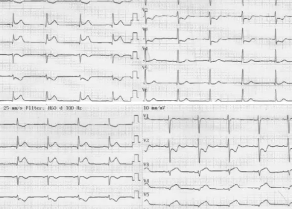
was 46/min with Kussmaul’s sign. An initial electrocardiogram showed definite ST-segment elevation in the inferior leads (II, III, aVF) with reciprocal changes (Fig. 1A), as well as ST-segment elevation in the reverse precordial leads (V3R, V4R, V5R, V6R) (Fig. 1B). Trans- thoracic echocardiography demonstrated akinesia of the right ventricular free wall, inferior wall, and posterior-lateral wall from the left ventricle base to the apex. Also, echocardiography showed 43.6%

of the left ventricular ejection fraction. The initial cardiac enzyme test was elevated (creatine kinase-MB 6.7 ng/mL). Emergency CAG was performed with the assumption of right ventricular myocardial in- farction related to the proximal RCA. The CAG showed total occlu- sion of the distal LCX (Fig. 2) and absence of the RCA ostium, despite repeated attempts at RCA catheterization and ascending aortogra- phy. Therefore, we concluded that the coronary artery related with the right ventricular infarction was the RCA originating from the distal LCX. Immediately after thrombus aspiration with a thrombus aspiration catheter (Thrombuster II®, Kaneka, Osaka, Japan) at the distal LCX, percutaneous coronary intervention (PCI) was followed by balloon angioplasty (Ikazuchi® 3.0×15 mm, Kaneka, Osaka, Japan) and stent insertion (Biomatrix® 4.0×18 mm, Biosensors, Morges, Switzerland) at the distal LCX. After stent insertion, CAG showed no residual stenosis and good distal flow at the distal LCX. Also, CAG showed that the distal LCX extended to the course of the RCA (Fig. 3).

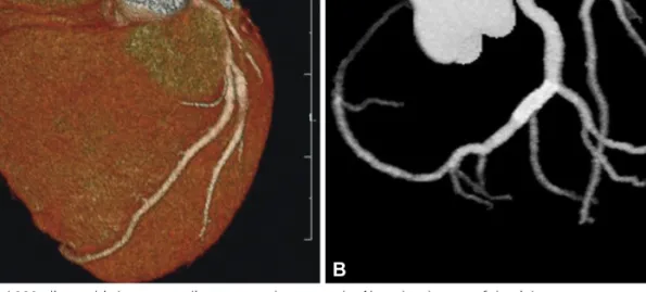
Before discharge, contrast enhanced 320-slice multi-detector car- diac computed tomography showed that the RCA ostium was ab- sent (Fig. 4A), and the distal LCX was extended to the RCA territory while supplying the right ventricle and patent distal LCX stent (Fig. 4B).

After PCI, the patient had no chest pain and was discharged without

any significant complications.

(2)

566 RV Infarction Related to Coronary Artery Anomaly

http://dx.doi.org/10.4070/kcj.2012.42.8.565 www.e-kcj.org

Discussion

The definition of SCA is that the blood flow of all coronary arteries

is provided by a single aortic ostium.

3)

Coronary artery anomalies are rare and SCA is present in 0.04-0.4% of the population who undergo CAG.

4)

This anomaly can be associated with a congenital

A  

B  

Fig. 1. Electrocardiography showing ST-segment elevation in the inferior leads (II, III, aVF) (A), and ST-segment elevation in the reverse precordial leads (V3R, V4R, V5R, V6R) (B).

Fig. 2. Coronary angiography showing total occlusion at the distal left cir-

cumflex artery. Fig. 3. Coronary angiography after stent insertion showed no residual ste-

nosis with good distal flow and extended the distal left circumflex artery

belong to course of the right coronary artery.

(3)

567 Sung Ho Ma, et al.

http://dx.doi.org/10.4070/kcj.2012.42.8.565 www.e-kcj.org

heart anomaly such as Tetralogy of Fallot, coronary arterio-venous fistula, or a bicuspid aortic valve.

2)

If this anomaly is not accompanied by atherosclerosis or aortic stenosis, the prognosis of SCA is relatively good. However, the more distal the SCA, the higher the blood flow resistance. This may cause a myocardial ischemia of the distal portion of the coronary artery.

4)

Sudden death of exercising men with this anomaly has often been reported and the cause is known to be insufficient collateral blood supply compared to that of a normal coronary artery. However, the cli- nical course can deteriorate as a myocardial infarction in the SCA.

5)

In this case, the RCA originated from the distal LCX and this an- omaly was classified as type L-1 according to the Lipton classifica- tion.

4)

Total occlusion of the RCA originating from the distal LCX caused right ventricular myocardial infarction with inferior myocar- dial infarction. Thus, we made a successful PCI and the patient’s con- dition improved.

Through a literature review, we searched for other types of RCA abnormalities originating from the left coronary sinus. There were 3 cases of the RCA arising from the LAD.

6-8)

These cases were associ- ated with myocardial infarction and PCI was conducted. However, these cases were not associated with right ventricular myocardial infarction. In another case with right ventricular myocardial infarc- tion, the RCA arising from the aorta passed between the ascending aorta and the pulmonary outflow tract, and the occlusion of the proximal RCA caused right ventricular myocardial infarction.

9)

Also, there was a similar case of the RCA arising from the LCX. This case was associated with right ventricular infarction and the initial CAG showed severe stenosis in the proximal LCX and a visible RCA arising from the distal LCX.

10)

However, our case involved a total occlusion of the RCA and the initial CAG showed no visible RCA. After PCI, we could observe the RCA originating from the distal LCX. Thus, our case is the first report of right ventricular infarction due to total oc-

clusion of an RCA originating from the distal LCX, followed by a suc- cessful PCI in Korea. We also report here a review of literature rel- evant to this case.

References

1. Chaitman BR, Lespérance J, Saltiel J, Bourassa MG. Clinical, angio- graphic, and hemodynamic findings in patients with anomalous origin of the coronary arteries. Circulation 1976;53:122-31.

2. Sharbaugh AH, White RS. Single coronary artery: analysis of the ana- tomic variation, clinical importance, and report of five cases. JAMA 1974;230:243-6.

3. Ogden JA, Goodyer AV. Patterns of distribution of the single coronary artery. Yale J Biol Med 1970;43:11-21.

4. Lipton MJ, Barry WH, Obrez I, Silverman JF, Wexler L. Isolated single coronary artery: diagnosis, angiographic classification, and clinical sig- nificance. Radiology 1979;130:39-47.

5. Fraisse A, Quilici J, Canavy I, Savin B, Aubert F, Bory M. Images in car- diovascular medicine: myocardial infarction in children with hypoplas- tic coronary arteries. Circulation 2000;101:1219-22.

6. Takano M, Seimiya K, Yokoyama S, et al. Unique single coronary artery with acute myocardial infarction: observation of the culprit lesion by intravascular ultrasound and coronary angioscopy. Jpn Heart J 2003;

44:271-6.

7. Aziz S, Ramsdale DR. Successful stenting of the left anterior descend- ing artery in a patient with a single left coronary ostium. Heart 2004;

90:799.

8. Hsu LA, Chu PH, Ko YS, Ko YL, Chiang CW. Transluminal coronary an- gioplasty and stenting in a patient with single coronary artery and acute myocardial infarction. Changgeng Yi Xue Za Zhi 1997;20:299- 303.

9. Saremi F, Gurudevan SV, Harrison AT. Isolated right ventricular infarc- tion owing to anomalous origin of right coronary artery: role of MR and CT in diagnosis. J Thorac Imaging 2009;24:34-7.

10. Voyce SJ, Abughnia H. An unusual cause of right ventricular myocar- dial infarction. J Invasive Cardiol 2010;22:E172-5.

A   B  

Fig. 4. Contrast enhanced 320-slice multi-detector cardiac computed tomography. Note the absence of the right coronary artery ostium at right coronary

cusp (A) and the extended distal portion of the left circumflex artery with patent stent (B).

수치

Fig. 1. Electrocardiography showing ST-segment elevation in the inferior leads (II, III, aVF) (A), and ST-segment elevation in the reverse precordial leads  (V3R, V4R, V5R, V6R) (B).
Fig. 4. Contrast enhanced 320-slice multi-detector cardiac computed tomography. Note the absence of the right coronary artery ostium at right coronary  cusp (A) and the extended distal portion of the left circumflex artery with patent stent (B).

참조

관련 문서

B: LAO caudal protection marking the site of LAD stump (arrow). C: AP caudal view showing the separate LAD ostium di- rectly originating from the left coronary aortic sinus. D:

was identified in eight patients; seven presented with ACS, and three with inferior myocardial infarction. 6)7) In most of the reported double RCA cases with atherosclerosis,

Acute myocardial infarction (AMI) is a rare complication that can occur after blunt chest trauma. 1)2) Blunt chest trau- ma can lead to various cardiac complications ranging from

Percutaneous coronary intervention (PCI) of chronic total occlusion in coronary arteries (CTO) has a lower success rate than PCI of nonoccluded coronary stenosis. 1)2) A

Fig. Coronary angiographic findings of the patient. A: angiographically normal left coronary system. B: the inferior wall of the left ventricle was supplied with rich collaterals

6 Moreover; in the latter case, the obtuse marginal(OM) branch was so prominent it was mis- judged as being the main circumflex artery. In conclusion, when myocardial infarction in

Multidetector computed tomography shows left and right coronary arteries originate from the left sinus of Valsalva (arrow) and proximal stenosis of the right coronary artery

Anomalous origin of the right coronary artery from the left anterior descending artery in a patient with single left coronary artery: a rare coronary artery anomaly and review of