191
Copyrights © 2014 The Korean Society of Radiology
INTRODUCTION
The most cases of anomalous origins of the vertebral artery (VA) are incidental findings because these variations are clinically asymptomatic (1). Recently, technological advances in CT and MR angiography imaging allow a better delineation of anatomical variations as well as vascular pathologies in the major intracranial and cervical arteries. Although an anomalous origin of the right vertebral artery (RVA) is rare, the identification of this variation is important for the performance of endovascular procedures or cardiothoracic surgeries (2). In this paper, a rare case of an aber- rant RVA originated from the aortic arch distal to the left subcla- vian artery on CT angiography will be presented, the relevant lit- eratures on this variation will be reviewed and their embryologic development and clinical implications will be discussed also.
CASE REPORT
A 34-year-old man visited after a stabbing injury of the neck
the emergency department with a hemiparesis right. The patient was alert awake and had stable vital signs on arrival. An initial neurologic examination revealed decreased muscle strength of the right extremities as 1/5 lower extremity power and 2/5 right upper extremity power. Following laboratory findings were with- in normal ranges: complete blood count, serum electrolytes, liver function test, serum creatinine and routine urinalysis. The pa- tient underwent a contrast-enhanced CT angiography for the evaluation of combined vascular injuries. The CT scan revealed a linear foreign body with low density (chopstick) in the right central spinal canal at C4–5 level without an associated vascular injury (Fig. 1A). Incidentally, it was also detected an anomalous origin of the RVA arising from the aortic arch distal to the left subclavian artery with Kummerell’s diverticulum (Fig. 1B, C).
The prevertebral segment of the RVA was located in the retro- esophageal and retrotracheal areas (Fig. 1D, E). Also it had an aberrant entrance to the C7 transverse foramen while the left VA showed a normal entrance to the C6 transverse foramen (Fig. 1F, G).
Case Report
pISSN 1738-2637 / eISSN 2288-2928 J Korean Soc Radiol 2014;70(3):191-194 http://dx.doi.org/10.3348/jksr.2014.70.3.191
Received November 18, 2013; Accepted December 29, 2013 Corresponding author: Hye Jin Baek, MD
Department of Radiology, Inje University College of Medicine, Haeundae Paik Hospital, 875 Haeundae-ro, Haeundae-gu, Busan 612-896, Korea.
Tel. 82-51-797-0366 Fax. 82-51-797-0379 E-mail: [email protected]
This is an Open Access article distributed under the terms of the Creative Commons Attribution Non-Commercial License (http://creativecommons.org/licenses/by-nc/3.0) which permits unrestricted non-commercial use, distri- bution, and reproduction in any medium, provided the original work is properly cited.
We present a rare case of an aberrant right vertebral artery originated from the distal aortic arch. This issue has been incidentally detected on a preoperative CT angiography after a stabbing injury of the cervical spinal cord. Normally, the right vertebral artery originates from the right subclavian artery. Therefore, in this case report we will review the incidence and the embryological mechanism of this aberrant course of the right vertebral artery and we will discuss as well the clinical importance of this variation.
Index terms Anomalous Origin Aberrant Vertebral Artery Subclavian Artery CT Angiography
Aberrant Right Vertebral Artery Originating from the Aortic Arch Distal to the Left Subclavian Artery: A Case Report
좌측 쇄골하 동맥 원위부 대동맥 궁에서 이상 기시하는 우측 척추 동맥:
증례 보고
Soo Heui Baek, MD, Hye Jin Baek, MD
Department of Radiology, Haeundae Paik Hospital, Inje University College of Medicine, Busan, Korea
Aberrant Right Vertebral Artery Originating from the Aortic Arch Distal to the Left Subclavian Artery
192
J Korean Soc Radiol 2014;70(3):191-194 jksronline.orgcostal longitudinal anastomosis which links the cervical inter- segmental arteries. The intersegmental arteries eventually oblit- erated with an exception of the seventh, which becomes the proximal subclavian artery and includes the VA point of origin in adults (3, 7, 8). An aberrant RVA originating from the aortic arch distal to the left subclavian artery is compatible with the persistence of the proximal dorsal aorta on the right side and with the segment regression of the right dorsal aorta between the sixth and seventh intersegmental arteries (3, 7, 8). The RVA is the only branch to stay connected to the persistent proximal dorsal aorta and arises distal to the left subclavian artery if the right subclavian artery originates from the seventh intersegmen- tal artery normally (1, 3).
With only 11 cases published in the literature previously the aberrant RVA as the last branch of the aortic arch is a very rare
DISCUSSION
Although the VA is classically the first branch of the ipsilateral subclavian artery, multiple anomalous origins of the VA have been reported in the literature (1). With a reported prevalence of 2.4–5.8% in a large series of autopsies is the left VA arising from the aortic arch between the left common carotid artery and left subclavian artery the most common variation of a VA origin (2- 4). An aberrant RVA is an extremely rare anomaly and may con- fuse an aberrant right subclavian artery by its retroesophageal course, especially when it is stenotic (5, 6). This variant is divid- ed into three categories: first, those directly originating from the aorta; second, those originating from the carotid or brachioce- phalic arteries; and third, those with duplicated origin (2, 4).
Embryologically, the VA is formed by the development of post-
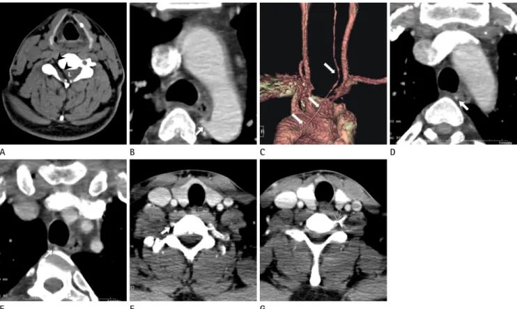
Fig. 1. A 34-year-old man with an incidentally diagnosed aberrant right vertebral artery.
A. Unenhanced axial CT image reveals a linear, hypodense foreign body (chopstick, arrowheads) in the right central spinal canal (L) at C4–5 level caused by stabbing injury.
B. Enhanced axial CT image reveals an aberrant origin of the right vertebral artery (RVA) with a Kummerell’s diverticulum at its origin (arrow).
C. Three dimensional volume rendering image demonstrates an anomalous origin of the RVA (arrows) that originating from the aortic arch distal to the left subclavian artery.
D, E. Enhanced axial CT images shows an unusual course of the RVA. The prevertebral segment (arrows) is located in the retroesophageal and retrotracheal areas.
F, G. Enhanced axial CT images reveal an aberrant entrance to the C7 transverse foramen of the RVA (arrow) which is compared with a normal entrance to the C6 transverse foramen of the left vertebral artery (open arrow).
E A
F B
G
C D
Soo Heui Baek, et al
193
jksronline.org J Korean Soc Radiol 2014;70(3):191-194
208-210
3. Albayram S, Gailloud P, Wasserman BA. Bilateral arch ori- gin of the vertebral arteries. AJNR Am J Neuroradiol 2002;
23:455-458
4. Ka-Tak W, Lam WW, Yu SC. MDCT of an aberrant right subclavian artery and of bilateral vertebral arteries with anomalous origins. AJR Am J Roentgenol 2007;188:W274- W275
5. Karcaaltincaba M, Haliloglu M, Ozkan E, Kocak M, Akinci D, Ariyurek M. Non-invasive imaging of aberrant right sub- clavian artery pathologies and aberrant right vertebral ar- tery. Br J Radiol 2009;82:73-78
6. Karcaaltincaba M, Strottman J, Washington L. Multidetec- tor-row CT angiographic findings in the bilateral aortic arch origin of the vertebral arteries. AJNR Am J Neurora- diol 2003;24:157
7. Newton TH, Mani RL. The vertebral artery. In Newton TH, Potts DG. Radiology of skull and Brain. St. Louis, MO:
Mosby, 1974:1659-1672
8. Moore KL. The Developing Human: Clinically Oriented Em- bryology, 3rd ed. Philadelphia, PA: Saunders, 1982 9. Goray VB, Joshi AR, Garg A, Merchant S, Yadav B, Mahesh-
wari P. Aortic arch variation: a unique case with anoma- lous origin of both vertebral arteries as additional branch- es of the aortic arch distal to left subclavian artery. AJNR Am J Neuroradiol 2005;26:93-95
10. Dabus G, Walker MT. Right vertebral artery arising from the aortic arch distal to the left subclavian artery diag- nosed with magnetic resonance angiography. Arch Neurol 2010;67:508
variation (6, 9, 10). In the most cases described in the literature, there were no clinical signs or symptoms related with this variant (2-4). In the present case the aberrant RVA showed an anoma- lous course of the prevertebral segment located in the retro- esophageal and retrotracheal areas with an aberrant entrance to the C7 transverse foramen. In contrast to previous reports, the patient presented a neurological deficit associated with a spinal cord injury after a stabbing accident.
A detailed knowledge about aberrations of the VA origin is potentially important to avoid an inadvertent vascular injury and an associated ischemic event of the brainstem during the endovascular procedures or a cardiothoracic surgery (1, 2, 4, 5).
Therefore, the possibility of such a variant must be considered if a VA cannot be found in the usual position.
In conclusion, we provide a rare case of an aberrant RVA as the last branch of the posterior aspect of the aortic arch in a pa- tient with a stabbing injury. With this report it will be suggested that an awareness of anomalous origins of the VA and its em- bryologic mechanism may be helpful for an identification of this variant on CT or MR angiographies in clinical practices.
REFERENCES
1. Lemke AJ, Benndorf G, Liebig T, Felix R. Anomalous origin of the right vertebral artery: review of the literature and case report of right vertebral artery origin distal to the left sub- clavian artery. AJNR Am J Neuroradiol 1999;20:1318-1321 2. Nasir S, Hussain M, Khan SA, Mansoor MA, Sharif S.
Anomalous origin of right vertebral artery from right ex- ternal carotid artery. J Coll Physicians Surg Pak 2010;20:
Aberrant Right Vertebral Artery Originating from the Aortic Arch Distal to the Left Subclavian Artery
194
J Korean Soc Radiol 2014;70(3):191-194 jksronline.org좌측 쇄골하 동맥 원위부 대동맥 궁에서 이상 기시하는 우측 척추 동맥: 증례 보고
백수희 · 백혜진
본 저자들은 경부 척수 자상으로 내원하여 시행한 수술 전 CT 혈관조영술에서 우연히 발견된 우측 미입 척추 동맥에 관한 증례를 경험하였다. 정상적으로 우측 척추 동맥은 우측 쇄골하 동맥의 근위부에서 기시하나 본 증례에서는 우측 척추 동 맥이 좌측 쇄골하 동맥의 원위부 대동맥 궁에서 이상 기시를 보였다. 우측 척추 동맥의 이상 기시는 대동맥 궁과 그 분지 들의 발달 과정에서 생기는 발생학적 변이로, 저자들은 본 증례 보고를 통하여 척추 동맥 변이의 발생학적 과정과 이러한 혈관 변이의 임상적 중요성을 강조하고자 한다.
인제대학교 의과대학 해운대백병원 영상의학과