• 검색 결과가 없습니다.

An Extremely Rare Variety of Anomalous Coronary Artery: Right Coronary Artery Originating From the Distal Left Circumflex Artery

N/A
N/A
Protected

Academic year: 2022

Share "An Extremely Rare Variety of Anomalous Coronary Artery: Right Coronary Artery Originating From the Distal Left Circumflex Artery"

Copied!
3
0
0

로드 중.... (전체 텍스트 보기)

전체 글

(1)

465

Print ISSN 1738-5520 / On-line ISSN 1738-5555 Copyright © 2010 The Korean Society of Cardiology CASE REPORT

DOI 10.4070 / kcj.2010.40.9.465

Open Access

An Extremely Rare Variety of Anomalous Coronary Artery:

Right Coronary Artery Originating From the Distal Left Circumflex Artery

Seung-Kyu Chung, MD, Seung-Jin Lee, MD, Sang-Ho Park, MD, Se-Whan Lee, MD, Won-Yong Shin, MD and Dong-Kyu Jin, MD

Department of Internal Medicine, Soonchunhyang University College of Medicine, Cheonan Hospital, Cheonan, Korea ABSTRACT

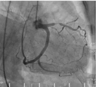
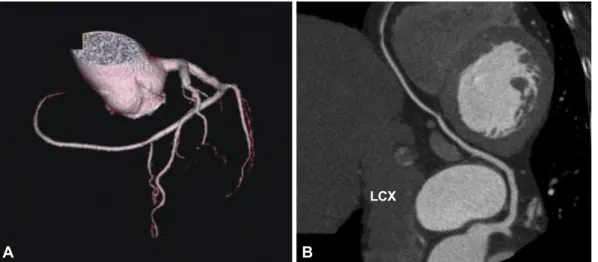
A single coronary artery (SCA) is a rare congenital anomaly of the coronary circulation, which is often associated with myo- cardial ischemia and other congenital cardiac anomalies. A 77-year-old woman visited our hospital complaining of typical chest pain. Coronary angiography revealed an isolated SCA. The right coronary artery did not originate from the aorta, but in- stead emerged from the distal left circumflex artery, with significant stenosis at the proximal portion of the left anterior de- scending artery. A stent was successfully implanted at the culprit lesion. There was no perfusion defect detected by a cardiac SPECT study. (Korean Circ J 2010;40:465-467)

KEY WORDS: Coronary vessel anomalies; Angina pectoris.

Received: January 3, 2010 Accepted: February 19, 2010

Correspondence: Seung-Jin Lee, MD, Department of Internal Medicine, Soonchunhyang University College of Medicine, Cheonan Hospital, 23- 20 Bongmyeong-dong, Dongnam-gu, Cheonan 330-721, Korea Tel: 82-41-570-3673, Fax: 82-41-574-5762

E-mail: [email protected]

cc

This is an Open Access article distributed under the terms of the Cre- ative Commons Attribution Non-Commercial License (http://creativecom- mons.org/licenses/by-nc/3.0) which permits unrestricted non-commer- cial use, distribution, and reproduction in any medium, provided the origi- nal work is properly cited.

Introduction

A single coronary artery (SCA) is an extremely rare con- genital anomaly of the coronary circulation, often associated with other congenital cardiac malformations and frequently presenting with angina.

1)

The incidence of SCA is only 0.024%

to 0.066% in the general population undergoing coronary an- giography.

2)

We report a case with a congenital anomalous right coronary artery, extending from the left circumflex ar- tery, with stenosis in the proximal left anterior descending ar- tery. The anomaly was successfully treated by stenting.

Case

A 77-year-old woman presented with complains of progres- sive effort-related chest pain for 20 days. She had no particu-

lar medical history. On physical examination, the blood pres- sure was 130/70 mmHg, and her pulse rate was 76 beats/min.

Her heart sounds were normal and no murmur was heard.

The electrocardiogram displayed normal sinus rhythm with- out any ST depression or T-wave inversion. The transthoracic echocardiography demonstrated normal hemodynamics with a normal pattern of contractility. The coronary angiography showed that the left coronary artery originated normally from the left sinus of Valsalva, but revealed a 90% tight stenosis at the proximal portion of the left anterior descending artery (Fig. 1). The figure of the left circumflex artery was unique, in that an aberrant branch extended from the distal left circum- flex artery, which crossed the crux and continued to the right atrioventricular groove, covering the territory of the right cor- onary artery (Fig. 2A). After several failed attempts to engage the right coronary artery, we performed aortography using a power injector to exclude high take-off of the right coronary artery. This also confirmed the absence of the right coronary ostium (Fig. 2B). Without delay, we successfully deployed a 4.0×12 mm zotarolimus-eluting stent (Endeavor

TM

, Medtronic Vascular, Santa Rosa, CA, USA) at the left anterior descend- ing artery, after intravenous injection of 5,000 IU heparin.

Contrast-enhanced 64-slice multi-detector cardiac comput-

ed tomography also showed absence of the right coronary ar-

tery and the extended left circumflex coronary artery to the

right coronary artery territory (Fig. 3). Before discharge, a te-

(2)

466 Single Coronary Artery

chnetium-99m single photon emission computerized to- mography (SPECT) study demonstrated noregional any re- gional perfusion defect. The stent was intact in an 8-month follow-up coronary angiography.

Discussion

The definition of an isolated SCA is when only one coro- nary artery originates from one of the sinus of Valsalvas, with this supplying the entire heart, regardless of its distribution.

This type of congenital anomaly is very rare, with a reported incidence of only 0.024% to 0.066% in the general popula- tion undergoing coronary angiography.

2)

In Korea, the inci- dence is even less. One retrospective study showed only 4 patients (0.00025%) diagnosed with SCA among 16,099 pa- tients, who underwent coronary angiography.

3)

In 1979, Lip- ton et al.

2)

proposed a very useful angiographic classification,

further modified in 1990 by Yamanaka and Hobbs.

4)

Accord- ing to the Lipton classification, our patient would be catego- rized in the L-I group, where L implies the ostium is located in the left sinus of Valsalva.

SCA is commonly associated with other congenital anom- alies, such as transposition of the great vessels, coronary ar- teriovenous fistula, or bicuspid aortic valve.

1)

Its prognosis is usually benign, and it is known that SCA does not interfere with coronary perfusion. However, Yamanaka and Hobbs

4)

ca- tegorized SCA as a potentially serious anomaly in the analy- sis of 1,686 patients undergoing coronary angiography. Some reports have also indicated that significant symptomatic isch- emic heart disease developed in 15% of SCA patients.

5)

It is reasonable to think that the clinical meaning and risk of SCA depends on the degree of stenosis and position of the stenotic lesion. If the stenosis develops at the proximal portion of the originated artery, it can cause grave clinical events, and cau- tion is needed when performing intervention.

An interesting case report, clinically presented as acute myo- cardial infarction at the middle portion of the anterior de- scending artery, revealed that the right coronary artery orig- inated from the middle portion of the left anterior descending artery, on the distal part of the culprit lesion, through the tr- ansverse branch.

6)

This patient was successfully treated with a stent. In another SCA case, the right coronary artery origi- nated from the distal left circumflex artery and had a signifi- cant stenosis just at the take-off of the aberrant right coronary artery. This patient was successfully treated with coronary by- pass surgery.

7)

However, myocardial ischemia can develop without definite stenosis, due to coronary vasospasm,

8)

with slow controlled ischemia caused by its long travel distance, as one of the pos- sible mechanisms. Consequently, if a patient complains of chest pain, but does not show causative lesions in SCA, the above possibility should be considered. It is necessary to check whether ischemia can develop by stress tests such as a tread- Fig. 1. Coronary angiography showed 90% stenosis at the proxi-

mal portion of the left anterior descending artery.

Fig. 2. Coronary angiography. An aberrant branch, extending from the distal left circumflex artery, continued to the right atrioventricular groove, covering the territory of the right coronary artery (A). Aortography showed the absence of the right coronary ostium (B).

A B

(3)

Seung-Kyu Chung, et al. 467

mill test or a SPECT scan. In our patient, coronary angiogra- phy revealed a relatively slow flow and delayed contrast me- dia washout in SCA, but did not show ischemia in a techne- tium-99m SPECT study.

REFERENCES

1) Ogden JA, Goodyer AV. Patterns of distribution of the single coro- nary artery. Yale J Biol Med 1970;43:11-21.

2) Lipton MJ, Barry WH, Obrez I, Silverman JF, Wexler L. Isolated sin- gle coronary artery: diagnosis, angiographic classification, and clini- cal significance. Radiology 1979;130:39-47.

3) Choi YH, Park JH, Kim YM, Kim YK, Kim MA, Park YB. Congeni- tal anomalies of the coronary arteries detected in adulthood. Korean Circ J 1997;27:287-95.

4) Yamanaka O, Hobbs RE. Coronary artery anomalies in 126,595 pa- tients undergoing coronary arteriography. Cathet Cardiovasc Diagn 1990;21:28-40.

5) Shirani J, Roberts WC. Solitary coronary ostium in the aorta in the ab- sence of other major congenital cardiovascular anomalies. J Am Coll Cardiol 1993;21:137-43.

6) Takano M, Seimiya K, Yokoyama S, et al. Unique single coronary ar- tery with acute myocardial infarction: observation of the culprit lesion by intravascular ultrasound and coronary angioscopy. Jpn Heart J 2003;44:271-6.

7) Vrolix MC, Geboers M, Sionis D, De Geest H, Van de Werf F. Right coronary artery originating from distal left circumflex: an unusual feature of single coronary artery. Eur Heart J 1991;12:746-7.

8) Yoshimoto K, Koiwaya Y, Tajimi T, et al. Anomalous right coronary artery originating from the distal left circumflex artery: single coronary artery with chronic atrial fibrillation. Jpn Heart J 2004;45:679-83.

Fig. 3. Contrast-enhanced 64-slice multi-detector cardiac computed tomography. It showed that the right coronary artery was absent (A) and the extended left circumflex coronary artery supplied the territory of the right coronary artery (B).

LCX

A B

수치

Fig. 2. Coronary angiography. An aberrant branch, extending from the distal left circumflex artery, continued to the right atrioventricular groove,  covering the territory of the right coronary artery (A)
Fig. 3. Contrast-enhanced 64-slice multi-detector cardiac computed tomography. It showed that the right coronary artery was absent (A) and  the extended left circumflex coronary artery supplied the territory of the right coronary artery (B).

참조

관련 문서

B: LAO caudal protection marking the site of LAD stump (arrow). C: AP caudal view showing the separate LAD ostium di- rectly originating from the left coronary aortic sinus. D:

The combination of coronary arteriovenous fistula to the coronary sinus (CS), dilatation of the entire length of coronary artery, coronary aneurysm and persistent left superior

Unreported Coronary Artery Anomaly: Association of Right Coronary Artery and Circumflex Coronary Artery with Single Ostium Originate from High Left Anterior Aorta.. Ferhat

how to diagnose correctly without withdrawing the guide- wire. Pseudo-stenosis caused by vessel wall invagina- tion during interventional treatment of 2 coronary vessels in a

On page 878, the title has been incorrectly marked as “RE: An Unusual Course of Right Coronary Artery Originating from Sinoatrial

Anomalous origin of the right coronary artery from the left anterior descending artery in a patient with single left coronary artery: a rare coronary artery anomaly and review of

Dual left anterior descending coronary artery (LAD) is a rare coronary anomaly and is incidentally detected during coronary angiography.. We report a case of a 65-year-old

An aberrant RVA originating from the aortic arch distal to the left subclavian artery is compatible with the persistence of the proximal dorsal aorta on the right side and with