• 검색 결과가 없습니다.

Acute Myocardial Infarction by Right Coronary Artery Occlusion Presenting as Precordial ST Elevation on Electrocardiography

N/A
N/A
Protected

Academic year: 2022

Share "Acute Myocardial Infarction by Right Coronary Artery Occlusion Presenting as Precordial ST Elevation on Electrocardiography"

Copied!
3
0
0

로드 중.... (전체 텍스트 보기)

전체 글

(1)

536

Print ISSN 1738-5520 / On-line ISSN 1738-5555 Copyright © 2010 The Korean Society of Cardiology CASE REPORT

DOI 10.4070 / kcj.2010.40.10.536

Open Access

Acute Myocardial Infarction by Right Coronary Artery Occlusion Presenting as Precordial ST Elevation on Electrocardiography

Sung Eun Kim, MD, Jun-Hee Lee, MD, Dae-Gyun Park, MD, Kyoo-Rok Han, MD and Dong-Jin Oh, MD

Division of Cardiology, Department of Internal Medicine, Kangdong Sacred Heart Hospital, Hallym University College of Medicine, Seoul, Korea

ABSTRACT

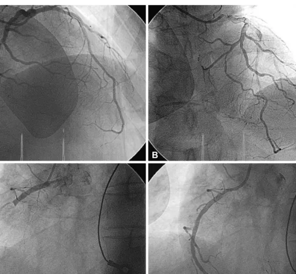
It is rare to observe ST-segment elevation in only the anterior leads and not the inferior leads during right coronary artery occlusion. We describe a case with acute myocardial infarction (MI) by right coronary artery occlusion who developed ST-seg- ment elevation only in the precordial leads V1 to V3. (Korean Circ J 2010;40:536-538)

KEY WORDS: Electrocardiography myocardial infarction.

Received: February 19, 2010 Accepted: March 26, 2010

Correspondence: Jun-Hee Lee, MD, Division of Cardiology, Depart- ment of Internal Medicine, Kangdong Sacred Heart Hospital, 445 Gil 1-dong, Gangdong-gu, Seoul 134-701, Korea

Tel: 82-2-2224-2405, Fax: 82-2-2225-2725 E-mail: [email protected]

cc

This is an Open Access article distributed under the terms of the Cre- ative Commons Attribution Non-Commercial License (http://creativecom- mons.org/licenses/by-nc/3.0) which permits unrestricted non-commer- cial use, distribution, and reproduction in any medium, provided the origi- nal work is properly cited.

Introduction

Acute ST-segment elevations in the precordial leads gen- erally suggest acute occlusion in the branches of the left cor- onary artery. Occlusion of the right coronary artery (RCA) may cause infarction of the inferior wall of the left ventricle with or without right ventricular (RV) myocardial infarction (MI), ma- nifested as ST-segment elevations in leads II, III, and aVF.

Anterior ST elevation due to occlusion of an RCA branch may be a mirror image of RV ischemia.

1-4)

We describe a patient with mild ST elevation in leads V1 to V3 caused by acute oc- clusion at the proximal segment RCA with rich collateral flow from the left coronary artery.

Case

A 52-year-old male smoker with no medical history pre- sented to the hospital because of substernal chest pain last- ing for forty minutes which was associated with shortness of breath and diaphoresis. On admission, his blood pressure was 170/100 mmHg, heart rate 76 beats per minute, and re-

spiratory rate of 20 breaths per minute. Electrocardiography (ECG) showed ST-segment elevations in the left precordial leads V1 through V3, and T-wave inversions in leads III and aVF (Fig. 1). The patient’s creatine kinase level was 287 IU/L (normal range 22-169) with an MB fraction of 8.0 ng/mL (nor- mal range 1-5) and troponin I of 3.4 ng/mL (normal range 0-1.5).

A diagnosis of acute anteroseptal wall MI was made, and the patient was immediately taken to the catheterization lab- oratory for percutaneous coronary intervention (PCI). First, RCA catheterization was done, and it showed a proximal complete occlusion. Angiography of the left coronary artery system demonstrated insignificant stenosis, and there was col- lateral flow of thrombolysis in myocardial infarction (TIMI) grade II to the RCA (Fig. 2). The patient underwent percu- taneous transluminal coronary angioplasty with coronary ar- tery stenting to the infarct-related RCA. Echocardiography performed after PCI demonstrated mild hypokinesis of the inferior wall of the left ventricle and a normal RV.

The in-hospital course of the patient was uneventful, and he was discharged from the hospital 5 days later. There remain- ed T-wave inversion in leads III and aVF without develop- ment of Q-wave.

Discussion

In cases of evolving Q-wave MI due to RCA occlusion, the

presence of ST elevation in V1 to V3-V4 occurs when the

occlusion is proximal to the branch that perfuses the RV.

5)

Acikel et al.

3)

reported ST-segment elevations in leads V1

through V3 caused by occlusion of the RV branch of the RCA

(2)

Sung Eun Kim, et al. 537

while performing primary PCI in a case of acute inferior MI.

Therefore, ST-segment elevations in V1 to V3 are explained by an occlusion proximal to the RV branch. Kida et al.

6)

studied

57 patients undergoing PCI of the RCA and observed pre- cordial ST-segment elevations when inferior wall blood flow was maintained by collaterals or codominant circulation de-

Fig. 2. Coronary angiographic findings of the patient. A: angiographically normal left coronary system. B: the inferior wall of the left ventricle was supplied with rich collaterals from left coronary system. C: coronary angiography showing complete luminal occlusion of the RCA proxi- mally. D: the RCA after percutaneous coronary intervention. RCA: right coronary artery.

B

D A

C

Fig. 1. Standard 12-lead electrocardiography recorded from a patient with chest pain. Note the presence of ST-segment elevations in leads from

V

1

to V

3

, and no ST-segment deviation in leads II, III, and aVF.

(3)

538 RCA Occlusion With ST Elevation in Precordial Leads spite total occlusion of the RCA. In our patient, rich collater- als from the left coronary artery supplied the inferior wall and the total occlusion of the RCA proximal to the RV bran- ch caused RV infarction despite mild inferior wall ischemia.

Although echocardiography performed after PCI showed normal RV size and contractility in our patient, it is probable that RV stunning occurred transiently.

In summary, this is an exceptional ECG showing ST eleva- tion in leads V1 to V3, secondary to occlusion of RCA.

REFERENCES

1) Celik T, Yuksel UC, Kursaklioglu H, Iyisoy A, Kose S, Isik E. Precor- dial ST-segment elevation in acute occlusion of the proximal right cor- onary artery. J Electrocardiol 2006;39:301-4.

2) Erol MK, Kocaturk H, Gurlertop Y. Images in cardiology: precordial ST segment elevation suggestive of anterior infarction caused by right coronary artery occlusion. Heart 2005;91:1134.

3) Acikel M, Yilmaz M, Bozkurt E, Gurlertop Y, Kose N. ST segment ele- vation in leads V1 to V3 due to isolated right ventricular branch occlu- sion during primary right coronary angioplasty. Catheter Cardiovasc Interv 2003;60:32-5.

4) Ryou NS, Cho MH, Shin DH, Cheong SS, Yoo SY. Precordial ST- segment elevation in acute right ventricular myocardial infarction.

Korean Circ J 2008;38:495-9.

5) Jim MH, On-On Chan A, Wong CP, et al. Clinical implications of pre- cordial ST-segment elevation in acute inferoposterior myocardial in- farction caused by proximal right coronary artery occlusion. Clin Car- diol 2007;30:331-5.

6) Kida M, Morishita H, Yokoi H, et al. Precordial ST-segment elevation

caused by right coronary artery occlusion. J Cardiol 1987;17:455-64.

수치

Fig. 1. Standard 12-lead electrocardiography recorded from a patient with chest pain. Note the presence of ST-segment elevations in leads from  V 1  to V 3 , and no ST-segment deviation in leads II, III, and aVF.

참조

관련 문서

1 John Owen, Justification by Faith Alone, in The Works of John Owen, ed. John Bolt, trans. Scott Clark, "Do This and Live: Christ's Active Obedience as the

core exercise program showed not only increasing peak torque of the right leg, left leg in Hip Extension of exercise group but also increasing peak torque of the right leg, left

Therefore, cTnT is a recommended biomarker for use in the detection of myocardial infarction (MI) and in acute coronary syndromes.[8] Indeed, several authors

The positive change pattern of CK-MB according to time on acute myocardiac infarction patient.. The positive change pattern of troponin-T according to time on

Sizes of left and right pupils Intensities of left and right pupils Ellipse ratios of left and right

void makeTree(Object root, Object left, Object right): Creates a binary tree with root as the root element, left as the left subtree, right as the right

Incident major cardiovascular events (coronary artery disease, ischemic stroke, hemorrhagic stroke and cardiovascular mortality) were set as primary end points.

SoS Investigators, Coronary Artery Bypass Surgery versus percutaneous coronary intervention with stent implantation in patients with multi-vessel coronary