• 검색 결과가 없습니다.

A Rare Coronary Anomaly: Atypical Double Right Coronary Artery With an Acute Inferior Myocardial Infarction

N/A
N/A
Protected

Academic year: 2022

Share "A Rare Coronary Anomaly: Atypical Double Right Coronary Artery With an Acute Inferior Myocardial Infarction"

Copied!
4
0
0

로드 중.... (전체 텍스트 보기)

전체 글

(1)

208 Copyright © 2012 The Korean Society of Cardiology Korean Circulation Journal

Introduction

Coronary artery anomalies (CAA) are rare angiographic findings.

The incidence of CAA is about 1-2% in angiographic studies of the adult population.

1)

Double right coronary artery (RCA) is a very rare coronary anomaly. Patients are mostly asymptomatic but acute cor- onary syndrome (ACS) is a possible, yet uncommon clinical present- ation. ACS is linked to CAA in some cases.

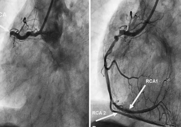
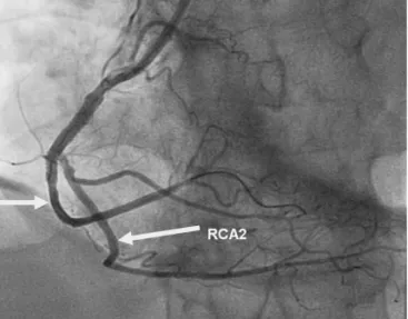
We report a rare anomaly, an A2 atypical double RCA, which pre- sented as an acute inferior wall myocardial infarction due to a th- rombotic occlusion in one of the two RCAs, which was successfully managed with primary percutaneous coronary intervention (PCI).

Case

A 61-year-old male patient was admitted to the emergency de- partment with retrosternal chest pain at rest for the past hour. He

Case Report

http://dx.doi.org/10.4070/kcj.2012.42.3.208 Print ISSN 1738-5520 • On-line ISSN 1738-5555

A Rare Coronary Anomaly: Atypical Double Right Coronary Artery With an Acute Inferior Myocardial Infarction

Halit Acet, MD 1 , Ferhat Ozyurtlu, MD 1 , Mehmet Zihni Bilik, MD 1 , and Faruk Ertas, MD 2

1

Department of Cardiology, Diyarbakir Training and Research Hospital, Diyarbakır,

2

Department of Cardiology, Dicle University Medicine Faculty, Diyarbakr, Turkey

Coronary artery anomalies are uncommon and often asymptomatic. A double right coronary artery (RCA) is an extremely rare coronary artery anomaly, and only a few cases of double RCA have been reported. We report on an atherosclerotic double RCA that appeared after primary percutaneous intervention in a patient with an acute inferior myocardial infarction. This is the second case in the literature in which coronary arteries can be accepted as a double RCA, which were hidden by a total atherosclerotic occlusion in the proximal part of the RCA. (Korean Circ J 2012;42:208-211)

KEY WORDS: Coronary vessel anomalies; Myocardial infarction; Atherosclerosis.

Received: May 25, 2011

Revision Received: August 3, 2011 Accepted: August 3, 2011

Correspondence: Halit Acet, MD, Department of Cardiology, Diyarbakir Training and Research Hospital, Diyarbakır 21090, Turkey

Tel: 90-412-252-3910 , Fax: 90-412-252-3910 E-mail: [email protected]

• The authors have no financial conflicts of interest.

This is an Open Access article distributed under the terms of the Creative Commons Attribution Non-Commercial License (http://creativecommons.

org/licenses/by-nc/3.0) which permits unrestricted non-commercial use, distribution, and reproduction in any medium, provided the original work is properly cited.

was a chronic smoker and had hypertension for 5 years. Blood pres- sure was 130/80 mm Hg, and pulse rate was 86 beats/minute. A phy- sical examination was completely normal. An ST segment elevation was noted in leads II, III, and aVF; and a reciprocal ST segment depres- sion in leads V 1-6 on electrocardiography. Coronary angiography showed total occlusion of the proximal RCA (Fig. 1A). The origin and course of the left coronary arteries were normal. Noncritical lesions were evident in the left anterior descending artery and circumflex artery. Primary PCI was performed on the RCA lesion. The lesion was predilated with a 2.0×15 mm balloon at 12 atm. Two different RCAs originating from a common main trunk appeared following a contrast injection to the right coronary orifice (Fig. 1B). Tubular ec- centric stenosis of 90% was noted in the proximal segment of the main RCA trunk, 70% tubular stenosis in the RCA1, and 40% ste- nosis in the osteal segment of RCA2 (Fig. 2A). Two bare metal stents (3.0×18 mm and 2.75×12 mm) were successfully deployed to the lesion at 15 atm for 30 seconds in the proximal segment of the main trunks of RCA and RCA1, respectively (Fig. 2B). A control coronary angiogram demonstrated two separate RCAs originating from a sin- gle ostium. So, we diagnosed the patient with an A2 atypical dou- ble RCA (Fig. 3). The patient was treated with a tirofiban infusion. The post-interventional period was uneventful, and the patient was fol- lowed up for 2 days at the intensive care unit and discharged with- out any complications.

Discussion

Most coronary anomalies are detected as incidental findings dur-

(2)

209 Halit Acet, et al.

http://dx.doi.org/10.4070/kcj.2012.42.3.208 www.e-kcj.org

ing coronary angiography. However, a double RCA is a very rare co- ronary anomaly. So far, only a few cases of double RCA have been re- ported.

2-5)

Since the second report, this rare anomaly has been ac- cepted as a benign entity. However, new reports in the last few years have indicated that a double RCA may be complicated with athero- sclerosis. Among 27 cases defined in the literature, atherosclerosis

was identified in eight patients; seven presented with ACS, and three with inferior myocardial infarction.

6)7)

In most of the reported double RCA cases with atherosclerosis, the atherosclerotic segment is usu- ally in the proximal part of the RCA that courses on the atrioven- tricular (AV) sulcus or in the posterior branch of the RCA going to the AV sulcus.

8)

In the submitted case of Akçay et al.

9)

and in our case,

A   B  

Fig. 1. Coronary angiography LAO view. A: totally occluded proximal RCA before percutaneous coronary intervention. B: the atypical double RCA appeared after predilatation with a balloon. LAO: left anterior oblique, RCA: right coronary artery.

A   B  

Fig. 2. Coronary angiography RAO view. A: the atypical double RCA appeared after predilatation with a balloon. B: angiography after implantation of a

bare metal stent. RAO: right anterior oblique, RCA: right coronary artery.

(3)

210 Atypical Double Right Coronary Artery

http://dx.doi.org/10.4070/kcj.2012.42.3.208 www.e-kcj.org

a lesion was evident in the ostial part of the anteriorly coursing RCA but not in other parts. Also, in their case, an atherosclerotic lesion was found in the posterior course of the RCA. Based on these two studies by Iacobellis et al.

10)

the authors suggested that an increased quantity of epicardial adipose tissue due to obesity may be a risk predictor for cardiovascular disease.

An attempt to classify a double RCA was tried (Table 1) by an au- thor from Turkey, where most of the studies concerning this anoma- ly have been reported.

11)

We diagnosed our case as an A2 atypical double RCA using this classification. Only one case has been report- ed that was admitted with ACS and identified as atypical RCA by coronary angiography.

11)

In that case, atherosclerotic lesions were only found in the proximal part of the RCA but not in the RCA1 or RCA2, and the stent was implanted in the main RCA. But in our case, atherosclerosis began proximally and was ongoing in RCA1 and

RCA2, so we implanted stents proximal to RCA and RCA1.

To our knowledge no A2 atypical double RCA case has been re- ported that was admitted with ACS and had atherosclerosis in the main RCA as well as in RCA1 and RCA2.

In conclusion, although controversy exists about the definition of a double RCA, it is generally considered a benign entity, but could be atherosclerotic and cause ACSs including myocardial infarction and may be associated with other anomalies.

12)13)

Coronary anoma- lies should be recognized to avoid problems during coronary inter- vention and cardiac surgery. Every operator should be familiar with coronary anomalies to perform an adequate examination.

References

1. Yamanaka O, Hobbs RE. Coronary artery anomalies in 126,995 patients undergoing coronary arteriography. Cathet Cardiovasc Diagn 1990;21:

28-40.

2. Altun A, Akdemir O, Erdogan O, Ozbay G. An interesting diagnostic dilem- ma: double right coronary artery or high take off of a large right ven- tricular branch. Int J Cardiol 2002;82:99-102.

3. Tuncer C, Batyraliev T, Yilmaz R, Gokce M, Eryonucu B, Koroglu S. Origin and distribution anomalies of the left anterior descending artery in 70,850 adult patients: multicenter data collection. Catheter Cardiovasc Interv 2006;68:574-85.

4. Sari I, Kizilkan N, Sucu M, et al. Double right coronary artery: report of two cases and review of the literature. Int J Cardiol 2008;130:e74-7.

5. Rohit M, Bagga S, Talwar KK. Double right coronary artery with acute inferior wall myocardial infarction. J Invasive Cardiol 2008;20:E37-40.

6. Sari I, Uslu N, Gorgulu S, Nurkalem Z, Eren M. Inferior myocardial infarc- tion and extensive atherosclerosis in a patient with double right coro- nary artery. Int J Cardiol 2006;111:321-3.

7. Tatli E, Buyuklu M, Altun A, Yilmaztepe M. A patient with double right coronary artery and acute inferior myocardial infarction due to the critical lesion in one of them. Int J Cardiol 2007;119:e30-1.

Table 1. Classification of double RCA

Classification Type Features

Real (G) Dual RCA G1 Double RCA, origin from the right aortic sinus with separate ostiums and continue in AV sulcus side by side until posterior interventricular sulcus

G2 Double RCA origin from the right aortic sinus. After a short main RCA, they are divided (in proximal), and continue in AV sulcus side by side until posterior interventricular sulcus

G3 Duble RCA origin from different aortic sinuses.

G4 RCA-conus branch or RCA-RV artery origin from separate ostiums

Atypical (A) Double RCA A1 Early PDA (PDA follows normal path from mid part of the RCA . This is also called high-output PLA) A2 Advanced RV branch origin from proximal or mid part of RCA ,and terminated as RCA and PDA A3 PDA and PLA separated from distal RCA before crux

A4 Double PDA separated from crux

Pseudo-Double (Y) RCA Y1 PDA or PLA is not reached to the left ventricle, advanced RV branch is not continued as RCA Y2 Other pseudo-dual RCA situations, such as over-dominant RCA

AV: atrioventricular, PDA: posterior descending artery, PLA: right ventricular posterior-lateral branches, RCA: right coronary artery, RV: right ventricle

Fig. 3. Left anterior oblique view of coronary angiography after implanta-

tion of a bare metal stent.

(4)

211 Halit Acet, et al.

http://dx.doi.org/10.4070/kcj.2012.42.3.208 www.e-kcj.org

8. Sacks HS, Fain JN. Human epicardial adipose tissue: a review. Am Heart J 2007;153:907-17.

9. Akçay A, Koroglu S, Kaya H, Koleoglu M, Acar G. An unusual appear- ance of double right coronary artery. Cardiol Res Pract 2010. pii:

123846.

10. Iacobellis G, Ribando MC, Assal F, et al. Echocardiographic epicardial adi- pose tissue is related to anthropometric and clinical parameters of metabolic syndrome: a new indicator of cardiovascular risk. J Clin Endo- crinol Metab 2003;88:5163-8.

11. Vural M. Suggestions for definition and classification of double right

coronary artery, which is frequently seen in Turkey despite its rarity in the literature. Anadolu Kardiyol Derg (Turkish) 2008;8:303-4.

12. Young-Hyman PJ, Tommaso CL, Singleton RT. A new double coronary artery anomaly: the right coronary artery originating above the coro- nary sinus giving off the circumflex artery. J Am Coll Cardiol 1984;4:

1329-31.

13. Garg N, Goel PK, Sinha N. Double right coronary artery with anomalous left main and septal arteries originating from the right coronary sinus.

Indian Heart J 2002;54:428-31.

수치

Fig. 1. Coronary angiography LAO view. A: totally occluded proximal RCA before percutaneous coronary intervention
Fig. 3. Left anterior oblique view of coronary angiography after implanta- implanta-tion of a bare metal stent.

참조

관련 문서

In addition, Wallace and Froum 20) reported that the bone formation rate was superior in the cases using a barrier membrane in comparison with the

Therefore, cTnT is a recommended biomarker for use in the detection of myocardial infarction (MI) and in acute coronary syndromes.[8] Indeed, several authors

Utility of T1-and T2-weighted high-resolution vessel wall imaging for the diagnosis and follow up of isolated posterior inferior cerebellar artery dissection with

Approved clinical use of bone marrow stem cells for myocardial infarction treatment... Cardiac

The purpose of this study was to analyze the impaction pattern of the impacted mandibular third molar and the relationship with the inferior alveolar nerve

(Background) Gallbladder wall thickening(GWT) and gallbladder contraction are often observed in patients with acute hepatitis.. The incidence of acute hepatitis A

 Although van der Waals equation is still less accurate at high

Incident major cardiovascular events (coronary artery disease, ischemic stroke, hemorrhagic stroke and cardiovascular mortality) were set as primary end points.