711
CASE REPORT Korean Circulation J 2004;34(7):711-714
A Case of Coronary Vessel Anomaly of the Left Circumflex Artery Originating from the Right Coronary Artery with Variant Angina
Joon-Seok Kim, MD, Jong-Min Lee, MD, Hee-Jeoung Yoon, MD, Young-Yong Ahn, MD, Ji-Young Kang, MD, Ji-Young Park, MD, Sun-Jong Jung, MD,
Seung-Won Jin, MD, Ki-Bae Seung, MD and Jae-Hyung Kim, MD
Department of Internal Medicine, College of Medicine, The Catholic University of Korea, Seoul, Korea ABSTRACT
Coronary vessel anomaly is a rare disease, with an incidence of about 0.6-1.3% of patients receiving coronary angiography. The ischemia in coronary vessel anomalies is due in most cases to atherosclerosis or compression of the coronary artery by a great vessel, but occasionally spasm of a coronary vessel anomaly is responsible for the pathogenesis of chest pain and myocardial ischemia. A 64-year-old female presented with a one-year history of effort angina. The left circumflex artery originated from the proximal right coronary artery. There was no athero- sclerotic lesion in the right and left coronary arteries, but a focal spasm in the right coronary artery by ergonovine.
In a patient with chest pain and coronary artery anomaly, if there is no coronary atherosclerosis, abnormal course or compression, the spasm test of the coronary artery should be documented. (Korean Circulation J 2004;34 (7):
711-714)
KEY WORDS:Coronary vessel anomalies;Angina pectoris, variant.
Introduction
Coronary vessel anomaly is a rare disease, which is discovered incidentally by coronary angiography. It pre- sents almost no symptoms, but occasionally causes symp- toms including, unconsciousness, tachycardia, chest pain and sudden death. It has been reported with incidences varying between 0.6 and 1.3% of adult patients under- going coronary angiography.1)2) A coronary vessel ano- maly of the right coronary artery is the most common, with an incidence of left circumflex anomaly of 0.3%.3)
This paper deals with a patient having a left circumflex anomaly originating from the right coronary artery and a coronary artery spasm in the right coronary artery.
Case
A 64-year-old female presented with a one-year history of effort angina. She had an 8 year history of hypertension and hyperlipidemia. On admission, her blood pressure, pulse rate, respiratory rate and body temperature were 150/80 mmHg, 76 beats per min, 20 per min and 36.6℃, respectively. There was no abnormal heart or respiration sounds. A laboratory examination showed that her tropo- nin T, CK-MB, total cholesterol, triglyceride and HDL- cholesterol were 0.01 ng/mL, 5.22 ng/mL, 187 mg/dL, 187 mg/dL and 43.7 mg/dL, respectively. Her thyroid function tests had been normal about 4 months earlier.
An electrocardiogram on admission showed regular sinus rhythm and no ischemic change (Figure 1). The chest radiograph was also normal. An echocardiogram revealed a normal left ventricular ejection fraction and mild tricu- spid regurgitation.
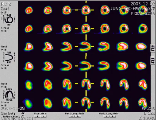
A cardiac perfusion SPECT using 50.4 mg of adeno- sine showed no redistribution (Figure 2). Diagnostic co- Received:April 29, 2004
Accepted:June 15, 2004
Correspondence:Jong-Min Lee, MD,Department of Internal Medicine, College of Medicine, The Catholic University of Korea, 520-2 Daeheung-dong, Jung-gu, Daejeon 301-723, Korea Tel:82-42-220-9504, Fax:82-42-253-9505
E-mail:[email protected]
Coronary Vessel Anomaly with Variant Angina
Korean Circulation J 2004;34(7):711-714 712
ronary angiography revealed a normal left anterior de- scending artery and its branch shows no abnormality or stenosis (Figure 3A). There was no arterial flow in the left circumflex artery during injection into the left coro- nary artery. Afterwards, the right coronary artery was
engaged, showing the left circumflex artery originating from the proximal right coronary artery, with no atheros- clerotic lesion in the right or left coronary arteries (Figure 3B). An ergonovine spasm test showed a focal spasm in the mid-portion of the right coronary artery (Figure 3C),
Figure 1. Electrocardiogram showing no ST-T abnormalities.
Figure 2. Myocardial SPECT-99mTc showing no perfusion defect in either the resting or stress phases.
Joon-Seok Kim, et al
713 although she noted chest pain, and an electrocardiogram
showed ST elevation (not available). She was treated with a calcium channel blocker, nitrate and aspirin after which the chest pain subsided.
Discussion
Coronary vessel anomaly is a rare disease, with an in- cidence of about 0.6-1.3% of patients receiving coronary angiograms and between 0.04-0.4% of the whole popu- lation.1)2)4) In most cases, a congenital cardiac anomaly is accompanied with great vessel transposition or coro- nary arteriovenous fistula.4)5) A coronary vessel anomaly of a right coronary artery anomaly is the most common, with a left circumflex anomaly having an incidence of 0.3% of coronary artery vessel anomalies.4) In our case, there was no cardiac anomaly and the left circumflex artery originated from the proximal part of the right coro- nary artery.
Mavi and his colleague reported that coronary vessel anomalies of the left circumflex artery originated from the left valsalva sinus (55.5%), the right coronary artery (36.9%) and right sinus of valsalva (25.9%).6)
Most coronary vessel anomalies cause no symptoms, but in some cases can cause chest pain, sudden death and myocardial infarction.7)8) The ischemia in coronary vessel anomalies is mostly due to atherosclerosis or compression
of the coronary artery by a great vessel, but occasionally a spasm of a coronary vessel anomaly is responsible for the pathogenesis of chest pain and myocardial ischemia.9) Os- hima and his colleague reported a case suspected as a sp- asm of a coronary vessel anomaly by an electrogram, which they treated with a calcium channel blocker (dil- tiazem, 120 mg per day).10) Kubota and his colleague reported a 68-year-old woman showing an abnormal left circumflex artery originating from the right sinus of the valsalva from coronary angiography and a focal spasm in an ergonovine injection test.11) Additionally, Wojtna and his colleague reported on a 45-year-old woman showing a focal spasm in a coronary vessel anomaly, but without stenosis in a coronary artery. She died due to an inferior wall myocardiac infarction.12)
In our case, the chest pain was due to a coronary artery spasm, as there were no abnormal or ischemic features in an electrocardiogram and myocardiac perfusion scan and with no atherosclerotic stenosis on coronary angiogra- phy. Chest pain arises when a right coronary artery spasm occurs during an ergonovine spasm test.
A coronary vessel anomaly is a rare disease, which is occasionally complicated by chest pain, unconsciousness and myocardiac infarction due to coronary atherosclero- sis, compression by a great vessel or coronary artery spasm.
In a patient with chest pain and a coronary artery anomaly, if there is no coronary atherosclerosis, abnormal course Figure 3. Coronary angiography. A: left coronary angiography: a left anterior descending artery showing no stenosis or arterial flow in the left circumflex artery. B: right coronary artery angiography showing the left circumflex artery origi- nating from the right coronary artery, with no stenosis. C: focal spasm (arrow) of mid portion of the right coronary artery during an ergonovine 50 μg infusion.
A B C
Coronary Vessel Anomaly with Variant Angina
Korean Circulation J 2004;34(7):711-714 714
or compression, the spasm test of the coronary artery should be documented.
REFERENCES
1) Chaitman BR, Lesperance J, Saltiel J, Bourassa MG. Clinical angiographic and haemodynamic finding in patient with ano- malous origin of coronary arteries. Circulation 1976;53:
122-31.
2) Garg N, Tewari S, Kapoor A, Gupta DK, Sinha N. Primary congenital anomalies of coronary arteries: a coronary arte- riographic study. Int J Cardiol 2000;74:39-46.
3) Mavi A, Sercelik A, Ayalp R, Pestemalci T, Batyraliev T, Gumusburun E. Variants origin of left circumflex coronary artery with angiograph. Saudi Med J 2002;23:1390-3.
4) Sharbaugh AH, White RS. Single coronary artery: analysis of the anatomic variation, clinical importance and report of five cases. JAMA 1974;230:243-6.
5) Lipton MJ, Barry WH, Obrez I, Silverman JF, Wexler L.
Isolated single coronary artery: diagnosis, angiographic cla- ssification and clinical significance. Radiology 1979;130:
39-47.
6) Mavi A, Sercelik A, Ayalp R, Pestemalci T, Batyraliev T, Gumusburun E. Variants in origin of the left circumflex coro-
nary artery with angiography. Saudi Med J 2002;23:1390-3.
7) Taylor AJ, Rogan KM, Virmani R. Sudden cardiac death as- sociated with isolated congenital coronary artery anomalies.
J Am Coll Cardiol 1992;20:640-7.
8) Choi KL, Kwon JI, Jung WH, Kim EA, Choi SJ, Jin DK, et al. Stenting of an anomalous coronary artery in acute myo- cardial infarction. Korean Circ J 1998;28:1378-81.
9) Mustafa I, Gula G, Radely-Smith R, Durrer S, Yacoub M.
Anomalous origin of left coronary artery from anterior aortic sinus: potential cause of sudden death. J Thorac Cardiovasc Surg 1981;82:297-300.
10) Ohsima M, Takizawa A, Watanabe K, Tamura Y, Yamazoe M, Izumi T, et al. A case of dual origin of the left anterior descending coronary artery from the left and right coronary arteries with variant angina. Kokyu To Junkan 1993;41:
667-71.
11) Kubota Y, Monji T, Nakagawa H, Uwatoko H, Kitamura K.
Anomalous origin of the left main coronary artery from the right aortic sinus of valsalva with vasospastic angina. Chest 1991;100:1167-8.
12) Wojtyna W, Wiese KH, Strauss P, Walter J. Prinzmetal phe- nomenon and a rare coronary anomaly as infarct and death cause of a 45-year-old patient. Z Kardiol 1982;71:106-11.