747
Print ISSN 1738-5520 / On-line ISSN 1738-5555 Copyright © 2011 The Korean Society of Cardiology CASE REPORT
http://dx.doi.org/10.4070/kcj.2011.41.12.747
Open Access
Periprocedural Myocardial Infarction After Retrograde Approach for Chronic Total Occlusion of Coronary Artery:
Demonstrated by Cardiac Magnetic Resonance Imaging
Sang Min Kim, MD1, Hyeon-Cheol Gwon, MD2, Hyun Jong Lee, MD3, Joon Hyouk Choi, MD4, Soo Hee Choi, MD2, Ji Hyun Yang, MD2, Sang Yeub Lee, MD1, Young Bin Song, MD2,
Joo-Yong Hahn, MD2, Jin Ho Choi, MD2, Seung-Hyuk Choi, MD2, and Sang Hoon Lee, MD4
1Cardiovascular Center, Chungbuk National University Hospital, Cheongju,
2Cardiac and Vascular Center, Samsung Medical Center, Sungkyunkwan University School of Medicine, Seoul,
3Cardiovascular Center, Sejong General Hospital, Bucheon,
4Cardiovascular Center, Jeju Hanmaum Hospital, Jeju, Korea
ABSTRACT
A retrograde approach through the collateral channels was recently proposed as one of the most promising current techniques for percutaneous coronary intervention of chronic total occlusion in coronary arteries (CTO). This report describes the case of a 68-year-old man in whom CTO was successfully crossed with a wire by the retrograde approach using septal collateral, but the patient suffered from a complication with septal myocardial infarction demonstrated by cardiac magnetic resonance im- aging. (Korean Circ J 2011;41:747-749)
KEY WORDS: Coronary occlusion; Percutaneous transluminal coronary angioplasty; Complications; Myocardial infarction.
Received: December 7, 2010 Revision Received: April 13, 2011 Accepted: May 11, 2011
Correspondence: Hyeon-Cheol Gwon, MD, Cardiac and Vascular Center, Samsung Medical Center, Sungkyunkwan University School of Medicine, 50 Irwon-dong, Gangnam-gu, Seoul 135-710, Korea Tel: 82-2-3410-3419, Fax: 82-2-3410-3849
E-mail: [email protected]
• The authors have no financial conflicts of interest.
cc This is an Open Access article distributed under the terms of the Cre- ative Commons Attribution Non-Commercial License (http://creativecom- mons.org/licenses/by-nc/3.0) which permits unrestricted non-commer- cial use, distribution, and reproduction in any medium, provided the origi- nal work is properly cited.
Introduction
Percutaneous coronary intervention (PCI) of chronic total occlusion in coronary arteries (CTO) has a lower success rate than PCI of nonoccluded coronary stenosis.1)2) A retrograde ap- proach through the collateral channels was recently propos- ed as one of the most promising current techniques for PCI of CTO.3)4) Especially, the septal collaterals would be the pre- ferred retrograde access route.5) We present a case in which CTO was successfully crossed with a wire by the retrograde ap- proach using septal collateral, but the patient suffered from a complication with septal myocardial infarction demonstrat-
ed by cardiac magnetic resonance imaging (MRI).
Case
A 68-year-old male patient underwent elective coronary an- giography for exertional chest pain (the Canadian Cardiovas- cular Society Functional Class II) since last one year. Coronary angiography showed CTO lesion in the ostium of the left an- terior descending artery (LAD) with collateral flow (grade III) from the right coronary artery (RCA), and 70% stenosis of the proximal left circumflex artery (Fig. 1A and B). Cardiac MRI showed no delayed enhancement, and preserved left ven- tricular function (Fig. 2A).
Retrograde approach was planned because difficulty in wiring was expected without a visible stump at the ostium of the LAD and large left main coronary artery. Two 7 Fr guiding catheters, including short-tip AL1 (Amplatz, Cordis, Miami Lake, FL, USA) for the RCA and XB 3.5 (Cordis, Miami Lake, FL, USA) for the left coronary artery were engaged in the right and left femoral arteries, respectively (Fig. 1C). Fielder FC wire (Abbott Vascular, Santa Clara, CA, USA) through Fi- neCrossTM Micro-Guide Catheter (Terumo, Tokyo, Japan) was successfully inserted into the collateral artery and LAD
748 Periprocedural MI After Retrograde Approach for CTO
subsequently, and was exchanged to a Miracle 3 wire (Ab- bott Vascular, Santa Clara, CA, USA). It passed through the totally occluded segment. After dilatation of the septal artery with 1.25 mm Sprinter® Legend (Medtronic, Minneapolis, MN, USA), a microcatheter was advanced into the left guid- ing catheter (Fig. 1D) and the guidewire was replaced with 300 cm of 0.014 inch guidewire, which traveled and cap- tured onto Y-connector of the contralateral catheter (Fig. 1E).
The totally occluded segment was dilated with a 1.25 mm balloon, followed by a 2.0 mm balloon dilatation. The guide- wire was antegradely reinserted through the dilated lesion down to the distal LAD. A 3.0×33 mm Cypher stent (Cordis, USA) was successfully implanted in the CTO lesion (Fig.
1F). The procedure was conducted for 85 minutes using 350 mL of contrast agent (Visipaque®).
The patient complained of chest pain during the procedure, particularly after microcatheter insertion into the septal ar- tery. Several large septal collateral arteries were available, and LAD flow was maintained during the procedure. TIMI 3 flow was maintained in the wired septal artery after the procedure.
Chest pain completely disappeared in about 30 minutes after the procedure. Post-procedural peak CK-MB level was 109 ng/mL at 12 hours. Cardiac MRI, 5 days after the procedure
showed new delayed enhancement in the septal area (Fig. 2B).
The standard treatment using dual antiplatelet agents was applied for the septal infarction.
Discussion
The retrograde approach is considered to be one of the most promising techniques to facilitate the success rate of PCI for CTO lesions.5) For patients with CTO who have well-develop- ed collaterals, switching to a retrograde approach could be a treatment option, especially when there is difficulty in cross- ing the lesion with an antegrade wire.
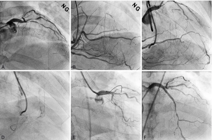
Because the retrograde approach requires an intercoronary channel between donor and recipient collateral artery, which enables to reach the distal CTO site retrogradely, CTO-op- erators should decide on the intercoronary channel to be ch- osen; epicardial collaterals or septal collaterals.5)6) Epicardial collaterals of a moderate size are present in about 50% of CTOs and those collaterals are often very tortuous. Also, bl- eeding secondary to perforation of an epicardial collateral co- uld be difficult to stop. By contrast, septal collaterals are more frequently seen and less tortuous than others.6) Moreover, the access route is shorter than the other routes and lies intramu- A
D
B
E
C
F
Fig. 1. Baseline left coronary angiography showed CTO lesion in the ostium of LAD (A). Right coronary angiography showed septal collateral from the RCA to LAD (B and C). Selective angiography using a microcatheter showed septal collateral (D). The guidewire was successfuly re- placed from the RCA to LAD (E). Final coronary angiography showed successful result (F). LAD: left anterior descending artery, CTO: chronic total occlusion, RCA: right coronary artery.
Sang Min Kim, et al. 749
scularly. Most importantly, only the septal channel is dilatable, therefore the septal channel is considered as the most suitable access route for a retrograde approach to CTOs, although the septal channel was dissected and complicated with peripro- cedural MI in our case.
Despite the retrograde approach being a novel technique for CTO lesions, it might potentially be accompanied with sev- eral unexpected complications; ischemic complications due to thrombus formation or from any damage to the collateral rou- tes, or dissection of the proximal part of the donor artery.7) In order to reduce this occurrence, excessive forceful attempts to cross a hydrophilic guidewire or a catheter through septal branch should be avoided and careful manipulation is re- quired.
In conclusion, this case suggests that the retrograde ap- proach using septal collateral as access route can cause myo- cardial infarction of the collateral territory and cardiac MRI can demonstrate this complication.
REFERENCES
1) Safian RD, McCabe CH, Sipperly ME, McKay RG, Baim DS. Initial
success and long-term follow-up of percutaneous transluminal coro- nary angioplasty in chronic total occlusions versus conventional ste- noses. Am J Cardiol 1988;61:G23-8.
2) Hamm CW, Kupper W, Kuck KH, Hofmann D, Bleifeld W. Recana- lization of chronic, totally occluded coronary arteries by new angio- plasty systems. Am J Cardiol 1990;66:1459-63.
3) Surmely JF, Tsuchikane E, Katoh O, et al. New concept for CTO re- canalization using controlled antegrade and retrograde subintimal tracking: the CART technique. J Invasive Cardiol 2006;18:334-8.
4) Chung SH, Kim MH, Yu LH, et al. Initial experience of retrograde wire approach in coronary chronic total occlusion intervention. Ko- rean Circ J 2009;39:228-35, doi: 10.4070/kcj.2009.39.6.228.
5) Surmely JF, Katoh O, Tsuchikane E, Nasu K, Suzuki T. Coronary sep- tal collaterals as an access for the retrograde approach in the percu- taneous treatment of coronary chronic total occlusions. Catheter Car- diovasc Interv 2007;69:826-32.
6) Werner GS, Ferrari M, Heinke S, et al. Angiographic assessment of collateral connections in comparison with invasively determined col- lateral function in chronic coronary occlusions. Circulation 2003;107:
1972-7.
7) Saito S. Different strategies of retrograde approach in coronary angio- plasty for chronic total occlusion. Catheter Cardiovasc Interv 2008;
71:8-19.
Fig. 2. Pre-procedural cardiac MRI showed no evidence of delayed enhancement (A). Post-procedural cardiac MRI showed new delayed en- huncement in the septal area (B). A: Pre-PCI: normal MRI. B: Post-PCI: microinfarction as indicated by circles. PCI: percutaneous coronary in- terrention.
A
B