322
D espite the strong antirestenotic efficacy of drug-eluting stents (DESs), the benefits of these devices are often atten- uated in patients with long or complex coronary artery lesions,
accompanied by an additional risk of adverse clinical out- comes.
1Furthermore, first-generation DESs are associated with delayed arterial healing and potential inflammation, as well as Background—Procedural and clinical outcomes still remain unfavorable for patients with long coronary lesions who
undergo percutaneous coronary intervention. The current study, therefore, evaluated 2 innovative drug-eluting stents for the management of long-lesion coronary artery disease.
Methods and Results—This randomized, multicenter, prospective trial, called the Long Drug-Eluting Stent (LONG-DES) V trial, compared the biodegradable polymer–based biolimus A9–eluting stent (BES) and the durable polymer–based platinum chromium everolimus-eluting stent (PtCr-EES) in 500 patients with long (≥25 mm) coronary lesions. The primary end point of the trial was in-segment late luminal loss at the 9-month angiographic follow-up. The BES and PtCr-EES groups had similar baseline characteristics, with a slightly shorter lesion length in the BES group versus the PtCr-EES group (29.24±12.17 versus 32.27±13.84 mm; P=0.016). In-segment late luminal loss was comparable between the 2 groups at the 9-month angiographic follow-up (BES, 0.14±0.38 versus PtCr-EES, 0.11±0.37 mm; difference, 0.031; 95% confidence interval, −0.053 to 0.091; P=0.03 for a noninferiority margin of 0.11, P=0.45 for superiority), as was in-stent late luminal loss (0.20±0.41 versus 0.24±0.38 mm;
P=0.29). The incidence of in-segment (6.1% versus 4.9%; P=0.63) and in-stent (3.7% versus 4.9%; P=0.59) binary restenosis was also similar between the groups. There was no significant between-group difference in the rate of composite outcome of death, myocardial infarction, and target vessel revascularization (41, 16.7% in BES versus 42, 16.5% in PtCr-EES; P=0.94).
Conclusions—BES and PtCr-EES implantation showed analogous angiographic and clinical outcomes for patients with de novo long coronary lesions.
Clinical Trial Registration—URL: http://www.clinicaltrials.gov. Unique identifier: NCT01186120.
(Circ Cardiovasc Interv. 2014;7:322-329.)
Key Words: angioplasty ◼ biolimus ◼ coronary artery disease ◼ everolimus ◼ stents
© 2014 American Heart Association, Inc.
Circ Cardiovasc Interv is available at http://circinterventions.ahajournals.org DOI: 10.1161/CIRCINTERVENTIONS.113.000841 Received September 5, 2013; accepted February 24, 2014.
From the Department of Cardiology (J.-Y. L., D.-W.P., Y.-H.K., J.-M.A., W.-J.K., S.-J.K., S.-W.L., C.W.L., S.-W.P., S.-J.P.) and Division of Biostatistics, Center for Medical Research and Information (S.-C.Y.), University of Ulsan College of Medicine, Asan Medical Center, Seoul, Korea; Department of Internal Medicine, Inje University Pusan Paik Hospital, Pusan, Korea (T.-H.Y.); Department of Internal Medicine, Kangwon National University Hospital, Chuncheon, Korea (B.-K.L.); Department of Internal Medicine, Soonchunhyang University Bucheon Hospital, Bucheon, Korea (N.-H.L.); Department of Internal Medicine, National Health Insurance Corporation Ilsan Hospital, Goyang, Korea (J.-Y.Y.); Department of Internal Medicine, Soonchunhyang University Hospital, Cheonan, Korea (W.-Y.S.); Department of Internal Medicine, Kyungpook National University Hospital, Daegu, Korea (H.S.P.);
Department of Internal Medicine, Daegu Catholic University Medical Center, Daegu, Korea (K.-S.K.); Department of Internal Medicine, Keimyung University Dongsan Medical Center, Daegu, Korea (S.H.H.); Department of Internal Medicine, Inje University Ilsan Paik Hospital, Goyang, Korea (S.Y.L.);
Department of Internal Medicine, Yeungnam University Medical Center, Daegu, Korea (J.-S.P.); Department of Internal Medicine, The Catholic University of Korea, Yeouido St. Mary’s Hospital, Seoul, Korea (Y.S.C.); Department of Internal Medicine, Kwangju Christian Hospital, Kwangju, Korea (S.U.L.);
and Department of Internal Medicine, The Catholic University of Korea, Daejeon St. Mary’s Hospital, Daejeon, Korea (S.-H.H.).
The Data Supplement is available at http://circinterventions.ahajournals.org/lookup/suppl/doi:10.1161/CIRCINTERVENTIONS.113.000841/-/DC1.
Correspondence to Seung-Jung Park, MD, Department of Cardiology, University of Ulsan College of Medicine, Cardiac Center, Asan Medical Center, 388-1 Poongnap-dong, Songpa-gu, Seoul 138-736, Korea. E-mail [email protected]
Comparison of Biolimus A9–Eluting (Nobori) and Everolimus-Eluting (Promus Element) Stents in Patients
With De Novo Native Long Coronary Artery Lesions
A Randomized Long Drug-Eluting Stent V Trial
Jong-Young Lee, MD, PhD; Duk-Woo Park, MD, PhD; Young-Hak Kim, MD, PhD;
Jung-Min Ahn, MD; Won-Jang Kim, MD, PhD; Soo-Jin Kang, MD, PhD; Seung-Whan Lee, MD, PhD;
Cheol Whan Lee, MD, PhD; Seong-Wook Park, MD, PhD; Sung-Cheol Yun, PhD;
Tae-Hyun Yang, MD, PhD; Bong-Ki Lee, MD, PhD; Nae-Hee Lee, MD, PhD; Joo-Young Yang, MD, PhD;
Won-Yong Shin, MD, PhD; Hun Sik Park, MD, PhD; Kee-Sik Kim, MD, PhD; Seung Ho Hur, MD, PhD;
Sung Yun Lee, MD, PhD; Jong-Seon Park, MD, PhD; Yun Seok Choi, MD, PhD;
Seung Uk Lee, MD, PhD; Sung-Ho Her, MD, PhD; Seung-Jung Park, MD, PhD
Coronary Interventions
by guest on February 11, 2016 http://circinterventions.ahajournals.org/
Downloaded from http://circinterventions.ahajournals.org/ by guest on February 11, 2016 Downloaded from http://circinterventions.ahajournals.org/ by guest on February 11, 2016 Downloaded from http://circinterventions.ahajournals.org/ by guest on February 11, 2016 Downloaded from http://circinterventions.ahajournals.org/ by guest on February 11, 2016 Downloaded from http://circinterventions.ahajournals.org/ by guest on February 11, 2016 Downloaded from http://circinterventions.ahajournals.org/ by guest on February 11, 2016 Downloaded from http://circinterventions.ahajournals.org/ by guest on February 11, 2016 Downloaded from http://circinterventions.ahajournals.org/ by guest on February 11, 2016 Downloaded from http://circinterventions.ahajournals.org/ by guest on February 11, 2016 Downloaded from
a propensity for late thrombosis, especially in high-risk lesions such as long coronary segments.
2Therefore, the development of newer-generation DESs that ensure both safety and efficacy has become a matter of intensive investigation. These newer- generation DESs use innovative stent platforms, polymers, and a variety of drugs. Several trials have indicated their potential advantage in interventions for long-lesion coronary artery dis- ease.
3,4Recently, several trials revealed that the outcomes of a newly developed biodegradable polymer–coated biolimus A9–eluting stent (BES) and a platinum chromium everolimus- eluting stent (PtCr-EES) were favorable.
1–5To date, no investigations have compared the benefits of the biodegradable polymer–based BES with the durable polymer PtCr-EES for the treatment of long coronary artery disease.
Because of their severity, long coronary artery lesions may be a practical target to assess the anticipated high antirestenotic efficacy and safety of these newer devices. Therefore, we con- ducted a prospective, multicenter, randomized study called the Long Drug-Eluting Stent (LONG-DES) V trial to evaluate the innovative BES and PtCr-EES for de novo native long coro- nary artery lesions.
Methods Study Design and Patient Population
The LONG-DES V trial was a prospective, randomized, single-blind, controlled study conducted in 14 major cardiac centers in South Korea between July 2010 and May 2012. The study protocol was ap- proved by an institutional review committee at each participating cen- ter and was conducted according to the principles of the Declaration of Helsinki regarding investigations on humans. All patients provided written, informed consent before participating in the trial. The spon- sors of this study contributed to the study design but had no role in the collection, monitoring, analysis, and interpretation of the data or in the preparation of the article.
The study consecutively enrolled 500 eligible patients, aged ≥18 years, with stable angina, unstable angina, non–ST-segment–eleva- tion myocardial infarction, or inducible ischemia. In addition, all pa- tients had ≥1 native long coronary lesion that was suitable for stent implantation. The inclusion criteria for angiographic eligibility were a target de novo lesion with a stenosis diameter ≥50%, visual ves- sel diameter ≥2.5 mm, visual lesion length ≥25 mm, and a planned total stent length ≥28 mm. The exclusion criteria were as follows:
acute ST-segment–elevation myocardial infarction (MI) necessitat- ing primary percutaneous coronary intervention (PCI) or cardio- genic shock; severely compromised ventricular dysfunction (ejection fraction <30%) or cardiogenic shock; allergy to antiplatelet drugs, heparin, stainless steel, contrast agents, biolimus, or everolimus; left main coronary artery disease (defined as >50% stenosis); renal dys- function (serum creatinine concentration ≥2.0 mg/dL) or dependence on dialysis; terminal illness with a life expectancy <1 year; active participation in another drug or device investigational study, without completion during the primary end point follow-up period; in-stent restenosis at the target vessel, with either a bare metal stent or a DES;
elective surgery planned within 6 months of the procedure, neces- sitating antiplatelet agent discontinuation; participation in a study on another coronary device; or inability to follow the study protocol.
Study Devices
Unlike certain drugs originally developed for other purposes and cur- rently used for DESs, biolimus A9 was specifically developed for local delivery to coronary arteries. Biolimus A9 is an integral com- ponent of the newly developed BES (Nobori, Terumo Corporation, Japan) with several unique features. The most important of these is its biodegradable polymer drug carrier coating (polylactic acid) found only on the abluminal stent surface. The BES has already shown promising clinical and angiographic outcomes.5–7
Newer PtCr-EESs (Promus Element, Boston Scientific, USA) use a durable, biocompatible, inert fluorocopolymer as the drug carrier.
These stents were developed to improve drug delivery, vessel con- formability, side branch access, radiopacity, radial strength, and frac- ture resistance. Like the BES, PtCr-EESs have also shown favorable clinical and angiographic outcomes in various trials.8,9
Randomization, Procedures, and Adjunct Pharmacotherapy
Patients who met the inclusion and exclusion criteria were random- ized 1:1 after diagnostic angiography and before PCI for treatment with BES or PtCr-EES by means of an interactive web response sys- tem. The randomization was performed via a central Internet-based allocation with stratification according to the participating center and blocked with random block sizes of 4 and 6. Patients, but not investi- gators, were unaware of the treatment assignment.
Stent implantation was performed according to standard tech- niques.10 The BES was available in diameters of 2.5, 2.75, 3.0, and 3.5 mm and in lengths of 14, 18, 24, and 28 mm, whereas the PtCr-EES was available in diameters of 2.5, 2.75, 3.0, 3.5, and 4.0 mm and in lengths of 12, 16, 20, 24, 28, 32, and 38 mm. In patients with multiple lesions who fulfilled the inclusion and exclusion criteria, the operator determined the hierarchy of the lesions and declared the target lesion for each patient before the procedure. The same randomly assigned stent was implanted in all lesions in patients requiring multilesion in- terventions, except when the assigned stent could not be inserted. In the latter case, crossover to another device was allowed. Full-lesion cover- age was attempted by implanting 1 or several stents without limitations.
Before or during the procedure, all patients received ≥200 mg as- pirin and a 300 to 600 mg loading dose of clopidogrel. Heparin was administered throughout the procedure to maintain an activated clot- ting time of ≥250 seconds. After the procedure, all patients received aspirin at a dosage of 100 mg/d indefinitely, as well as clopidogrel at a dosage of 75 mg/d for ≥12 months.
Study End Points and Definitions
The primary end point of the trial was in-segment late luminal loss within 9 months of the index procedure, defined as the difference
WHAT IS KNOWN
• Despite the strong antirestenotic efficacy of drug- eluting stents, the benefits of these devices are often attenuated in patients with long or complex coronary artery lesions.
• To date, there are no investigations comparing the use of the biodegradable polymer–based biolimus A9–eluting stent with the durable polymer platinum chromium everolimus-eluting stent for the treatment of long coronary artery disease.
WHAT THE STUDY ADDS
• In this prospective, multicenter, randomized trial in- volving patients with long coronary artery lesions, biolimus A9–eluting stent implantation was noninfe- rior to platinum chromium everolimus-eluting stent implantation as assessed by 9-month angiographic in-segment late luminal loss.
• Both stent platforms were associated with compa-
rable low rates of clinical end points at 12 months,
suggesting that both stents are equally effective in
the treatment of long coronary artery lesions.
324 Circ Cardiovasc Interv June 2014
between the minimal luminal diameter, assessed immediately after the procedure and at angiographic follow-up, measured within the margins, and at 5 mm proximal and distal to the stent. Secondary angiographic end points were in-stent and in-segment binary resteno- sis and in-stent late loss at 9 months. Secondary clinical end points included death, MI, ischemia-driven target lesion revascularization, ischemia-driven target vessel revascularization, stent thrombosis, a composite of major adverse cardiac events (ie, death, MI, and target vessel revascularization) within 12 months, and device success.
All deaths were considered to be from cardiac causes unless a non- cardiac cause could be identified. The diagnosis of MI was based on the presence of new Q waves in ≥2 contiguous leads on an ECG or an elevation in the creatine kinase (CK)–muscle-brain (MB) isoenzyme fraction or troponin I concentration >3× above the normal upper limit in ≥2 blood samples. Periprocedural MI was defined as an elevation of CK-MB >3× above the normal upper limit in ≥2 blood samples with a normal range in the baseline value within 48 hours of the procedure.
If the pre-PCI CK-MB values were above the normal upper limits, as in the case of patients initially presenting with acute MI, a CK-MB re- elevation ≥50% greater than the most recent preprocedure concentra- tion, with documentation that the values were stable or falling before PCI, was required for the diagnosis of periprocedural MI in this setting.
Revascularization of the target lesion and vessel was considered to be ischemia driven if (1) the diameter of the treated lesion (or vessel) was characterized by ≥50% stenosis, as assessed by quantitative coronary analysis accompanied by ischemic signs (ie, positive functional tests) or symptoms; or (2) the diameter of the target lesion (or vessel) was characterized by ≥70% stenosis, with or without documented isch- emia.11 Stent thrombosis was described as definite or probable accord- ing to the definitions set forth by the Academic Research Consortium.12 Device success was defined as a final stenosis of the vessel diameter
<30% by visual estimation after implantation of the assigned stent only.
Patient Follow-Up and Data Management
A 12-lead ECG was obtained for each patient, and serum concen- trations of CK and its MB isoenzyme were measured before stent- ing, 8 to 16 hours after the procedure, and again 18 to 24 hours after the procedure. Clinical follow-up visits were scheduled 30 days, 9 months, and 12 months after the procedure, and all eligible patients were asked to return for an angiographic follow-up 9 months after the procedure, or earlier, if anginal symptoms occurred. Figure 1 shows the flow of patients during follow-up.
Clinical, angiographic, procedural, and outcome data were col- lected by specialized personnel using a dedicated electronic case re- port form at the Clinical Data Management Center. Personnel were unaware of treatment assignments. All outcomes of interest were confirmed by source documentation collected at each of the 14 ma- jor cardiac centers and were centrally adjudicated by an independent Clinical Events Committee whose members were blinded to the as- signed stent. An independent Data and Safety Monitoring Board peri- odically reviewed the data to identify potential safety issues.
Quantitative Coronary Angiography
Coronary angiograms were digitally recorded at baseline, immedi- ately after the procedure, and at the 9-month angiographic follow-up.
Experienced assessors who were unaware of the identity of the al- located stent assessed the angiograms off-line in the Angiographic Core Laboratory (Asan Medical Center, Seoul, Korea) via a CASS V automated edge-detection system (Pie Medical Imaging, Maastricht, The Netherlands). All measurements were performed on angiograms recorded after the intracoronary administration of nitroglycerin.
Standard qualitative and quantitative analyses and definitions were used for the quantitative coronary angiographic analysis.13 The refer- ence diameter was determined by interpolation.
Figure 1. Patient flow and follow-up in the Long Drug-Eluting Stent (LONG-DES) V trial. No reliable data are available on the assessment criteria for patient eligibility.
All quantitative coronary angiographic analyses were performed within the stented segment (in-stent analysis) and over the entire seg- ment, including the stent and its 5-mm proximal and distal margins (in-segment analysis). Angiographic variables included absolute le- sion length, stent length, reference vessel diameter, minimum lumen diameter, percent diameter stenosis, binary restenosis rate, immediate gain, late loss, and patterns of recurrent restenosis. Binary restenosis was defined as ≥50% diameter stenosis on follow-up angiography.
The Mehran classification was used to quantitatively assess patterns of angiographic restenosis.14
Statistical Analysis
The primary objective of this study was to assess whether the angio- graphic outcomes of the biodegradable BES were comparable (ie, not inferior) with those of the durable PtCr-EES. To calculate the study sample size, an in-segment late luminal loss of 0.24±0.38 mm was assumed for the PtCr-EES, an assumption based on previously pub- lished results,15,16 because of a lack of specified data. Calculation of the study sample size was based on a margin of noninferiority for in- segment late luminal loss of 0.11 mm, which was equivalent to 40% of the assumed mean value (±SD) for late luminal loss of the PtCr-EES.
In each group, 180 patients were required to demonstrate noninferior- ity of the BES, as estimated by using an α level of 0.05 and a statisti- cal power of 80%. Furthermore, 500 patients (250 per group) were required to fulfill the primary end point based on the expectation that
≈30% of patients would not undergo follow-up angiography. Sample size was calculated by using PASS software (NCSS, Kaysville, UT).
All analyses were based on the intention-to-treat principle.
Differences between treatment groups were evaluated by using the Student t test for continuous variables and the χ2 or Fisher exact test for categorical variables. Cumulative event curves were generated by applying the Kaplan–Meier method. The noninferiority hypothesis
was statistically assessed via a Z test, in which the P values for non- inferiority were calculated to compare differences between groups with margins of noninferiority.17 Trial data were held by the Trial Coordination Center at the Asan Medical Center. Analyses were per- formed with SAS software, version 9.1 (SAS Institute, Cary, NC), by a statistical analyst who was unaware of the identity of the implanted stent. All P values were 2-sided, apart from those used for noninferi- ority testing of the primary end point.
Results
Baseline Characteristics and Procedural Results Between July 2010 and May 2012, 500 patients were randomized to receive PCI with the BES (n=245) or the PtCr-EES (n=255).
Baseline clinical characteristics, lesions, and procedural charac- teristics are shown in Tables 1 and 2. Most of these characteristics were similar between the BES group and the EES group, except for the length of stents implanted into the target lesion and the maximal pressure. The overall rate of device success was 99.8%, taking both groups of patients into account. Only 1 device failure with the allocated stent was observed in the BES group.
Table 1. Baseline Clinical Characteristics of Enrolled Patients*
Characteristics
BES (245 Patients)
PtCr-EES (255 Patients) P Value
Age, y 63.1±10.5 63.5±10.6 0.65
Male sex, n (%) 167 (68.2) 184 (72.2) 0.38
Body mass index 25.3±2.9 24.7±2.9 0.02
Diabetes mellitus, n (%) 79 (32.2) 89 (34.9) 0.33 Hypertension, n (%) 161 (65.7) 154 (60.4) 0.23 Hyperlipidemia, n (%) 131 (53.5) 145 (56.9) 0.36 Current smoker, n (%) 63 (25.7) 74 (29.0) 0.42 Family history of CAD, n (%) 11 (4.5) 22 (8.6) 0.16 Previous coronary angioplasty,
n (%)
16 (6.5) 26 (10.2) 0.15
Previous bypass surgery, n (%) 1 (0.4) 0 (0) 0.31
Previous MI, n (%) 6 (2.4) 11 (4.3) 0.32
Left ventricular ejection fraction, %
60.3±7.6 60.2±7.5 0.93
Multivessel disease, n (%) 124 (50.6) 140 (54.9) 0.37
Clinical indication, n (%) 0.80
Stable angina or silent ischemia
142 (58.0) 145 (56.9)
Unstable angina 68 (27.8) 74 (29.0)
NSTEMI 35 (14.3) 36 (14.1)
BES indicates biolimus A9–eluting stent; CAD, coronary artery disease;
MI, myocardial infarction; NSTEMI, non–ST-segment–elevation myocardial infarction; and PtCr-EES, platinum chromium everolimus-eluting stent.
*Plus–minus values are means±SDs. Data are provided for the intention-to- treat population.
Table 2. Baseline Lesions and Procedural Characteristics*
Characteristics
BES (245 Patients)
PtCr-EES (255 Patients) P Value Lesion characteristics
Target vessel, n (%) 0.72
Left anterior descending 159 (64.9) 171 (67.1) Left circumflex 33 (13.5) 32 (12.5) Right coronary 53 (21.6) 52 (20.4) TIMI flow grade=0 or 1, n (%) 25 (10.2) 21 (8.2) 0.46 Bifurcation lesion, n (%) 79 (32.2) 63 (24.7) 0.04
Thrombus, n (%) 11 (4.5) 8 (3.1) 0.48
Severe tortuosity, n (%) 3 (1.2) 4 (1.6) 0.93 Severe calcification, n (%) 26 (10.6) 32 (12.5) 0.27
Ulceration, n (%) 16 (6.5) 16 (6.2) 0.95
Procedural characteristics No. of stents used at the target
lesion (%)
0.20
One stent 115 (46.9) 117 (45.9)
Two stents 116 (47.3) 110 (43.1)
Three stents 13 (5.3) 26 (10.2)
Four stents 1 (0.4) 2 (0.8)
Mean 1.6±0.6 1.7±0.7 0.25
Length of stents used at the target lesion, mm
36.1±11.5 39.3±13.8 0.005 Average stent diameter at the
target lesion, mm
3.2±0.3 3.2±0.4 0.41
Maximal pressure, atm 12.1±4.0 13.5±3.5 <0.001 Direct stenting, n (%) 16 (6.5) 12 (4.7) 0.44 Postadditional balloon inflation 190 (77.6) 178 (69.8) 0.06 Intravascular ultrasound
guidance, n (%)
189 (77.1) 188 (73.7) 0.41
Glycoprotein IIb/IIIa antagonists, n (%)
5 (2.0) 6 (2.4) 0.99
BES indicates biolimus A9–eluting stent; PtCr-EES, platinum chromium everolimus-eluting stent; and TIMI, thrombolysis in myocardial infarction.
*Plus–minus values are means±SDs.
326 Circ Cardiovasc Interv June 2014
Angiographic Outcomes
Quantitative angiographic results at baseline, immediately after the procedure, and at the 9-month follow-up are shown in Table 3. Angiographic measurements of the lesions
before and immediately after the procedure were similar in the 2 groups. Follow-up angiography was performed in 164 patients (66.9%) in the BES group and 164 patients (64.3%) in the PtCr-EES group (P=0.54). The median duration of the angiographic follow-up was 9.1 months (interquartile range, 8.1–10.3 months). Patients undergo- ing angiographic follow-up were younger (P=0.003), more likely to have hyperlipidemia (P=0.002), less likely to have previous coronary angioplasty (P=0.026), more likely to have non–ST-segment–elevation myocardial infarction (P=0.037), and more likely to have undergone intravascular ultrasound-guided PCI (P=0.001) than those who did not return for angiographic follow-up (Tables I and II in the Data Supplement).
At the 9-month angiographic follow-up, in-segment late luminal loss (the primary study end point) of the BES was similar to that of the PtCr-EES (0.14±0.38 versus 0.11±0.37 mm; P for noninferiority=0.03, P for superiority=0.45;
Figure 2; Table 3). The rates of in-segment binary restenosis in the 2 groups were 6.1% and 4.9%, respectively (P=0.63), and the patterns of restenosis were similar between the groups (Table 4). The extent of in-stent late luminal loss (0.20±0.41 versus 0.24±0.38 mm; P=0.29) and the rates of in-stent binary restenosis (3.7% versus 4.9%; P=0.59) were also similar in the BES and PtCr-EES groups.
Clinical Outcomes
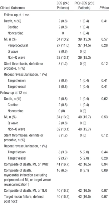
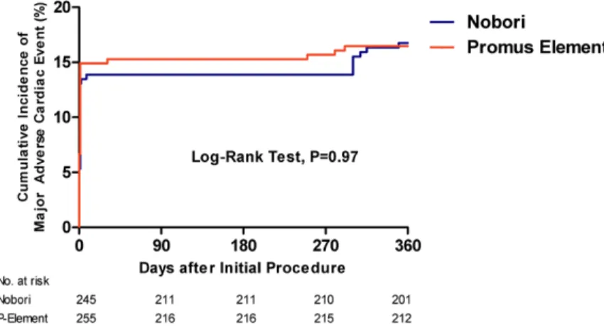
Major clinical events during follow-up are summarized in Table 5. All patients completed the 12-month clinical follow- up. At 1 and 12 months, the incidence of individual and com- posite clinical outcomes did not significantly differ between the 2 groups (Figure 3). The most common clinical event dur- ing the 12-month period was periprocedural MI, and no sig- nificant difference in its incidence was observed between the 2 groups (27, 11.0% in BES versus 37, 14.5% in PtCr-EES;
P=0.28). After excluding periprocedural MI, the incidence
of other composite outcomes also did not differ (16, 6.5% in BES versus 8, 3.1% in PtCr-EES; P=0.09). Periprocedural MI could not predict the future occurrence of adverse clinical out- comes (hazard ratio 2.45, 95% confidence interval, 0.20–4.81,
P=0.98 for major adverse cardiac events).Figure 2. Cumulative rates of in-segment late luminal loss at follow-up angiography. Late luminal loss was defined as the dif- ference between the minimal luminal diameter at the end of the procedure and the minimal luminal diameter at follow-up.
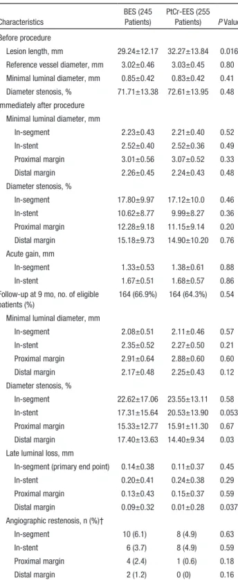
Table 3. Quantitative Angiographic Analysis*
Characteristics
BES (245 Patients)
PtCr-EES (255 Patients) P Value Before procedure
Lesion length, mm 29.24±12.17 32.27±13.84 0.016 Reference vessel diameter, mm 3.02±0.46 3.03±0.45 0.80 Minimal luminal diameter, mm 0.85±0.42 0.83±0.42 0.41 Diameter stenosis, % 71.71±13.38 72.61±13.95 0.48 Immediately after procedure
Minimal luminal diameter, mm
In-segment 2.23±0.43 2.21±0.40 0.52
In-stent 2.52±0.40 2.52±0.36 0.49
Proximal margin 3.01±0.56 3.07±0.52 0.33
Distal margin 2.26±0.45 2.24±0.43 0.48
Diameter stenosis, %
In-segment 17.80±9.97 17.12±10.0 0.46
In-stent 10.62±8.77 9.99±8.27 0.36
Proximal margin 12.28±9.18 11.15±9.14 0.20 Distal margin 15.18±9.73 14.90±10.20 0.76 Acute gain, mm
In-segment 1.33±0.53 1.38±0.61 0.88
In-stent 1.67±0.51 1.68±0.57 0.86
Follow-up at 9 mo, no. of eligible patients (%)
164 (66.9%) 164 (64.3%) 0.54
Minimal luminal diameter, mm
In-segment 2.08±0.51 2.11±0.46 0.57
In-stent 2.35±0.52 2.27±0.50 0.21
Proximal margin 2.91±0.64 2.88±0.60 0.60
Distal margin 2.17±0.48 2.25±0.43 0.12
Diameter stenosis, %
In-segment 22.62±17.06 23.55±13.11 0.58
In-stent 17.31±15.64 20.53±13.90 0.053
Proximal margin 15.33±12.77 15.91±11.30 0.67 Distal margin 17.40±13.63 14.40±9.34 0.03 Late luminal loss, mm
In-segment (primary end point) 0.14±0.38 0.11±0.37 0.45
In-stent 0.20±0.41 0.24±0.38 0.29
Proximal margin 0.13±0.43 0.15±0.37 0.59
Distal margin 0.09±0.32 0.01±0.28 0.037
Angiographic restenosis, n (%)†
In-segment 10 (6.1) 8 (4.9) 0.63
In-stent 6 (3.7) 8 (4.9) 0.59
Proximal margin 4 (2.4) 1 (0.6) 0.18
Distal margin 2 (1.2) 0 (0) 0.16
BES indicates biolimus A9–eluting stent; and PtCr-EES, platinum chromium everolimus-eluting stent.
*Plus–minus values are means±SDs.
†In 1 case in the PtCr-EES group, angiographic restenosis was detected concomitantly in the in-stent area and proximal to the margins.
During the 12-month period, only 3 BES-treated patients experienced stent thrombosis, with 2 definite cases observed at 1 and 10 days and 1 probable case observed at 8 days dur- ing the dual antiplatelet regimen. However, none of the EES- treated patients experienced stent thrombosis. Three patients initially presented with non–ST-segment–elevation myocar- dial infarction with high thrombotic burden and long lesion length. Procedures were performed without intravascular ultrasound guidance or the use of high-pressure adjunctive balloon dilatation.
Discussion
This prospective, randomized trial compared the efficacy of biodegradable polymer BES implantation with durable PtCr- EES implantation for the management of de novo native long coronary lesions. The BES and PtCr-EES demonstrated simi- lar rates of in-segment late luminal loss at the 9-month angio- graphic follow-up. Furthermore, both stent platforms exhibited similar outcomes for clinical end points at the 12-month fol- low-up visit, suggesting that both devices were equally effec- tive in the treatment of long coronary artery lesions.
Long coronary artery lesions comprise >20% of current PCI practice and are a major determinant of poor prognostic out- comes after stent implantation.
1,18,19Therefore, investigating the relative efficacy and safety of newer-generation DESs in these high-risk lesion subsets is of paramount clinical impor- tance. The LONG-DES III trial found that the sirolimus-elut- ing stent significantly lowered in-segment late loss compared with the EES, with a particularly beneficial effect at the proxi- mal margin.
19Recently, the LONG-DES IV trial demonstrated comparable angiographic and clinical outcomes for the siro- limus-eluting stent and a resolute zotarolimus-eluting stent.
3In keeping with the continuing concept of the previous LONG-DES trial series, we performed the LONG-DES V trial to evaluate 2 stents with different characteristics in terms of angiographic late loss and clinical outcomes. In several ran- domized trials, the BES showed fairly similar angiographic outcomes to those of the sirolimus-eluting stent, with compa- rable clinical outcomes.
20,21In those trials, in-stent late loss at
the 9-month angiographic follow-up was between 0.10 and 0.12 mm for a relatively short lesion length. However, the current LONG-DES V trial showed an in-stent late loss of 0.20 mm for lesions with a mean length of 30 mm. Recently, large clinical tri- als also showed good clinical outcomes for the BES compared with a cobalt chromium-EES or the sirolimus-eluting stent.
6,7,22After the launch of the PtCr-EES, the PLATINUM work- horse trial was devised to compare this new stent with a cobalt chromium-EES and found similar clinical outcomes for the 2 devices.
9In the PLATINUM Quantitative Coronary
Table 4. Angiographic Pattern of Restenosis*Characteristics
BES (245 Patients)
PtCr-EES (255
Patients) P Value Overall number of in-stent
restenosis cases
10 8 0.63
Focal, n (%)
IA (gap) 1 (10.0) 1 (12.5)
IB (margin) 2 (20.0) 0
IC (focal body) 4 (40.0) 5 (62.5)
ID (multifocal) 0 0
Diffuse, n (%)
II (intrastent) 1 (10.0) 1 (12.5) III (proliferative) 1 (10.0) 1 (12.5)
IV (total occlusion) 0 0
BES indicates biolimus A9–eluting stent; and PtCr-EES, platinum chromium everolimus-eluting stent.
*Classified according to the Mehran criteria.14
Table 5. Clinical Events at Follow-Up*
Clinical Outcomes
BES (245 Patients)
PtCr-EES (255 Patients) P Value Follow-up at 1 mo
Death, n (%) 2 (0.8) 1 (0.4) 0.41
Cardiac 2 (0.8) 1 (0.4)
Noncardiac 0 1 (0.4)
MI, n (%) 34 (13.9) 39 (15.3) 0.57
Periprocedural 27 (11.0) 37 (14.5) 0.28
Q wave 2 (0.8) 0 (0)
Non–Q wave 32 (13.1) 39 (15.3)
Stent thrombosis, definite or probable, n (%)
3 (1.2) 0 (0) 0.12
Repeat revascularization, n (%)
Target lesion 2 (0.8) 1 (0.4) 0.41
Target vessel 2 (0.8) 1 (0.4) 0.41
Follow-up at 12 mo
Death, n (%) 2 (0.8) 1 (0.4) 0.62
Cardiac 2 (0.8) 1 (0.4)
Noncardiac 0 (0) 0 (0)
MI, n (%) 34 (13.9) 40 (15.7) 0.53
Q wave 2 (0.8) 0 (0)
Non–Q wave 32 (13.1) 40 (15.7)
Stent thrombosis, definite or probable, n (%)
3 (1.2) 0 (0) 0.12
Repeat revascularization, n (%)
Target lesion 8 (3.3) 5 (2.0) 0.44
Target vessel 9 (3.7) 5 (2.0) 0.28
Composite of death, MI, or TVR† 41 (16.7) 42 (16.5) 0.94 Composite of death,
myocardial infarction excluding periprocedural MI, or target vessel revascularization†
16 (6.5) 8 (3.1) 0.09
Composite of death, MI, or TLR 40 (16.3) 42 (16.5) 0.97 Target lesion failure, defined
post hoc‡
40 (16.3) 42 (16.5) 0.97
BES indicates biolimus A9–eluting stent; MI, myocardial infarction;
PtCr-EES, platinum chromium everolimus-eluting stent; TLR, target lesion revascularization; and TVR target vessel revascularization.
*Percentages are from the intention-to-treat analysis. P values were calculated by using the χ2 test or Fisher exact test, as appropriate.
†Prespecified major adverse cardiac events were defined as a composite of all-cause death, MI, and ischemia-driven TVR.
‡Target lesion failure, defined post hoc, was a composite of death from cardiac causes, any MI (not clearly attributable to a nontarget vessel), or ischemia-driven TLR.
328 Circ Cardiovasc Interv June 2014
Angiography (QCA) substudy, the efficacy end point of angi- ographic in-stent late luminal loss was 0.17±0.25 mm at 9 months using the PtCr-EES, with a mean lesion length of 15.4 mm. These results are in agreement with previously reported results for the cobalt chromium-EES.
8,23–25In our long lesion series (mean lesion length, 32.27 mm), in-stent late loss was 0.24±0.38 mm for the PtCr-EES, providing the first evidence of angiographic outcomes for the long lesion subset.
To the best of our knowledge, our investigation provides the first comparison of angiographic outcomes for 2 novel DES designs in the treatment of long-lesion coronary artery dis- ease. The current study showed that both angiographic and clinical outcomes using the biodegradable polymer–based BES and the biocompatible inert PtCr-EES were similar for complex coronary lesions. In addition, even in such complex lesions, the majority of events occurred shortly after stent- ing; thus, the standard use of a 6-month dual antiplatelet regi- men could be effective in stable clinical situations with the advanced generation of DESs.
However, because this trial was powered to detect signifi- cant differences for angiographic surrogate markers but not clinical end points, our findings should be evaluated in larger clinical trials with clinical end points as the primary outcomes.
Certain issues associated with this trial should be consid- ered. First, although we assessed angiographic outcomes, especially in-segment late loss, the trial was not equipped to assess clinical outcomes. Although angiographic findings can be regarded as surrogate markers for clinical outcomes, they do have limitations. Larger, long-term clinical comparative studies are, therefore, required to compare the efficacy of the BES and the PtCr-EES further. An additional limitation of our study was the relatively short follow-up period of 12 months.
Different types of polymers used to construct novel DESs will affect the long-term safety and efficacy of the stent. Therefore, a longer follow-up period is essential to confirm the continuing durability of the newer devices. Third, the angiographic follow- up rate in the current trial was only 65.6%, which was lower than the protocol-based estimated rate. A nonangiographic follow-up rate could be high and seriously compromise the consistency and validity of the results. Fourth, this trial was not based on an all-comer design, which would engender selection bias, such as a relatively lower complex lesion subset. Fifth, data from the different participation centers and operators were not independently analyzed, which could potentially lead
to skewed outcomes. Finally, there were some imbalances in baseline and procedural characteristics, as exemplified by dif- ferences in lesion length, total stent length, maximal pressure, and the use of postadditional balloon inflation for achieving a similar poststent lumen diameter. Furthermore, the length of available stents was different between the groups, and these differences were too great to make a distorted conclusion. In addition, we assumed that the sample size was not based on a clinical outcome, which could, therefore, lead to an underesti- mation because of inadequate statistical power; larger clinical trials with sufficient statistical power for clinical outcomes are required. Nevertheless, considering the directionality of these potential effects, our overall findings are not likely to change.
In conclusion, implantation of a biodegradable polymer–
coated BES versus a durable polymer–coated PtCr-EES yielded comparable angiographic outcomes in patients with native de novo long coronary artery lesions, without signifi- cant differences in death rates, MI, angiographic restenosis, or stent thrombosis during a 12-month follow-up.
Sources of Funding
This study was supported by funds from the Korea Healthcare Technology Research and Development Project, Ministry of Health and Welfare (A120711), and CardioVascular Research Foundation, Seoul, Korea. The study was also supported by a grant from Terumo Korea, Seoul, Korea, and Terumo, Tokyo, Japan.
Disclosures
Dr S.-J. Park reports receiving consulting and lecture fees from Terumo and Boston Scientific and research grant support from Terumo and Boston Scientific; Dr Y.-H. Kim reports receiving lecture fees from Boston Scientific; Dr S.-J. Kang reports receiving lecture fees from Boston Scientific; Dr S.-W. Lee reports receiving lecture fees from Terumo and Boston Scientific; and Dr S.-W. Park reports receiving research grant support from Boston Scientific. The other authors report no conflicts.
References
1. Kastrati A, Dibra A, Mehilli J, Mayer S, Pinieck S, Pache J, Dirschinger J, Schömig A. Predictive factors of restenosis after coronary implantation of sirolimus- or paclitaxel-eluting stents. Circulation. 2006;113:2293–2300.
2. Suh J, Park DW, Lee JY, Jung IH, Lee SW, Kim YH, Lee CW, Cheong SS, Kim JJ, Park SW, Park SJ. The relationship and threshold of stent length with regard to risk of stent thrombosis after drug-eluting stent implanta- tion. JACC Cardiovasc Interv. 2010;3:383–389.
3. Ahn JM, Park DW, Kim YH, Song H, Cho YR, Kim WJ, Lee JY, Kang SJ, Lee SW, Lee CW, Park SW, Yun SC, Han S, Lee SY, Lee BK, Cho JH, Yang TH, Lee NH, Yang JY, Park JS, Shin WY, Kim MH, Bae JH,
Figure 3. Kaplan–Meier 12-month actuarial inci- dence of major adverse cardiac events. Major adverse cardiac events were defined as the com- posite of death, myocardial infarction, or ischemia- driven target vessel revascularization.
Kim MK, Yoon J, Park SJ. Comparison of resolute zotarolimus-eluting stents and sirolimus-eluting stents in patients with de novo long coronary artery lesions: a randomized LONG-DES IV trial. Circ Cardiovasc Interv.
2012;5:633–640.
4. Park DW, Kim YH, Song HG, Ahn JM, Kim WJ, Lee JY, Kang SJ, Lee SW, Lee CW, Park SW, Yun SC, Seung KB, Yang TH, Lee SG, Lee JH, Seong IW, Cheong SS, Lee BK, Lee NH, Lee SW, Lee SW, Lee K, Kim HS, Jeon DS, Kim MK, Nah DY, Tahk SJ, Park SJ. Comparison of everolimus- and sirolimus-eluting stents in patients with long coronary artery lesions: a randomized LONG-DES-III (Percutaneous Treatment of LONG Native Coronary Lesions With Drug-Eluting Stent-III) Trial. JACC Cardiovasc Interv. 2011;4:1096–1103.
5. Chevalier B, Silber S, Park SJ, Garcia E, Schuler G, Suryapranata H, Koolen J, Hauptmann KE, Wijns W, Morice MC, Carrie D, van Es GA, Nagai H, Detiege D, Paunovic D, Serruys PW. Randomized compari- son of the nobori biolimus a9-eluting coronary stent with the taxus lib- erte paclitaxel-eluting coronary stent in patients with stenosis in native coronary arteries: The nobori 1 trial--phase 2. Circ Cardiovasc Interv.
2009;2:188–195.
6. Christiansen EH, Jensen LO, Thayssen P, Tilsted HH, Krusell LR, Hansen KN, Kaltoft A, Maeng M, Kristensen SD, Bøtker HE, Terkelsen CJ, Villadsen AB, Ravkilde J, Aarøe J, Madsen M, Thuesen L, Lassen JF;
Scandinavian Organization for Randomized Trials with Clinical Outcome (SORT OUT) V Investigators. Biolimus-eluting biodegradable polymer- coated stent versus durable polymer-coated sirolimus-eluting stent in unselected patients receiving percutaneous coronary intervention (SORT OUT V): a randomised non-inferiority trial. Lancet. 2013;381:661–669.
7. Meredith IT, Whitbourn R, Scott D, El-Jack S, Zambahari R, Stone GW, Teirstein PS, Starzyk RM, Allocco DJ, Dawkins KD. PLATINUM QCA:
a prospective, multicentre study assessing clinical, angiographic, and in- travascular ultrasound outcomes with the novel platinum chromium thin- strut PROMUS Element everolimus-eluting stent in de novo coronary stenoses. EuroIntervention. 2011;7:84–90.
8. Smits PC, Hofma S, Togni M, Vázquez N, Valdés M, Voudris V, Slagboom T, Goy JJ, Vuillomenet A, Serra A, Nouche RT, den Heijer P, van der Ent M. Abluminal biodegradable polymer biolimus-eluting stent versus du- rable polymer everolimus-eluting stent (COMPARE II): a randomised, controlled, non-inferiority trial. Lancet. 2013;381:651–660.
9. Stone GW, Teirstein PS, Meredith IT, Farah B, Dubois CL, Feldman RL, Dens J, Hagiwara N, Allocco DJ, Dawkins KD. A prospective, randomized evaluation of a novel everolimus-eluting coronary stent: the PLATINUM (a Prospective, Randomized, Multicenter Trial to Assess an Everolimus- Eluting Coronary Stent System [PROMUS Element] for the Treatment of Up to Two de Novo Coronary Artery Lesions) trial. J Am Coll Cardiol.
2011;57:1700–1708.
10. Levine GN, Bates ER, Blankenship JC, Bailey SR, Bittl JA, Cercek B, Chambers CE, Ellis SG, Guyton RA, Hollenberg SM, Khot UN, Lange RA, Mauri L, Mehran R, Moussa ID, Mukherjee D, Nallamothu BK, Ting HH. 2011 ACCF/AHA/SCAI Guideline for Percutaneous Coronary Intervention: executive summary: a report of the American College of Cardiology Foundation/American Heart Association Task Force on Practice Guidelines and the Society for Cardiovascular Angiography and Interventions. Circulation. 2011;124:2574–2609.
11. Park DW, Kim YH, Yun SC, Kang SJ, Lee SW, Lee CW, Park SW, Seong IW, Lee JH, Tahk SJ, Jeong MH, Jang Y, Cheong SS, Yang JY, Lim DS, Seung KB, Chae JK, Hur SH, Lee SG, Yoon J, Lee NH, Choi YJ, Kim HS, Kim KS, Kim HS, Hong TJ, Park HS, Park SJ. Comparison of zo- tarolimus-eluting stents with sirolimus- and paclitaxel-eluting stents for coronary revascularization: the ZEST (comparison of the efficacy and safety of zotarolimus-eluting stent with sirolimus-eluting and paclitaxel- eluting stent for coronary lesions) randomized trial. J Am Coll Cardiol.
2010;56:1187–1195.
12. Laskey WK, Yancy CW, Maisel WH. Thrombosis in coronary drug-elut- ing stents: report from the meeting of the Circulatory System Medical Devices Advisory Panel of the Food and Drug Administration Center
for Devices and Radiologic Health, December 7-8, 2006. Circulation.
2007;115:2352–2357.
13. Lansky AJ, Dangas G, Mehran R, Desai KJ, Mintz GS, Wu H, Fahy M, Stone GW, Waksman R, Leon MB. Quantitative angiographic methods for appropriate end-point analysis, edge-effect evaluation, and prediction of recurrent restenosis after coronary brachytherapy with gamma irradiation.
J Am Coll Cardiol. 2002;39:274–280.
14. Mehran R, Dangas G, Abizaid AS, Mintz GS, Lansky AJ, Satler LF, Pichard AD, Kent KM, Stone GW, Leon MB. Angiographic patterns of in-stent restenosis: classification and implications for long-term outcome.
Circulation. 1999;100:1872–1878.
15. Kim YH, Park SW, Lee SW, Park DW, Yun SC, Lee CW, Hong MK, Kim HS, Ko JK, Park JH, Lee JH, Choi SW, Seong IW, Cho YH, Lee NH, Kim JH, Chun KJ, Park SJ; Long-DES-II Study Investigators. Sirolimus- eluting stent versus paclitaxel-eluting stent for patients with long coronary artery disease. Circulation. 2006;114:2148–2153.
16. Grube E, Hauptmann KE, Buellesfeld L, Lim V, Abizaid A. Six-month results of a randomized study to evaluate safety and efficacy of a Biolimus A9 eluting stent with a biodegradable polymer coating. EuroIntervention.
2005;1:53–57.
17. Chow SC, Liu JP. Design and Analysis of Clinical Trials. 2nd ed. New York, NY: John Wiley and Son; 2004.
18. Serruys PW, Silber S, Garg S, van Geuns RJ, Richardt G, Buszman PE, Kelbaek H, van Boven AJ, Hofma SH, Linke A, Klauss V, Wijns W, Macaya C, Garot P, DiMario C, Manoharan G, Kornowski R, Ischinger T, Bartorelli A, Ronden J, Bressers M, Gobbens P, Negoita M, van Leeuwen F, Windecker S. Comparison of zotarolimus-eluting and everolimus-elut- ing coronary stents. N Engl J Med. 2010;363:136–146.
19. Romagnoli E, Godino C, Ielasi A, Gasparini G, Tzifos V, Sciahbasi A, Lioy E, Presbitero P, Colombo A, Sangiorgi G. Resolute Italian study in all comers: immediate and one-year outcomes. Catheter Cardiovasc Interv.
2012;79:567–574.
20. Kadota K, Muramatsu T, Iwabuchi M, Saito S, Hayashi Y, Ikari Y, Nanto S, Fujii K, Inoue N, Namiki A, Kimura T, Mitsudo K. Randomized com- parison of the Nobori biolimus A9-eluting stent with the sirolimus-elut- ing stent in patients with stenosis in native coronary arteries. Catheter Cardiovasc Interv. 2012;80:789–796.
21. Ostojic M, Sagic D, Beleslin B, Jung R, Perisic Z, Jagic N, Nedeljkovic M, Mangovski L, Milosavljevic B, Stojkovic S, Orlic D, Antonic Z, Miloradovic V, Topic D, Paunovic D. First clinical comparison of Nobori -Biolimus A9 eluting stents with Cypher- Sirolimus eluting stents:
Nobori Core nine months angiographic and one year clinical outcomes.
EuroIntervention. 2008;3:574–579.
22. Stefanini GG, Kalesan B, Serruys PW, Heg D, Buszman P, Linke A, Ischinger T, Klauss V, Eberli F, Wijns W, Morice MC, Di Mario C, Corti R, Antoni D, Sohn HY, Eerdmans P, van Es GA, Meier B, Windecker S, Jüni P. Long-term clinical outcomes of biodegradable polymer biolimus-elut- ing stents versus durable polymer sirolimus-eluting stents in patients with coronary artery disease (LEADERS): 4 year follow-up of a randomised non-inferiority trial. Lancet. 2011;378:1940–1948.
23. Serruys PW, Ong AT, Piek JJ, Neumann FJ, van der Giessen WJ, Wiemer M, Zeiher A, Grube E, Haase J, Thuesen L, Hamm C, Otto-Terlouw PC.
A randomized comparison of a durable polymer Everolimus-eluting stent with a bare metal coronary stent: the SPIRIT first trial. EuroIntervention.
2005;1:58–65.
24. Serruys PW, Ruygrok P, Neuzner J, Piek JJ, Seth A, Schofer JJ, Richardt G, Wiemer M, Carrié D, Thuesen L, Boone E, Miquel-Herbert K, Daemen J. A randomised comparison of an everolimus-eluting coronary stent with a paclitaxel-eluting coronary stent: the SPIRIT II trial. EuroIntervention.
2006;2:286–294.
25. Stone GW, Midei M, Newman W, Sanz M, Hermiller JB, Williams J, Farhat N, Mahaffey KW, Cutlip DE, Fitzgerald PJ, Sood P, Su X, Lansky AJ; SPIRIT III Investigators. Comparison of an everolimus-eluting stent and a paclitaxel-eluting stent in patients with coronary artery disease: a randomized trial. JAMA. 2008;299:1903–1913.